As healthcare practitioners who primarily see problems in the human brain, it is incumbent on us to ensure that foetuses' brains are protected as best as humanly possible, especially from iatrogenic h...
A paediatrician telephoned me about a nine-year-old who had a generalized tonic-clonic seizure in sleep, wanting to know whether an MRI and EEG should be requested. The child is intellectually and dev...
Herewith another example:
The above looks a little like a sharp and slow wave at F7-M1-F3-FP1.
Then look at it on the standard AP bipolar montage; see below.

It looks far less conspicu...
The patient had an episode of amnesia yesterday. He recalls seeing flashes of multi-coloured "circles and squiggles on the left" and then recalls telephoning his wife, who was not at home. He has no r...
Re this post, Frontal lobe seizures versus functional seizures? Teenager
A colleague asks, "could this be phantom spike-and-wave?"
The short answer is no. Why? Phantom spike-and-wave (also known as...
When reading on the common average montages, I am often reminded of the quote by WS Gilbert, "Things are seldom what the seem".
Take a look at the following page.
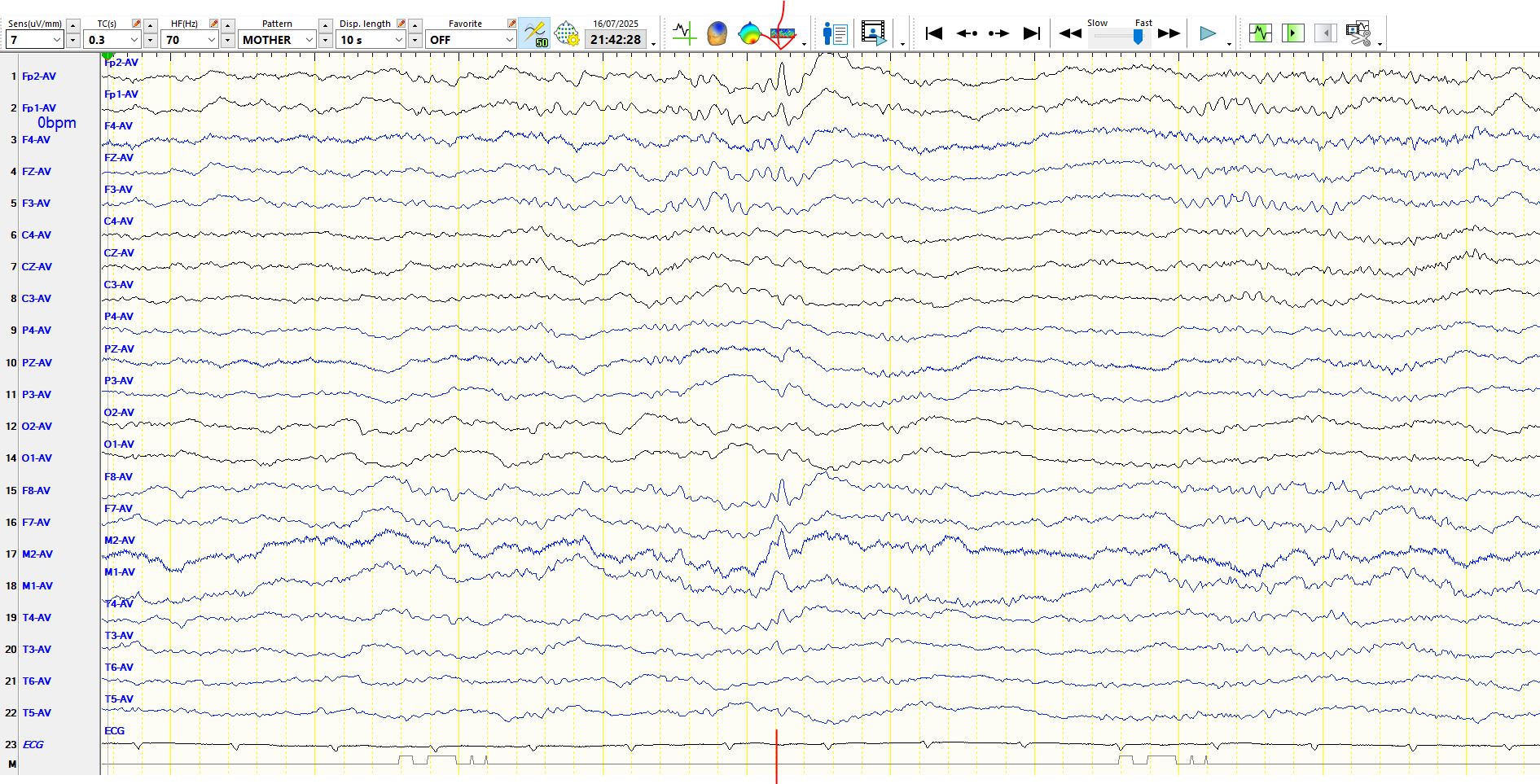
The discharge correspond...
Part of the vaccine against the (false positive) spike protein on the EEG is the use of amplitude as a criterion. However, no criterion is absolute, but in disregarding it you need to be very, very, v...
The asymmetry of the following rhythms during transient arousal resembles a seizure at T4-T6. The following are three consecutive pages.


In contradistinction to a seizure, the above...
The EEG is normal during wakefulness. What do you make of the following discharges during sleep?
The epoch is taken from sleep:
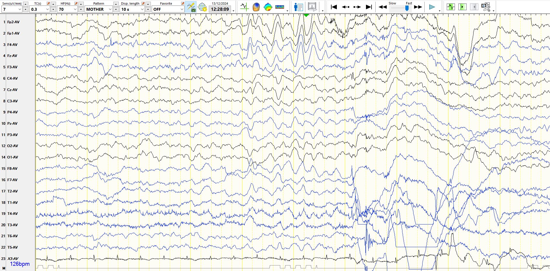
Here is the above page, represented on the anterior-posterior bi...
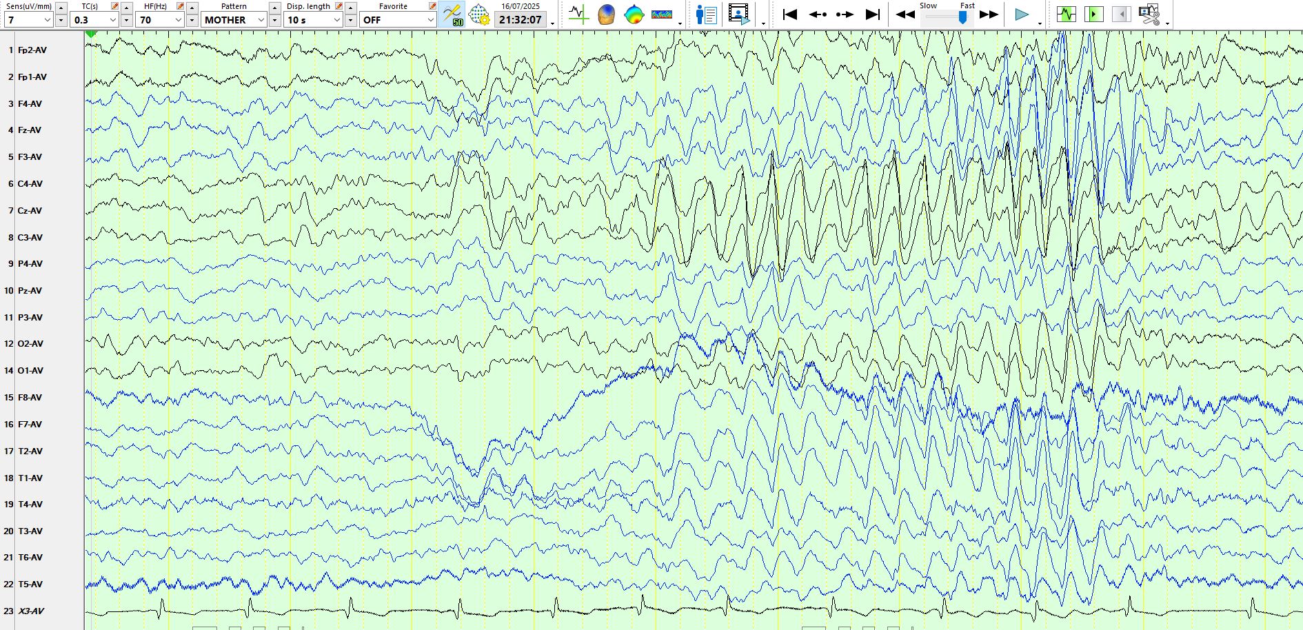
What you make of the following three consecutive pages of the EEG?
Figure 1.
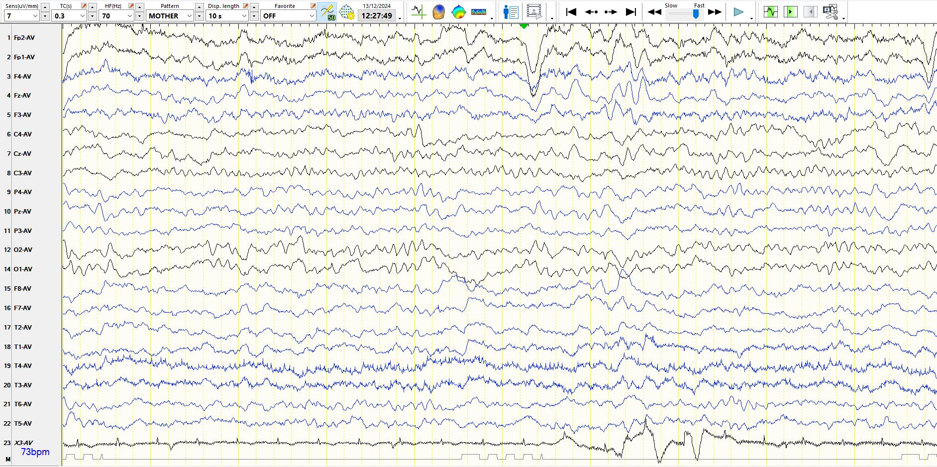
Figure 2: This follows the previous page.

Figure 3.
Here are a few other pages from the sam...
If you want to start an argument, wading into the territory of triphasic waves is sure to have the desired effect! Some authors suggest that spikes and triphasic waves rarely coexist, but I doubt that...