
Six years old, encephalopathy?
Jul 17, 2025What you make of the following three consecutive pages of the EEG?
Figure 1.
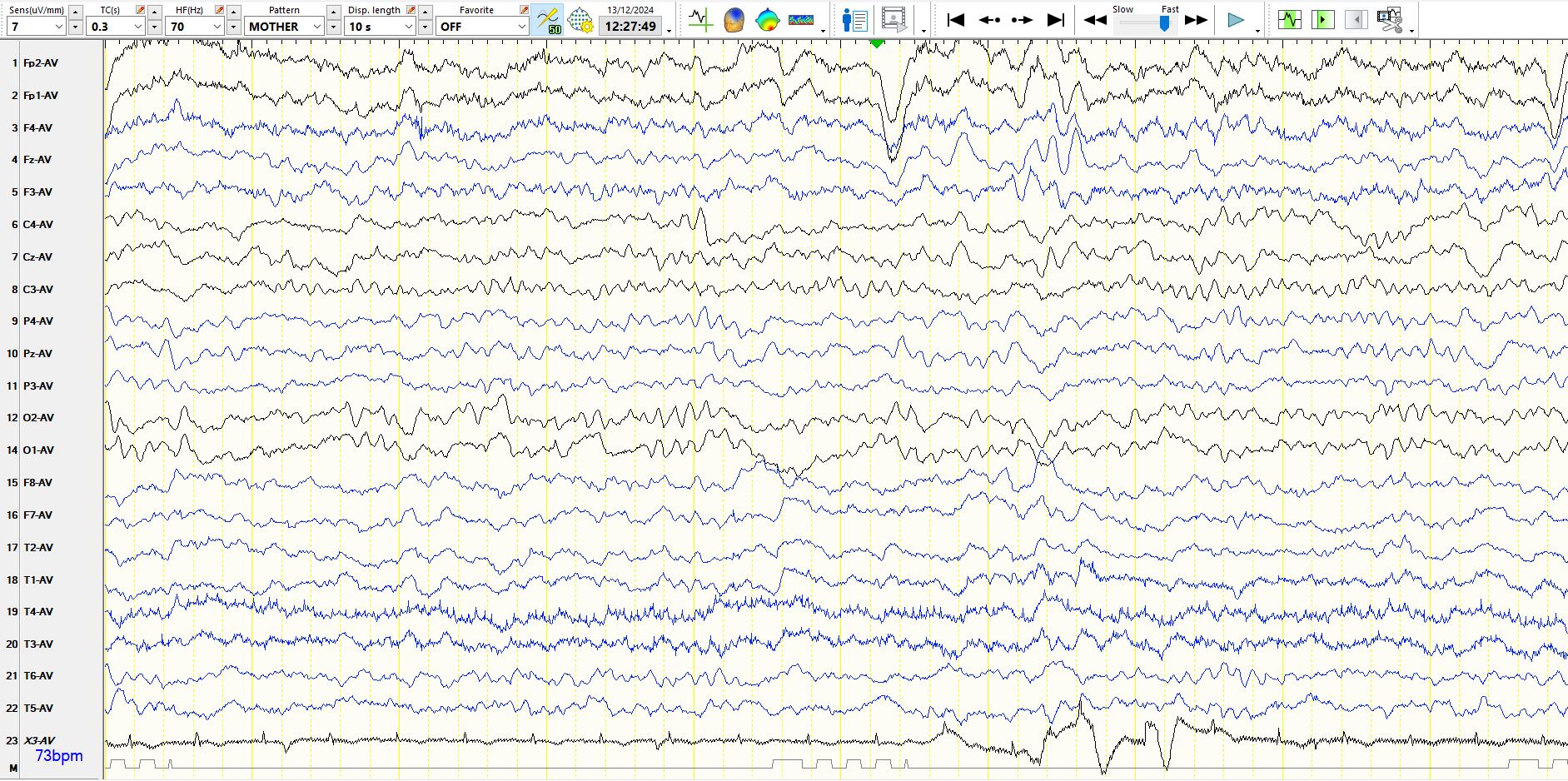
Figure 2: This follows the previous page.

Figure 3.
Here are a few other pages from the same patient's EEG:
Figure 4:
Have a careful look at CZ in the above image, as well as the second page from the top. What do you make of this?
Figure 5: Coronal montage

Elsewhere in the recording:
Figure 6:
In the image at the top of this page the patient is awake, with well-developed alpha rhythms over the occipital and parietal regions. During the last three seconds of the first page, occipital alpha becomes less conspicuous and parietal alpha become slightly more conspicuous, synchronous with the appearance of slower frequencies in the frontal and temporal regions, indicating early drowsiness.
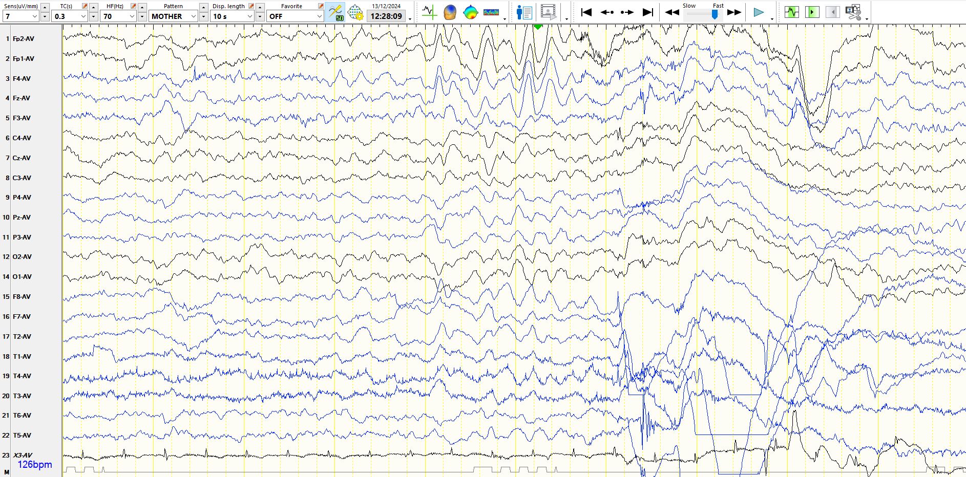
In figure 2 there is brief arousal, with the fleeting appearance of alpha rhythms over the occipital regions, followed by a second eyeblink, which heralds the end of the alpha rhythms. Following this eyeblink there is a mild diminution in the abundance of EMG, there is no movement and diffuse theta waves predominate. If anything, the background frequencies progressively slow subtly during the last seven seconds of this page. This is a sure sign that the patient is becoming drowsier.
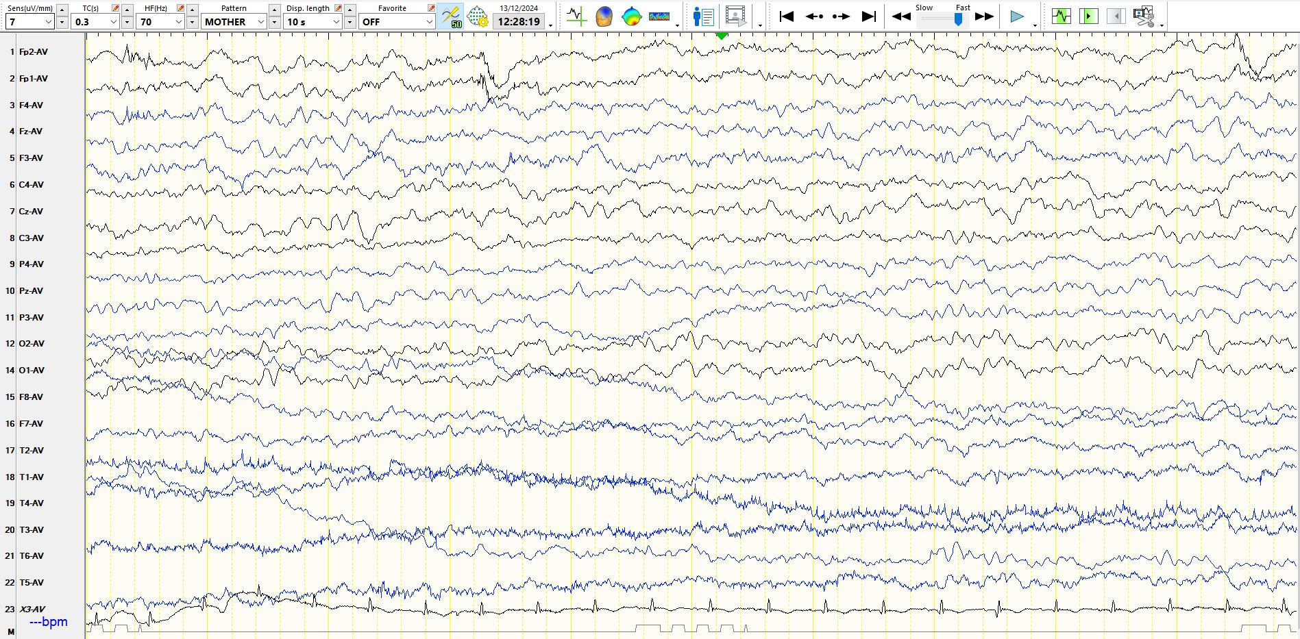
In figure 3 there is further slowing of the background rhythms, followed by a burst of generalised, bi-synchronous theta waves, characteristic of hypnagogic hypersynchrony. This is physiologically normal at this age. Patient moves at the end of this burst, indicating arousal and this is followed by an eye-blink.
In figure 4, as in figure 2, there are some unusual theta waves at CZ, in addition to the physiological waveforms in this region; the unusual waveforms are not present in contiguous electrodes. At times, it is difficult to spot electrode artifacts on the common average montage. In figure 5 the electrode artifact at CZ is apparent. As mentioned in previous posts, this is one of the weaknesses of the common average montage; it is considerably easier to mistake electrode artifacts for physiological and pathological waves, including spikes. You have been warned!
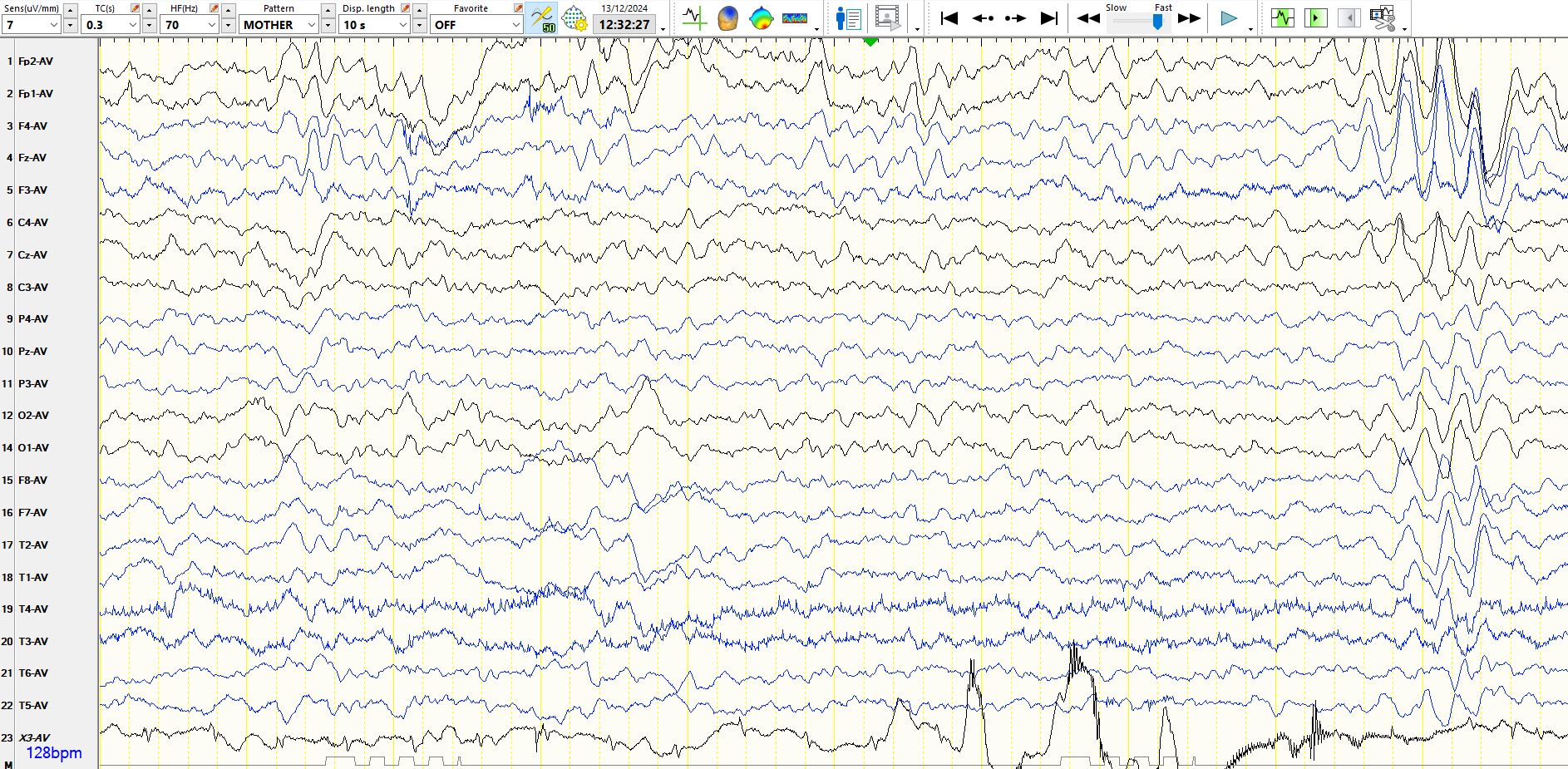
Figure 6 demonstrates another example of paroxysmal hypnagogic hypersynchrony.
Conclusion? The EEG is normal.