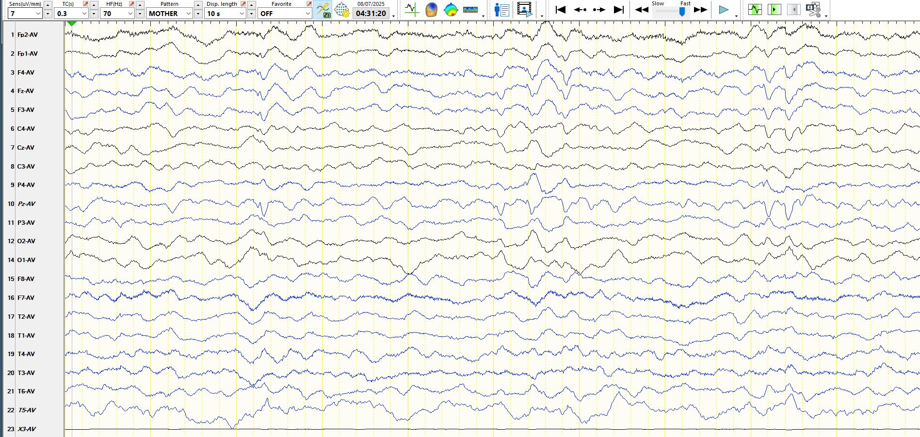
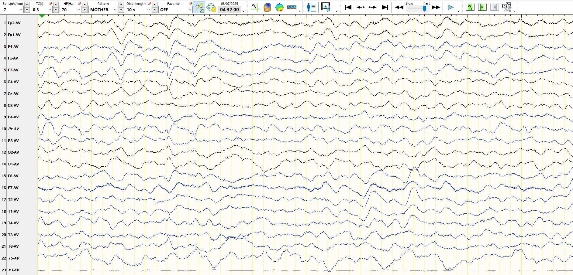
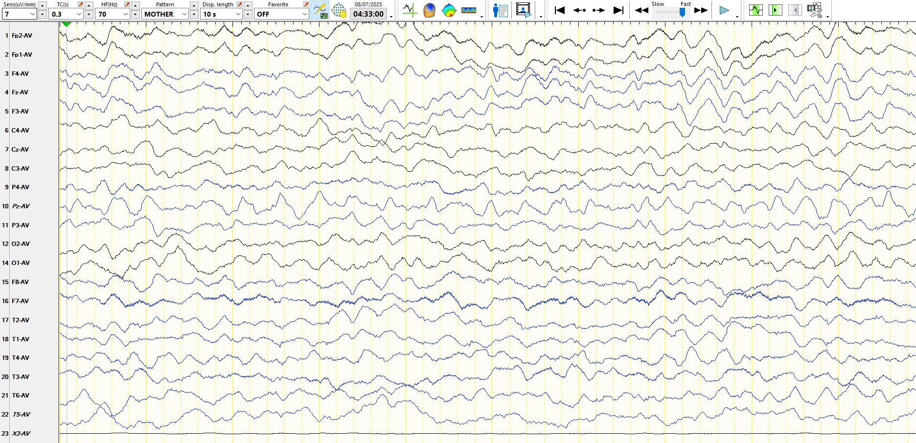
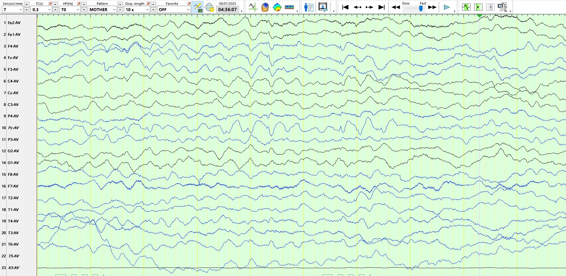
Parietal spike and wave versus generalized spike and wave?
Jul 08, 2025Have a look at the following few pages from an EEG of a patient who is asleep. Try to come to a conclusion before reading the comments at the bottom.
.
Page two:
Page 3:
Page 4:
Page 5:
Page 6:
Page 7:
Page 8:
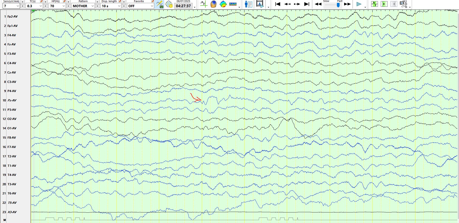
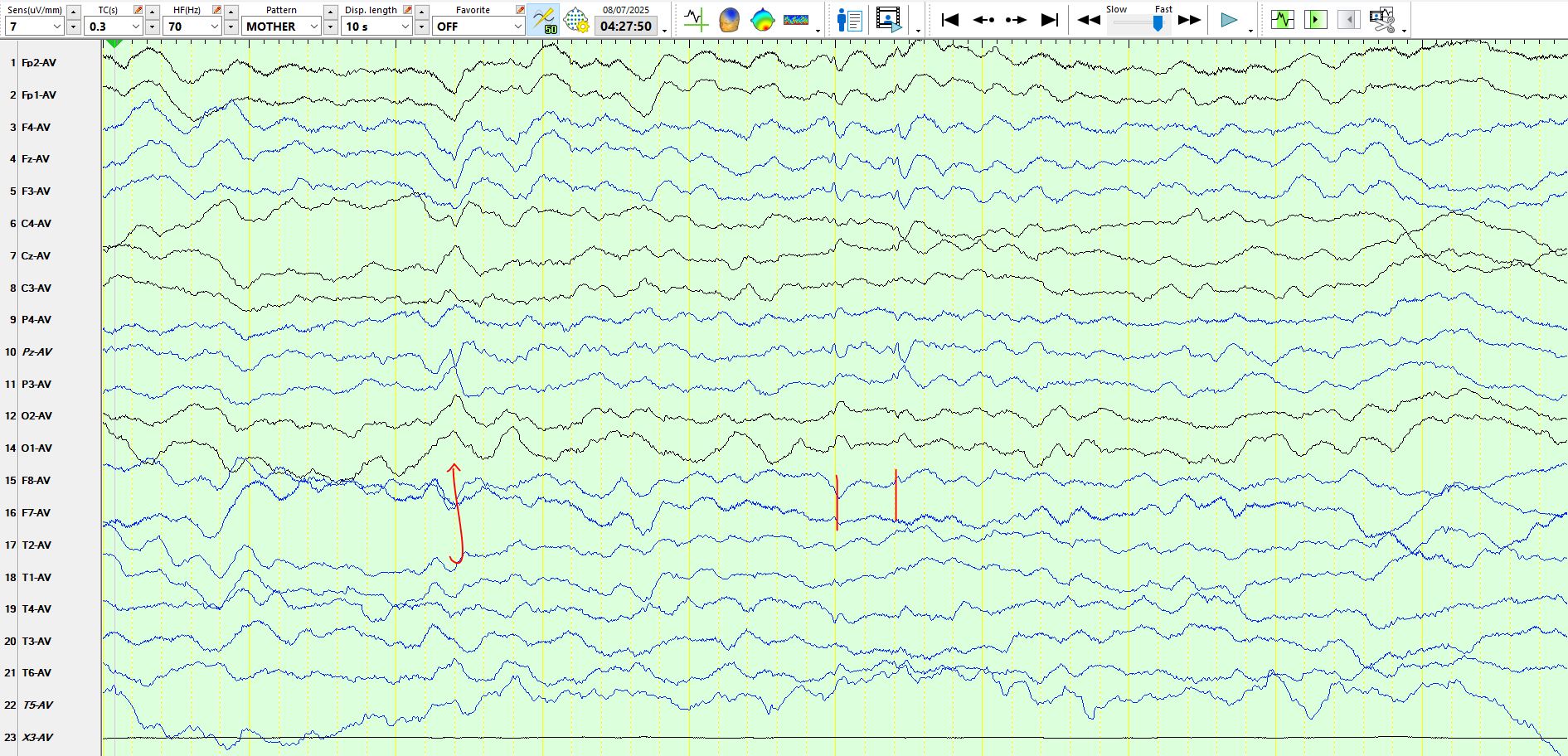
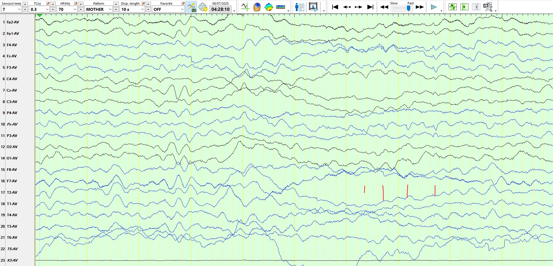
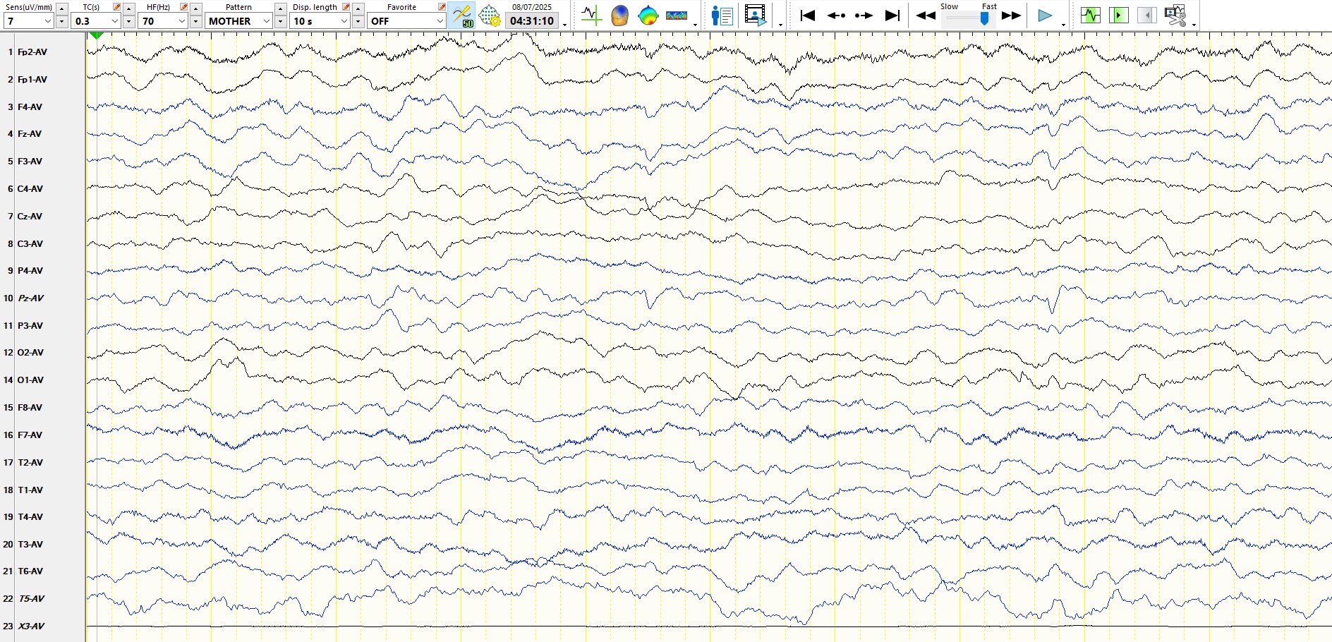
In each of the above pages notice that the spike and wave-like discharge in no instance resembles its "neighbours", P4 and P3. In the above pages, Pz is generally opposite in polarity to P3, while PZ most closely resembles FZ, F4, F3 and C4. Hence, PZ has been transposed, almost certainly with CZ. The patient is asleep and hence these waves likely are vertex waves. This example shows you how closely vertex waves may resemble spike and wave. Just for good measure, in Fig 7 there are a train of broader vertex waves at "PZ", which in reality must be CZ, as these waveforms always appear largely, if not exclusively at CZ; these are unmistakably vertex waves, and this proves that PZ is CZ! In Figure 3, the waves at CZ in the 3rd second of the recording resemble those at O1 and O2, settling the issue that CZ is in fact PZ. Similar analyses of other waves on different pages lead to the same conclusions.
After reading the EEG, as I always do when this occurs, I telephoned the technologist to ask him to confirm the transposition, but the electrodes had already been removed. Transpositions are very common in my experience; these relate to human error in setting up the patient, electrodes pulling out of headboxes and being put back incorrectly by patients, nurses and technologists and fading labels on the electrode wires causing people to plug the wires in in an incorrect manner. These transpositions keep me on my toes. When one is doing epilepsy surgery, getting these wrong is tantamount to operating on the wrong knee. Believe it or not, but I have even spotted transpositions of the entire set of left and right hemisphere electrodes on a few occasions over the years.

