
What is the point of these electrodes? Have a look at the following images from this study, Relationship Between EEG Electrode and Functional Cortex in the International 10 to 20 System - PubMed, and ...
I have always based my advice on UK legislation, as US legislation has varied amongst states and I would not want to be accused of cherry-picking regulations. This is not to say that UK law is the bes...
The American Epilepsy Society has recently updated its guidelines. You can find them here:
Seizures, Driver Licensure, and Medical Reporting Update | Neurology
Bottom line?
They advise a minimum...
The following illustrates a relatively common issue:
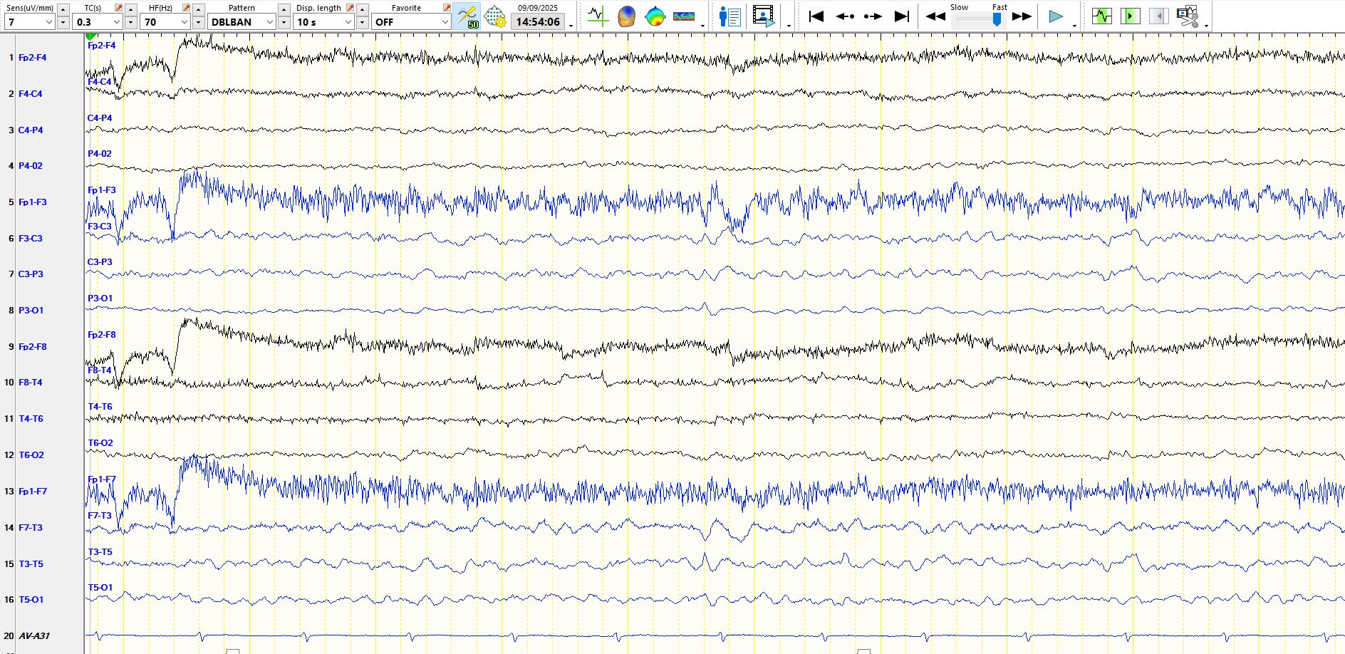
In the above image, the sharp wave and, less clearly, the slow wave, demonstrate phase reversal at F3 and T3. Because F7 is closer than T3...
This question pertains to the discussion yesterday Newly diagnosed focal epilepsy; be prepared for a battle
When selecting an antiseizure medication, there are a few issues that should enjoy priority...
https://jamanetwork-com.ez.sun.ac.za/journals/jamaneurology/article-abstract/2838122
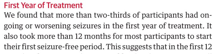
Key Points
Question Can treatment response to antiseizure medication (ASM) in people with newly diagnosed...
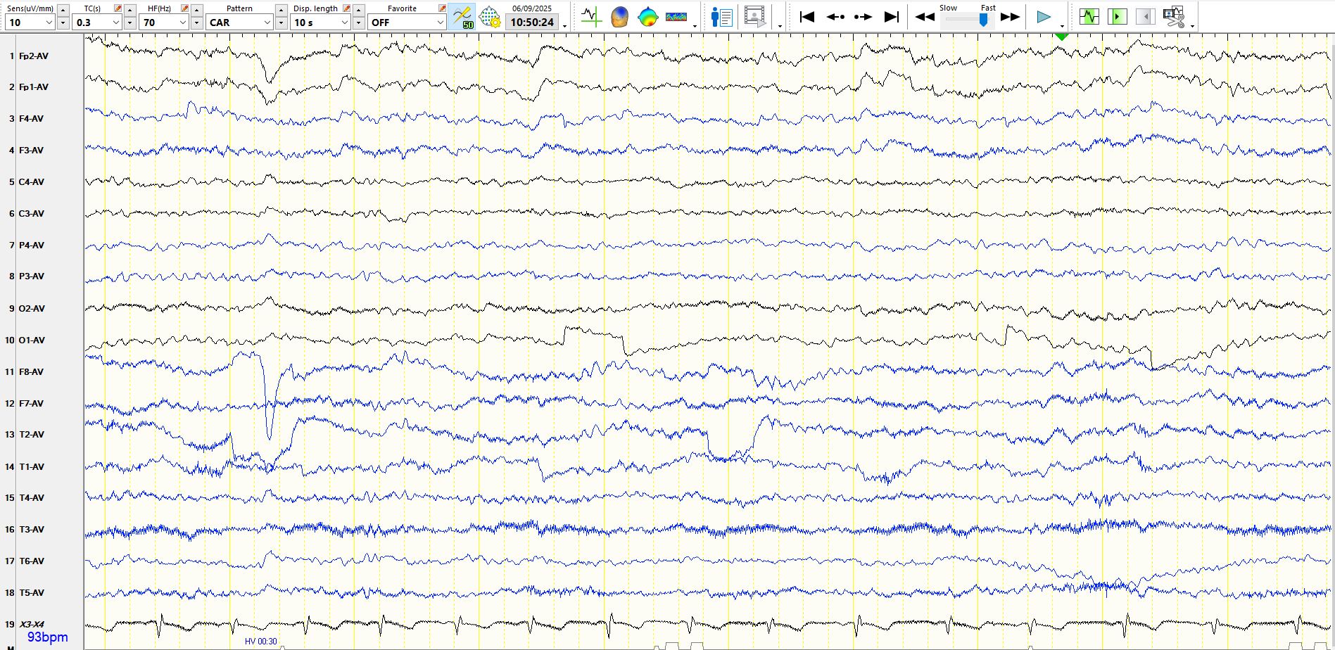
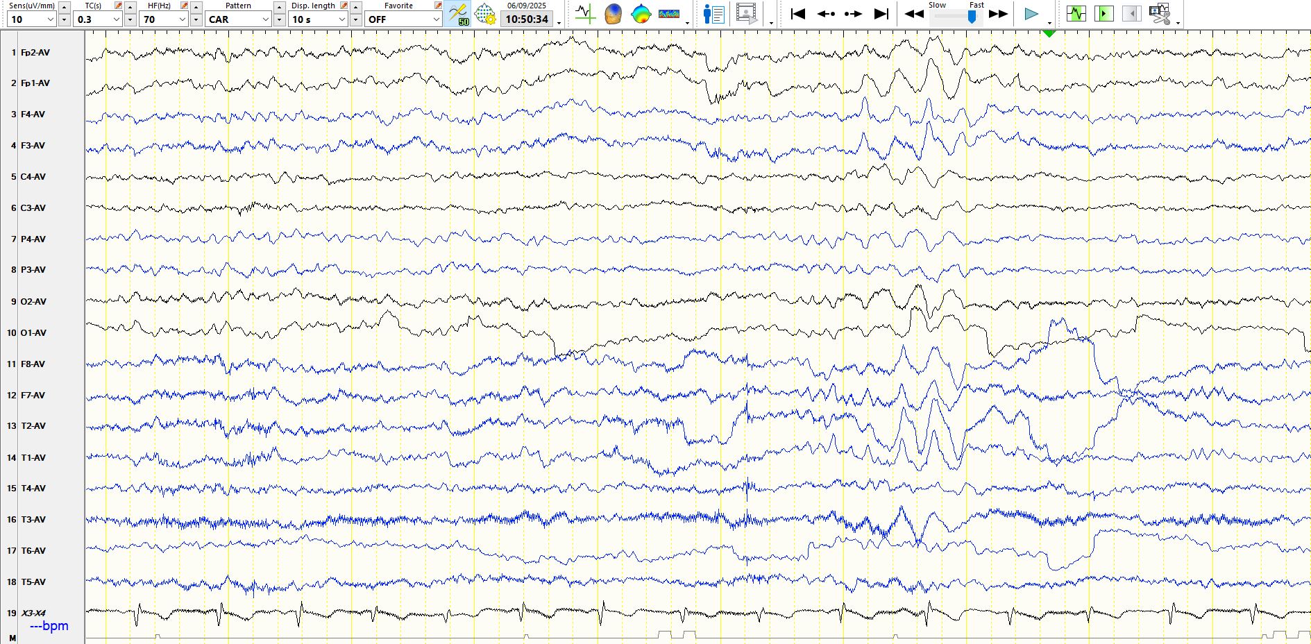
Have a look at the following series of pages, some of which are sequential epochs:
The first three are consecutive pages, recorded at 10 uV / mm:
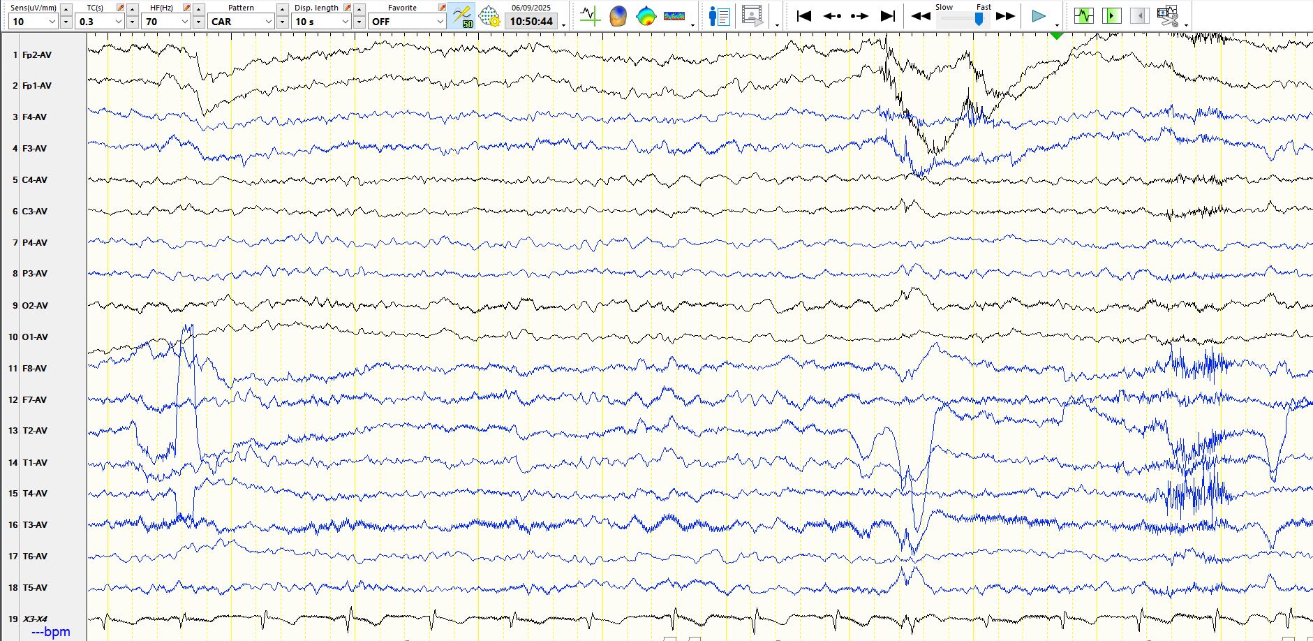
This is a short while later:
...
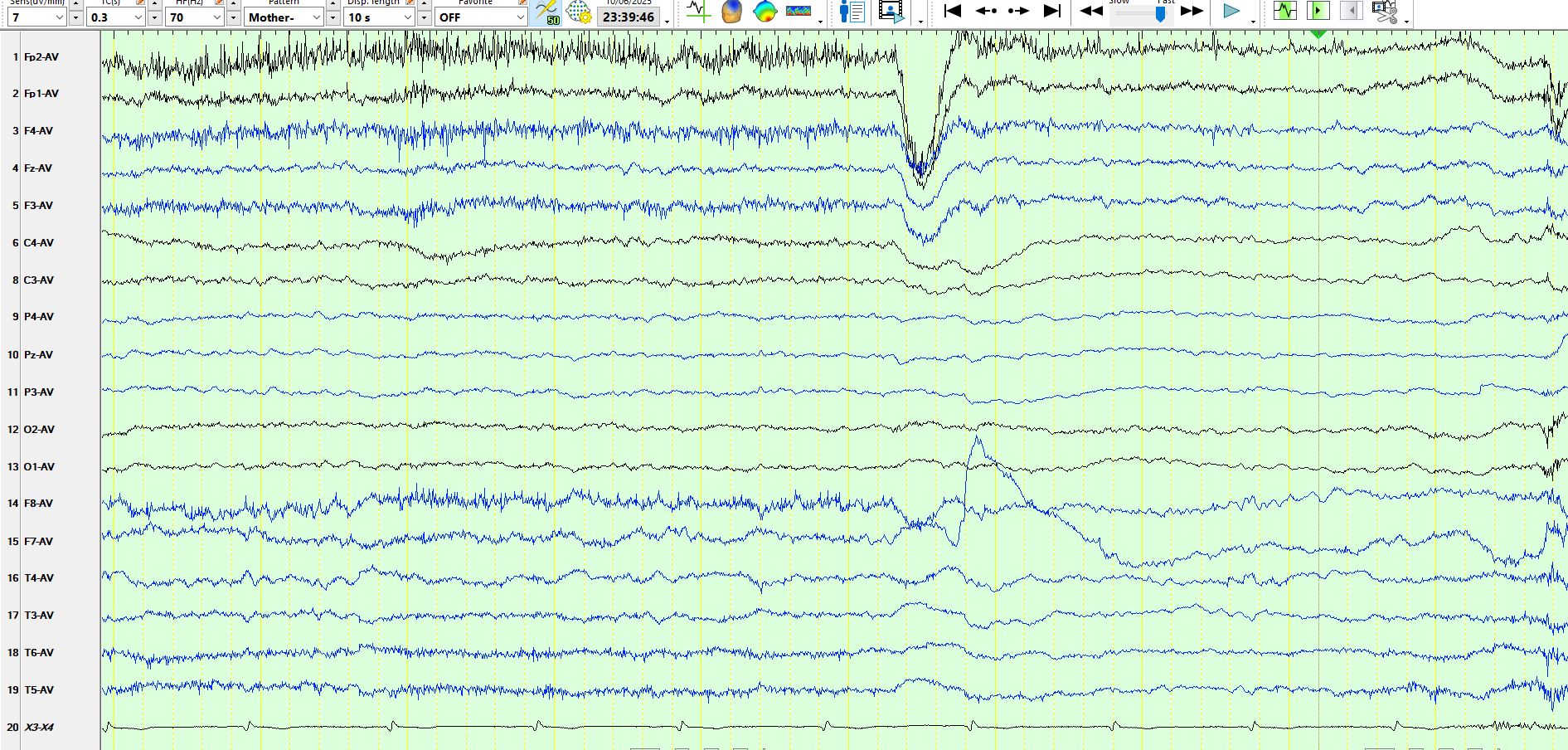
This is the EEG of a 60-year-old patient:
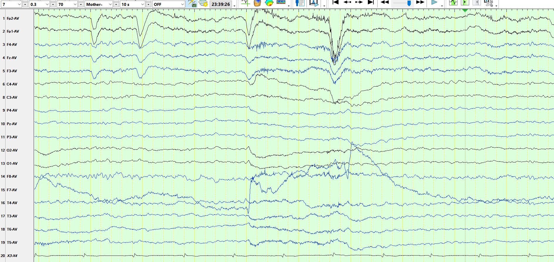
Figure 2:
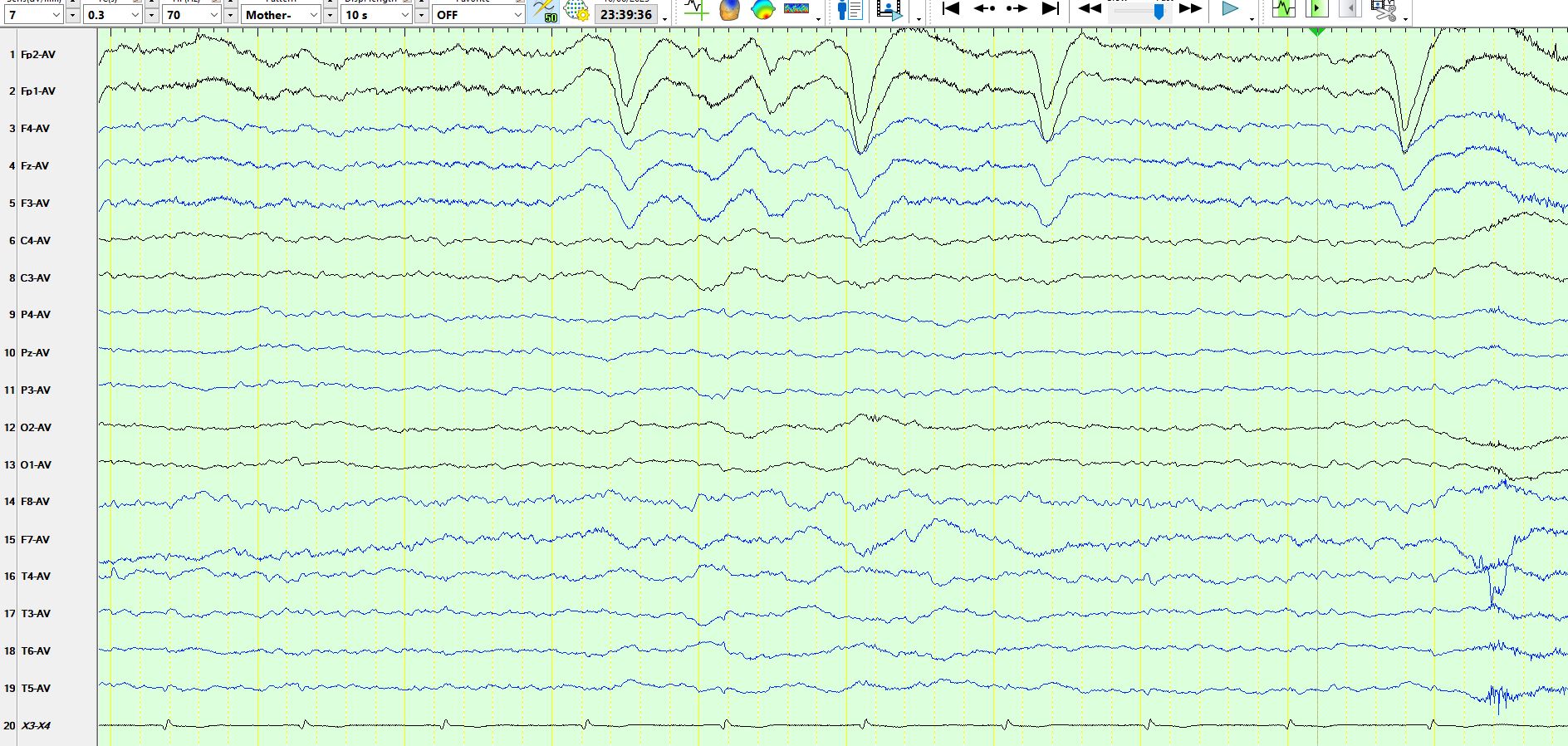
Figure 3, the AP bipolar montage of the same page:
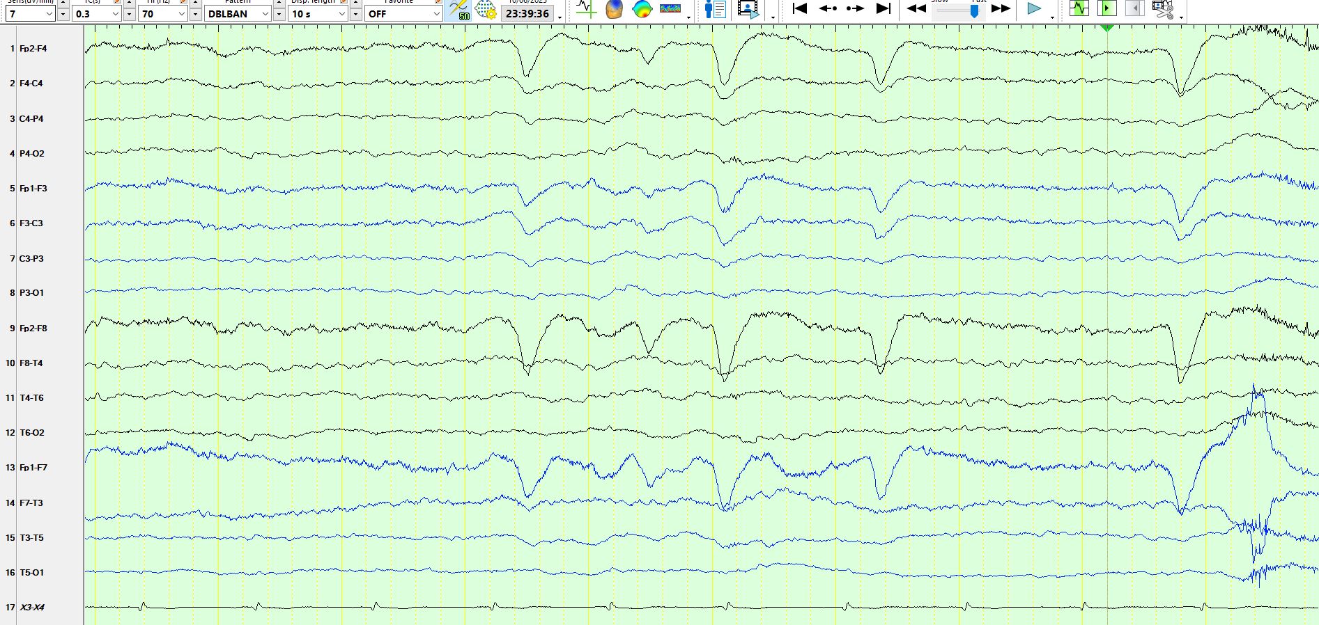
Figure 4, the coronal montage of the same page:
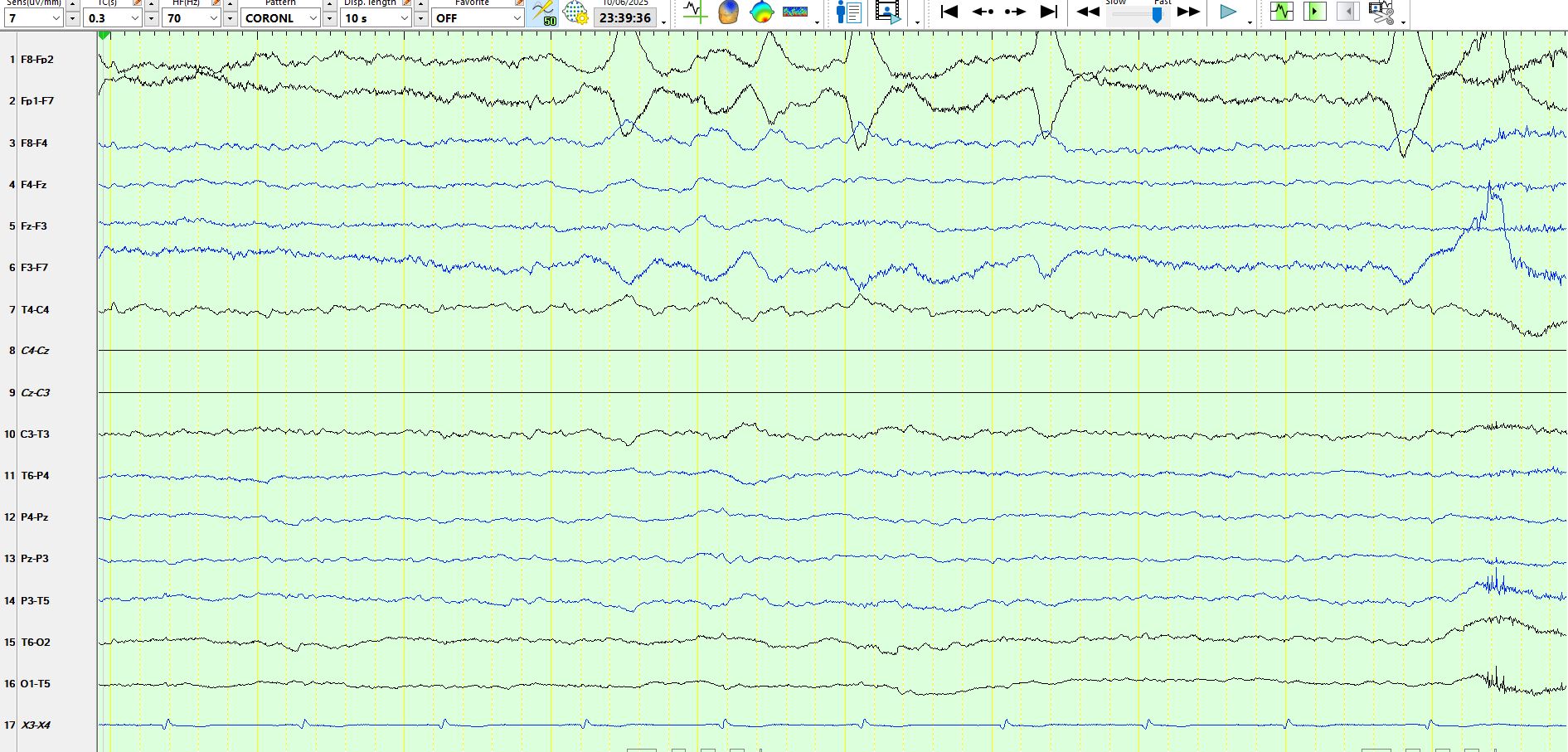
Figure 5:
F7 and F8 are 2 of the electrodes that are most prone to come adrift during prolonged recordings. There are various ways to deal with this problem when reviewing the recording, which can look even mor...
Do the following represent alpha rhythms that are propagating anteriorly? Or is this drowsiness? Could these waves be spikes? Could this be a benign phenomenon?
figure 1:
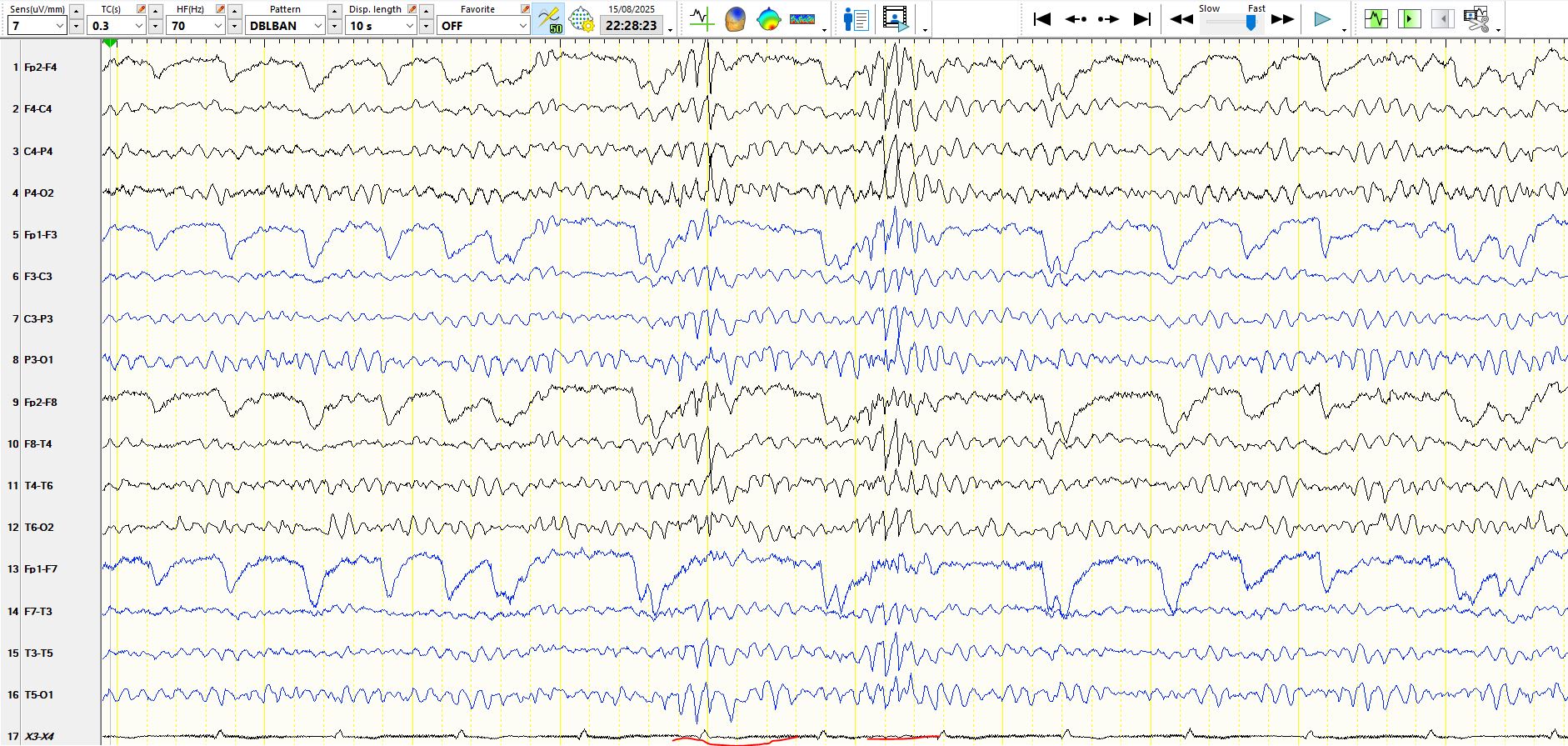
Figure 2:
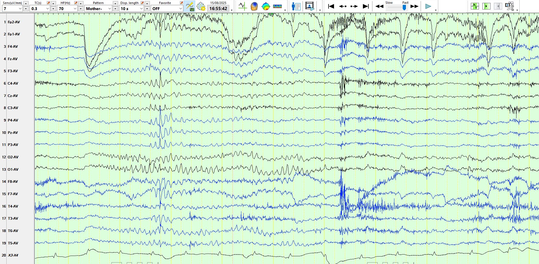
Fig...
Medial temporal sclerosis (or mesial temporal sclerosis) is essentially a pathological diagnosis; high quality MRI scans interpreted by experts who work in the field are strongly predictive of the pat...
This discussion related to an EEG performed on someone in ICU refers ICU, "Not waking up, seizures? Brain dead?".
To reiterate, judgements about suppression can be quite tricky, doubly so in the ...
