The history predicts non-dominant (right) temporal lobe epilepsy.
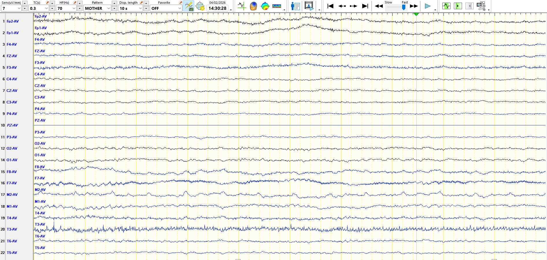
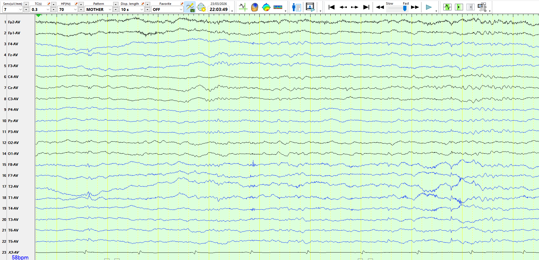
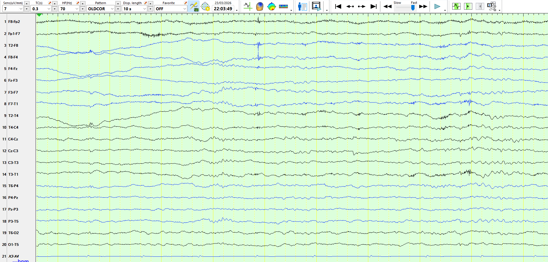
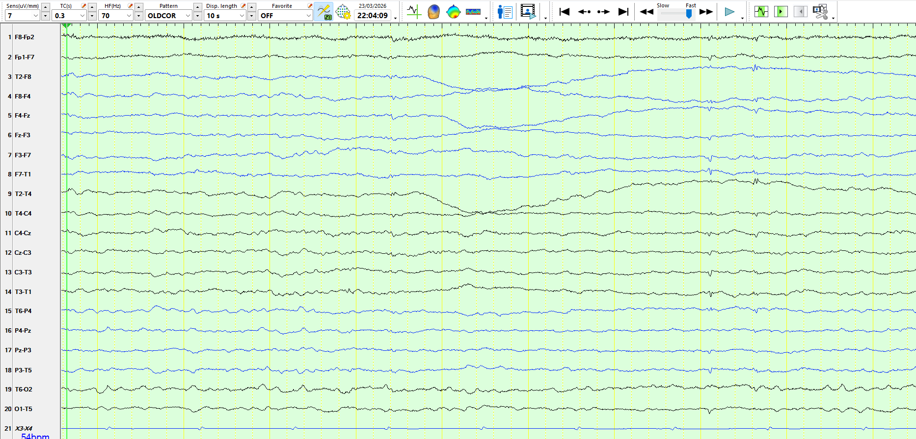
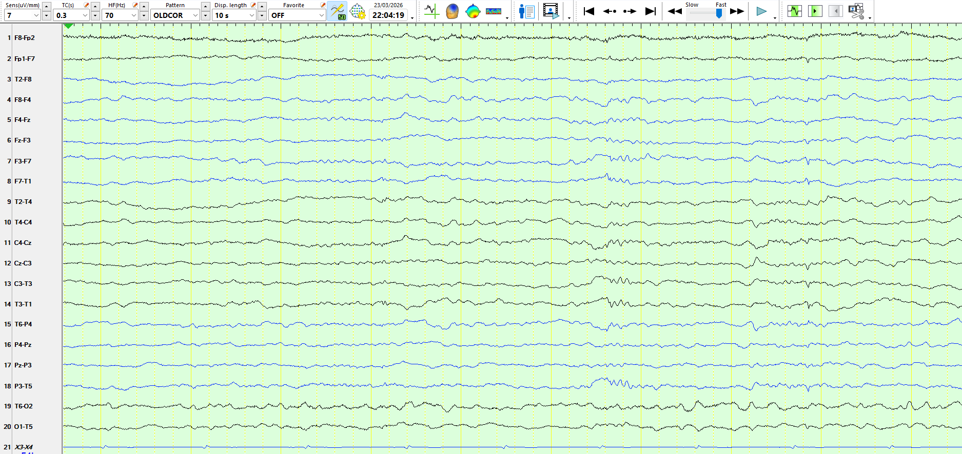
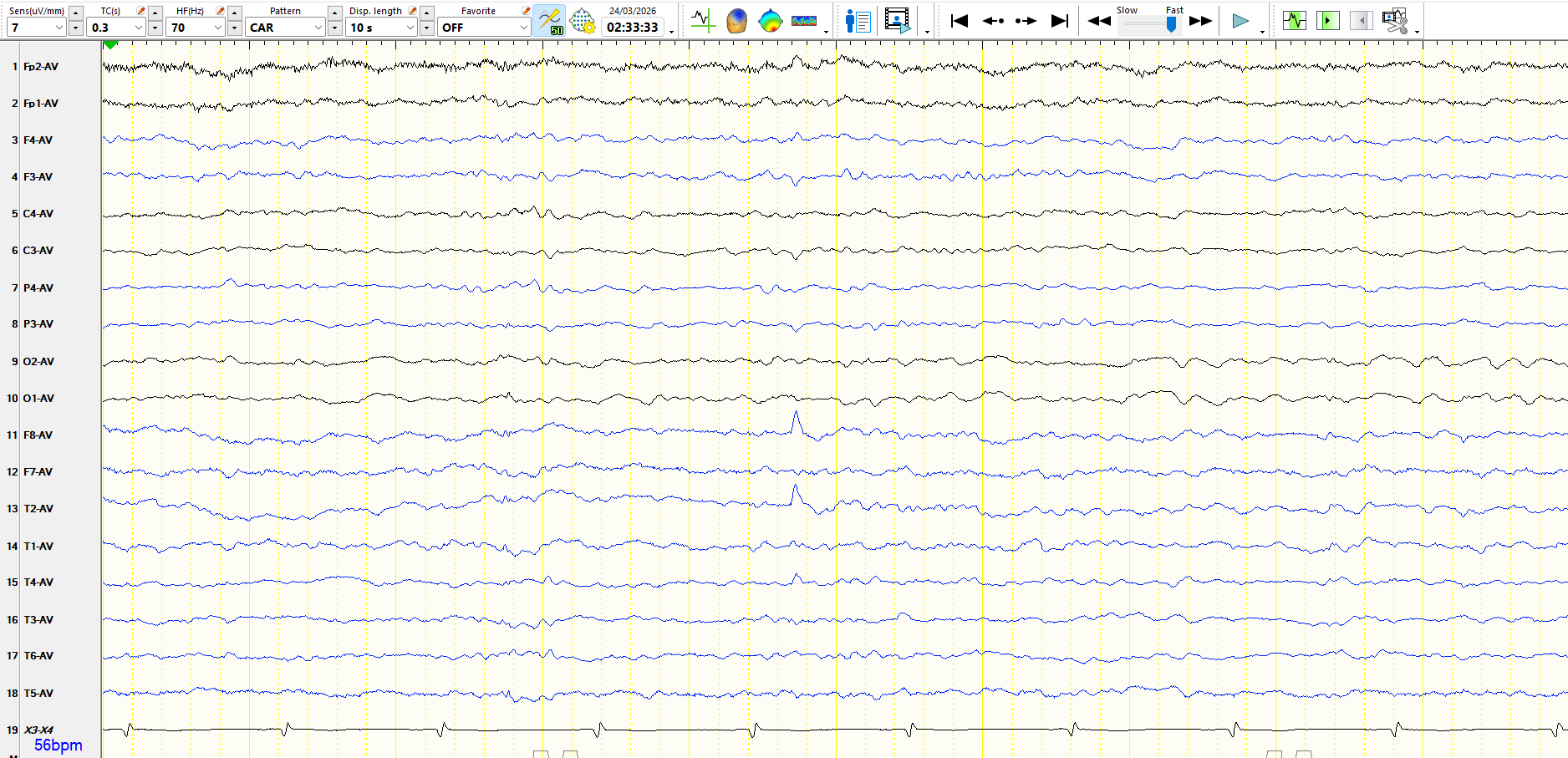
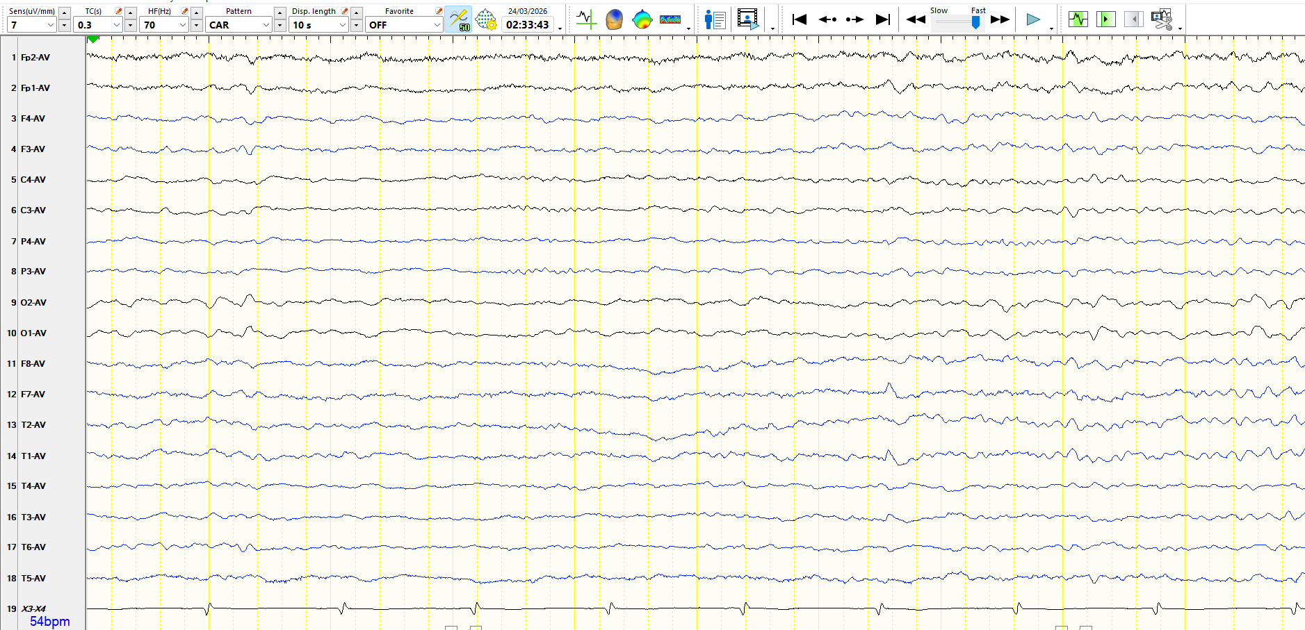
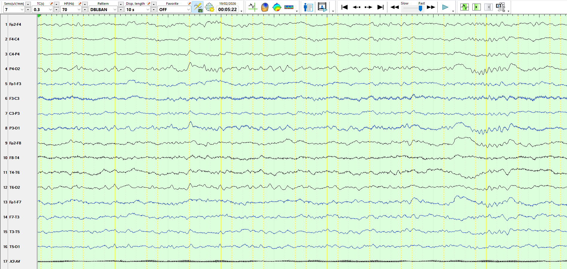
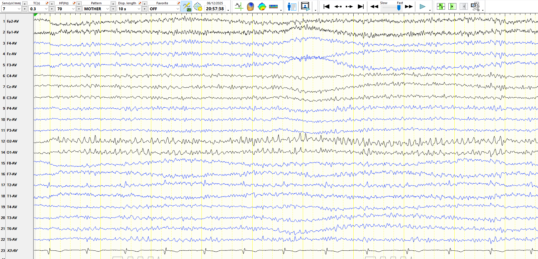
Awake:
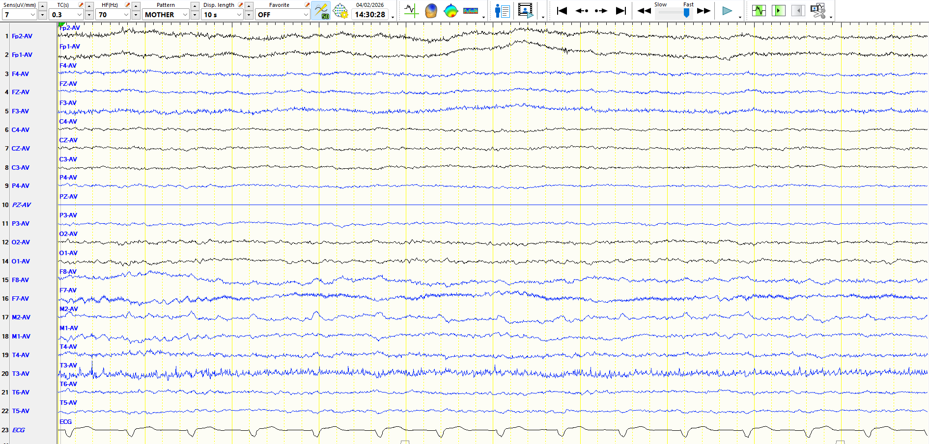
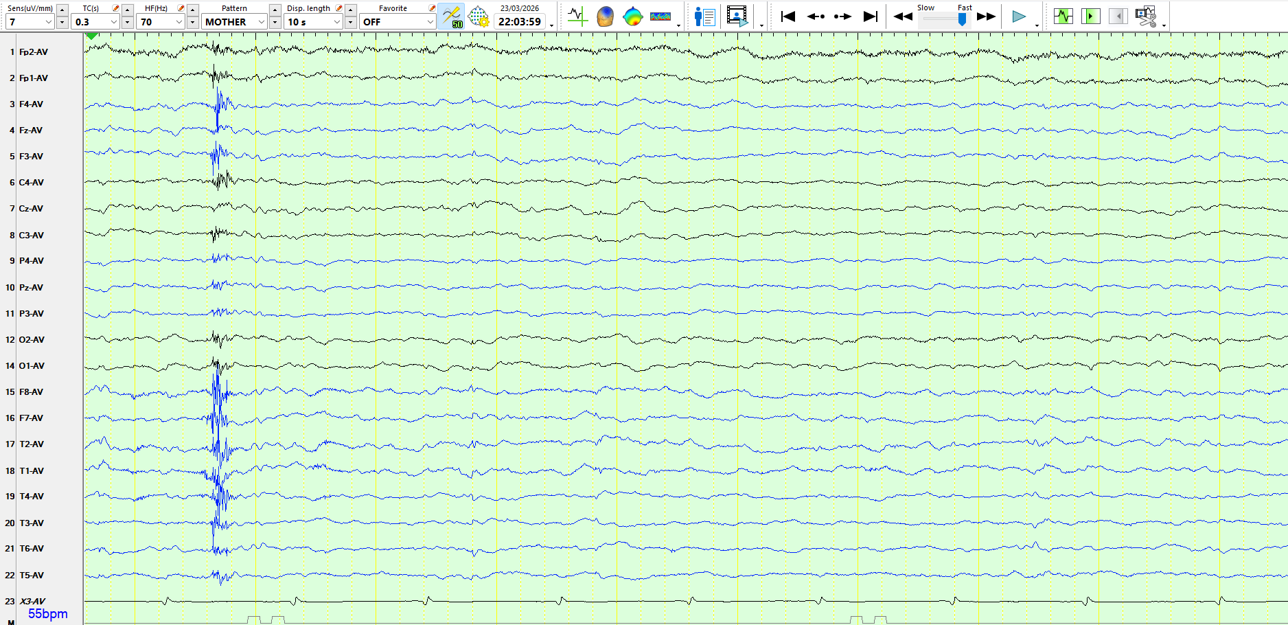
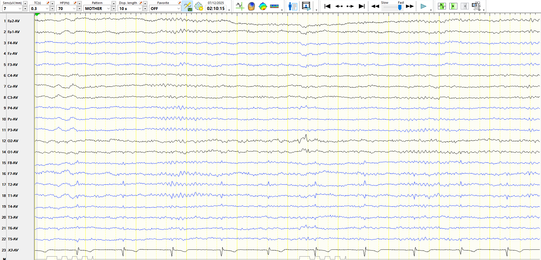
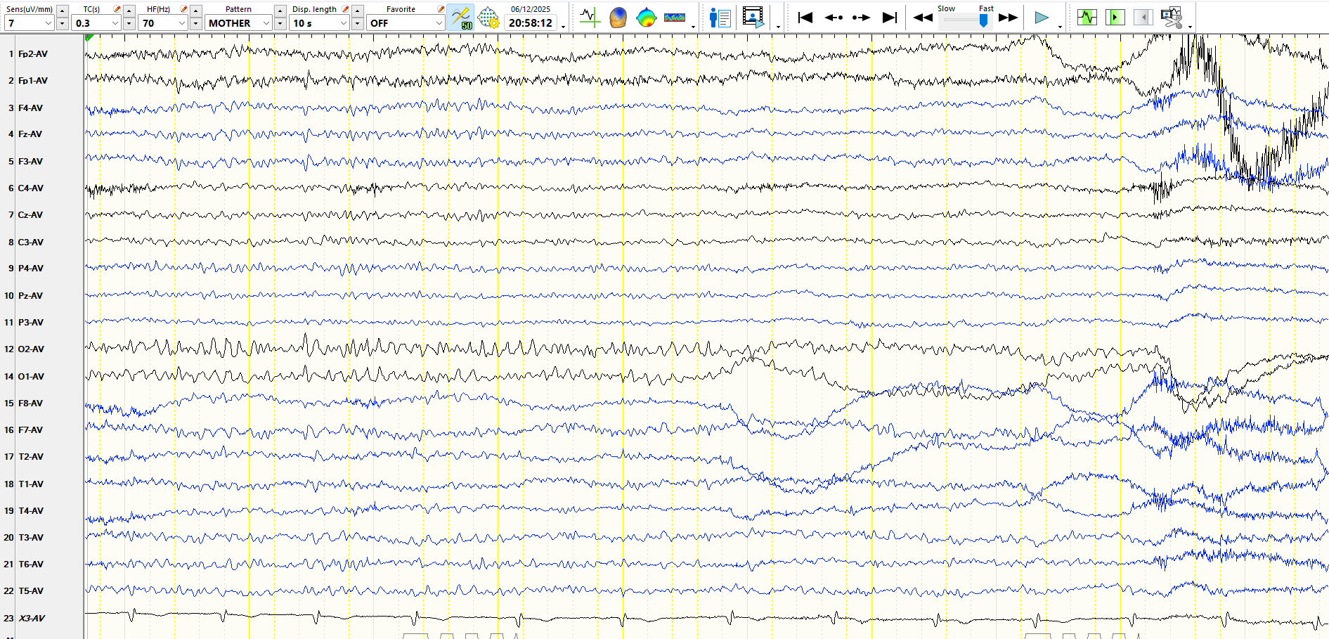
Drowsy:
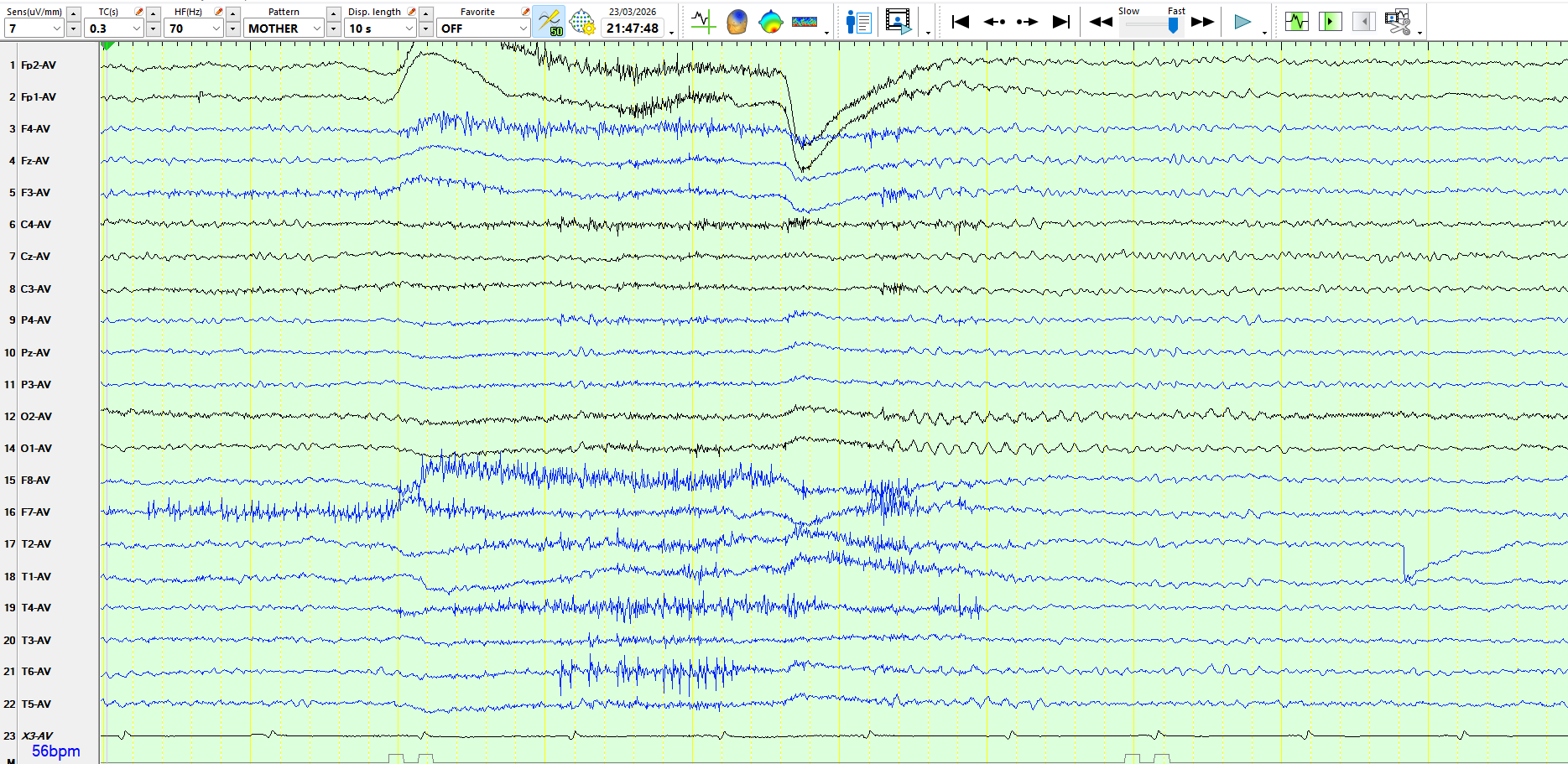
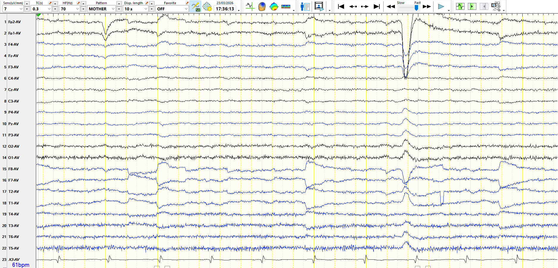
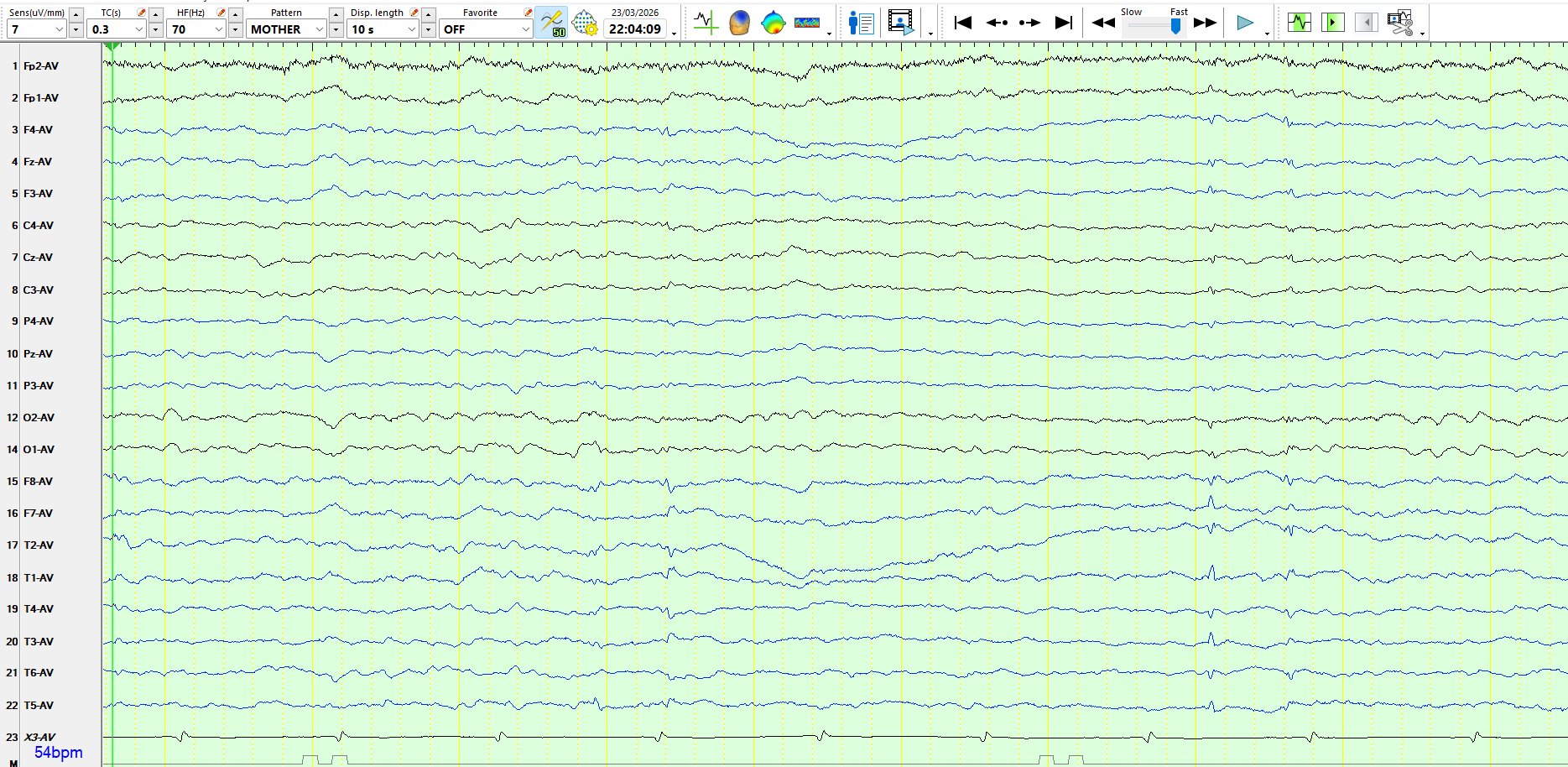
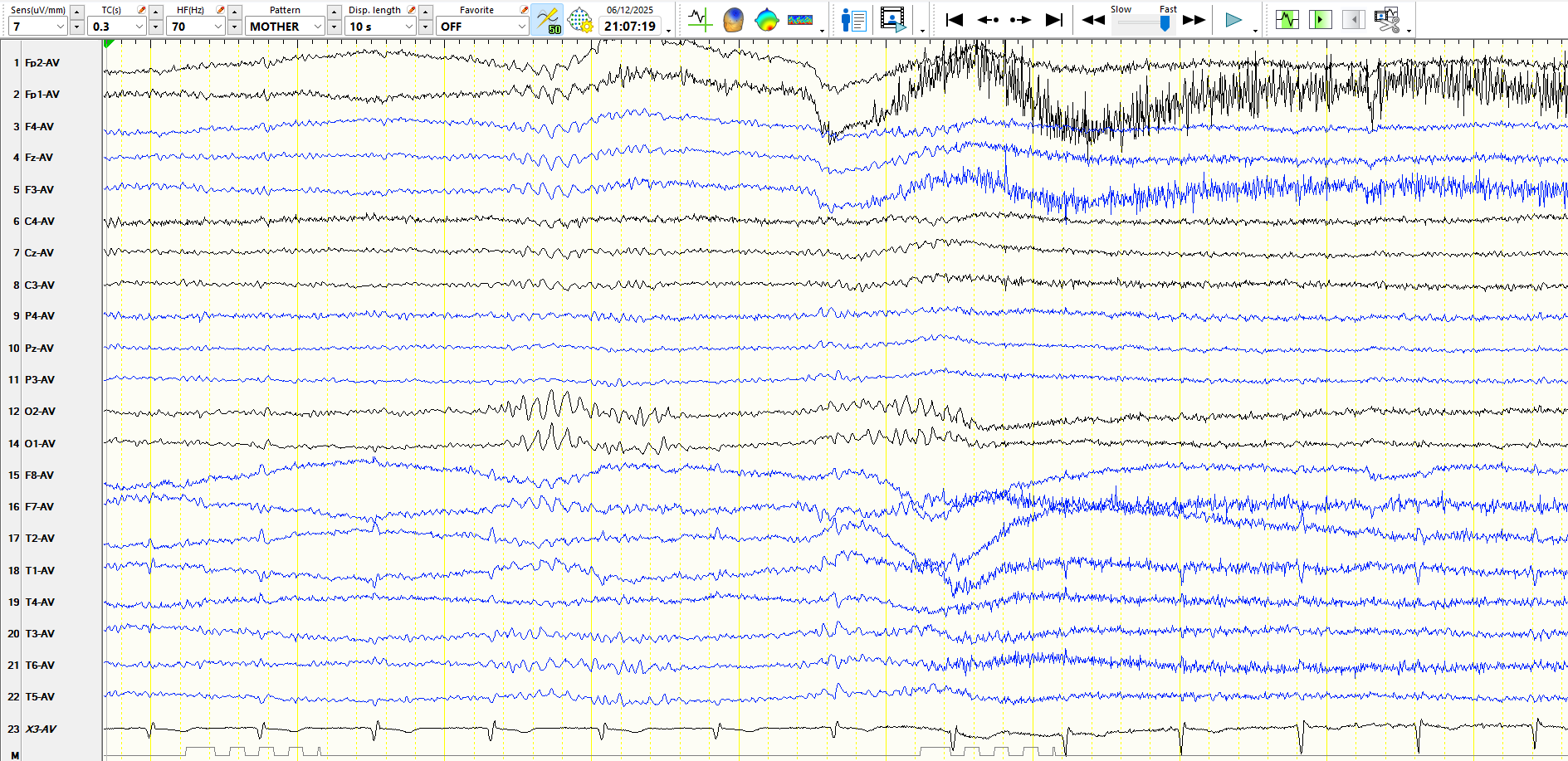
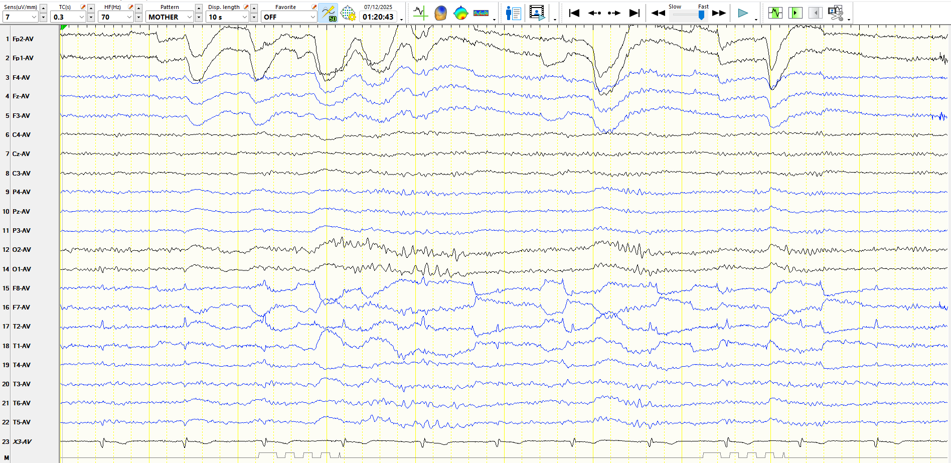
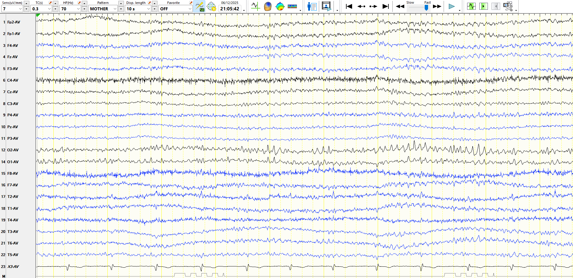
Sleep:

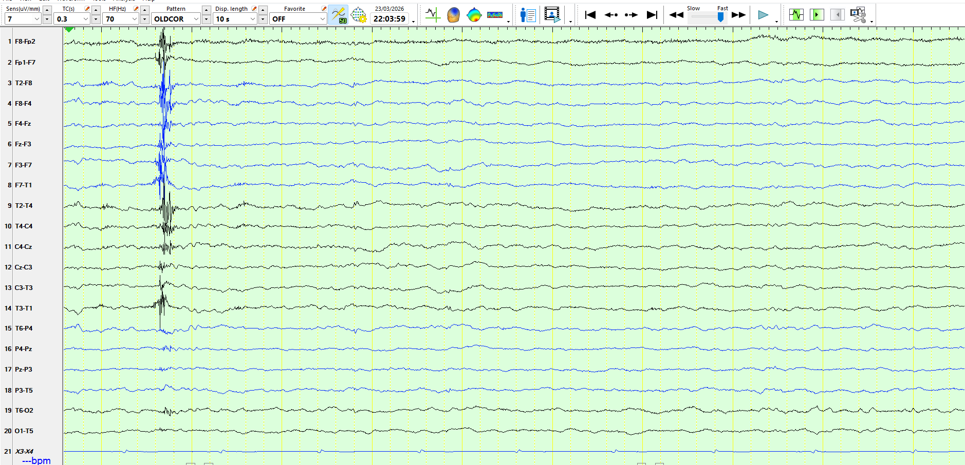
There have been 2 neurological events, approximately 2 years apart. These were virtually identical and both occurred while standing in the house. The patient had a sense of vertigo, as the world appea...
Young adult (20s)
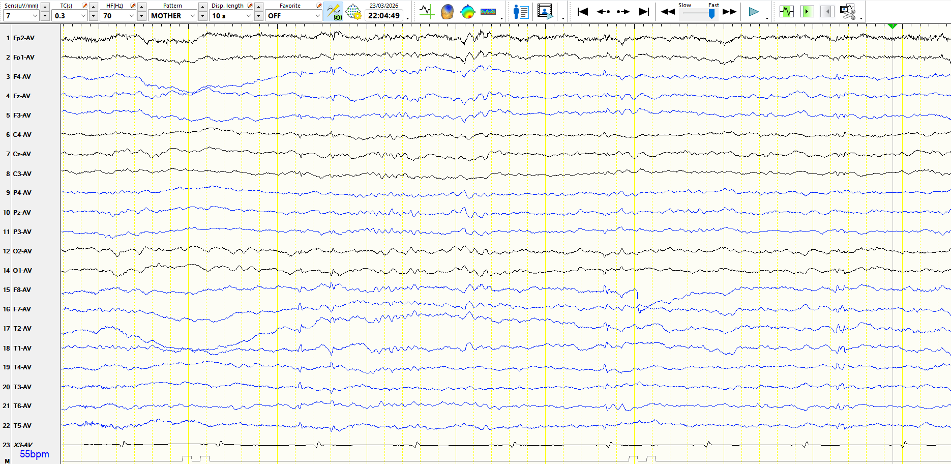
The following is from an adult patient:
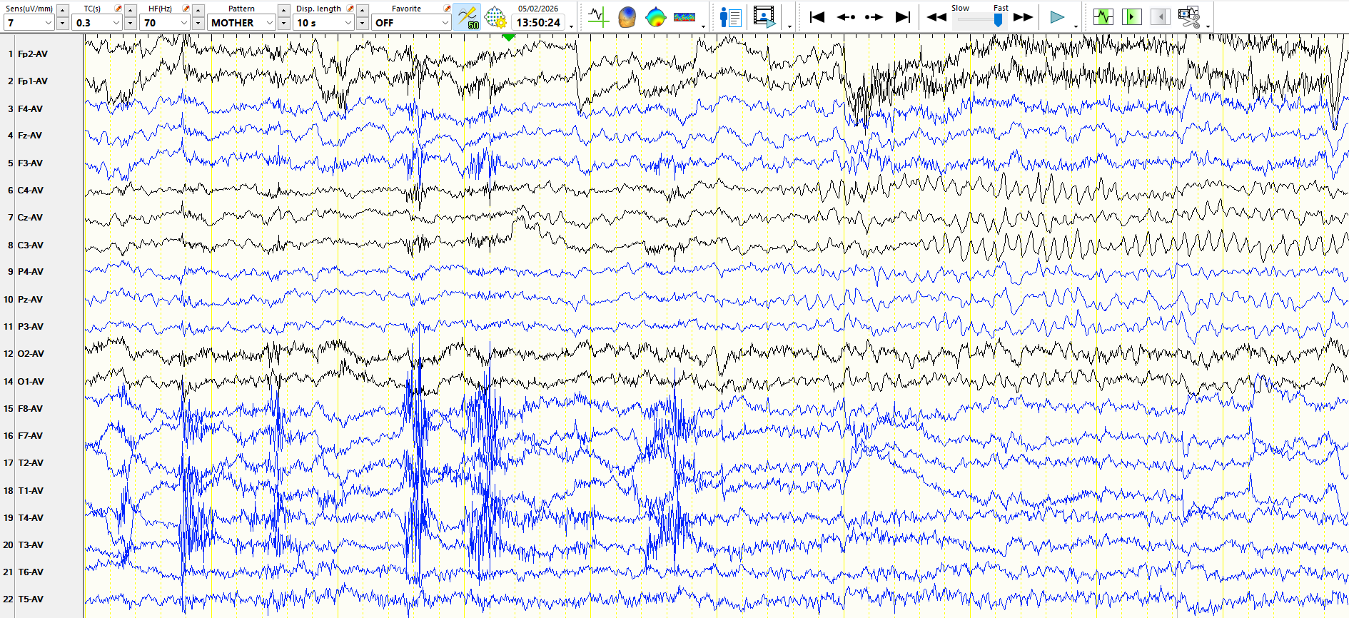
In the page above, rhythmic theta waves appear at CZ-PZ. The patient is drowsy; the alpha rhythms attenuate, as does temporal EMG, and there are tempora...
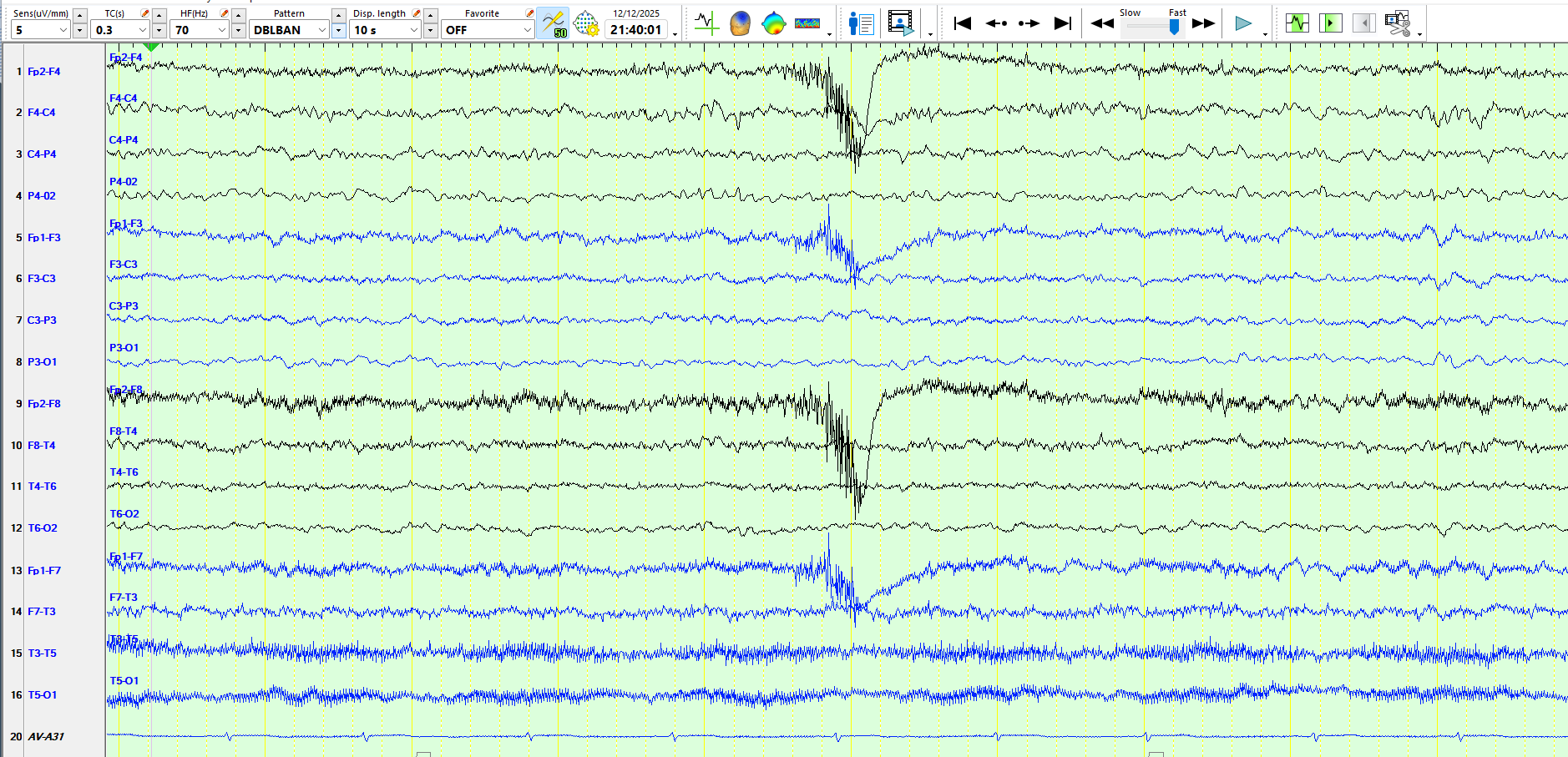
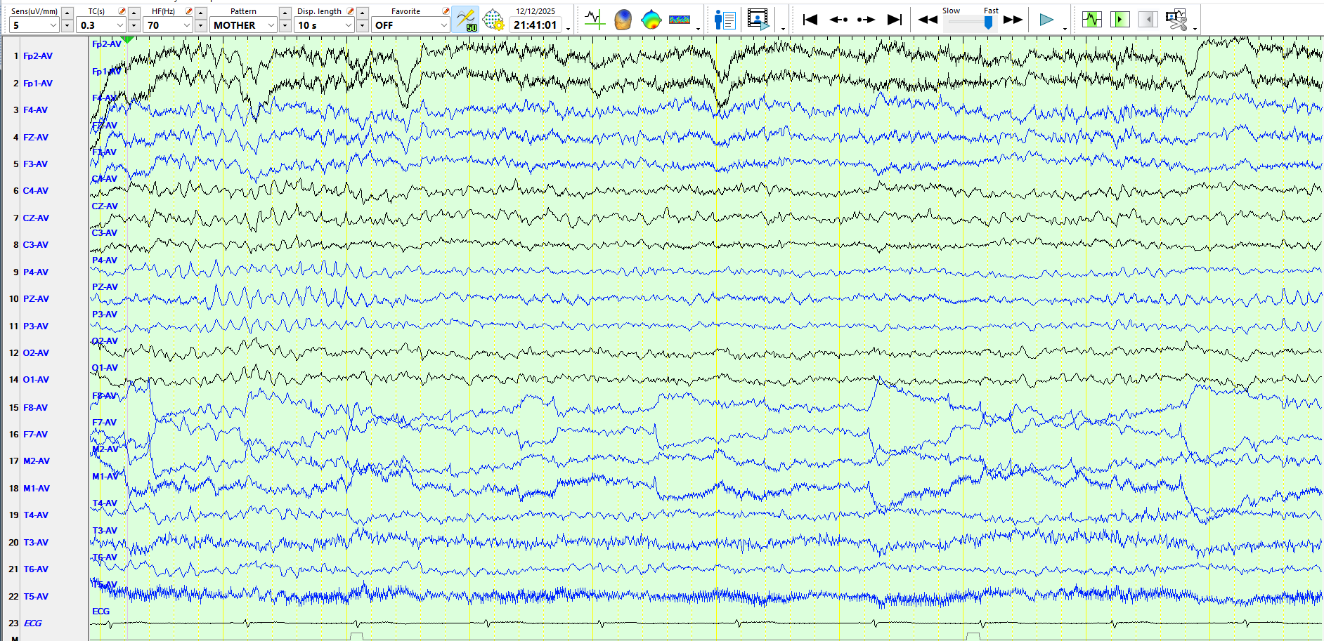
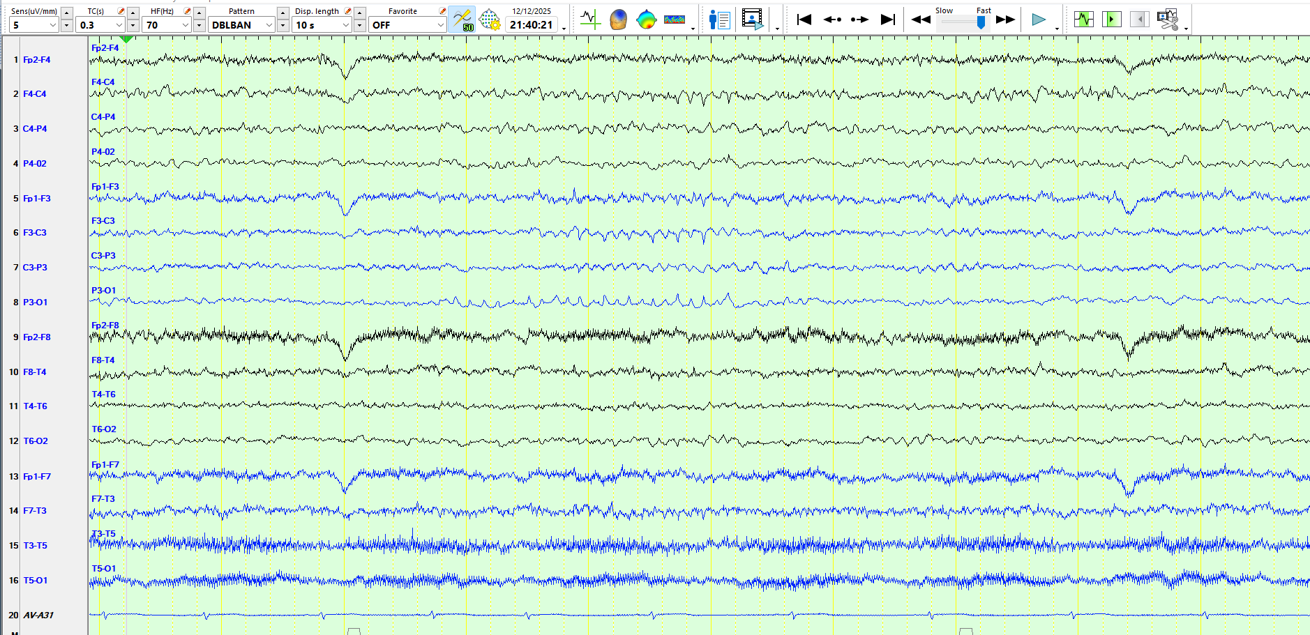
The EEG is normal during wakefulness. Here are a variety of pages from sleep, each represented in different montages. These discharges appeared profusely, typically at least once every 10 seconds dur...
What do these waves during wakefulness and sleep represent?
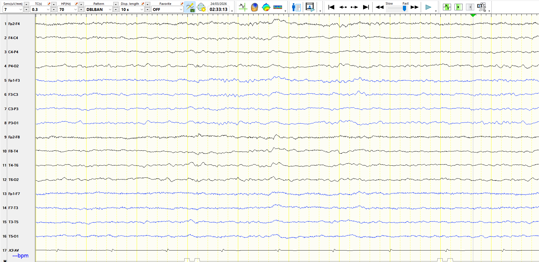
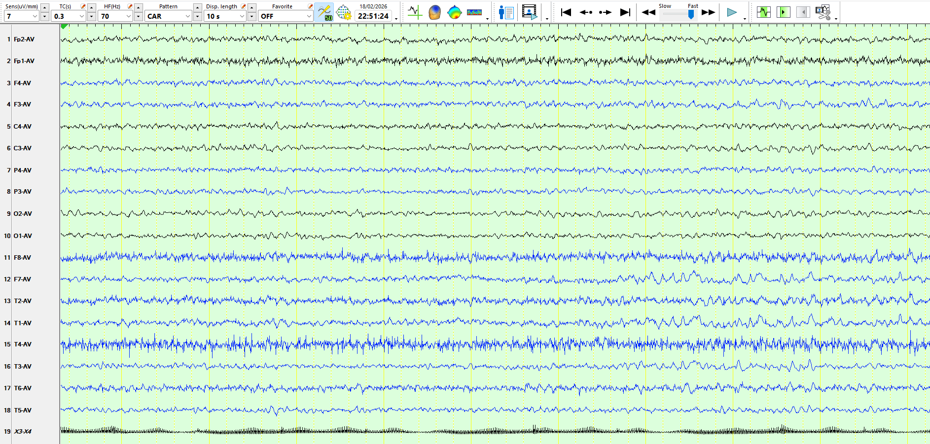
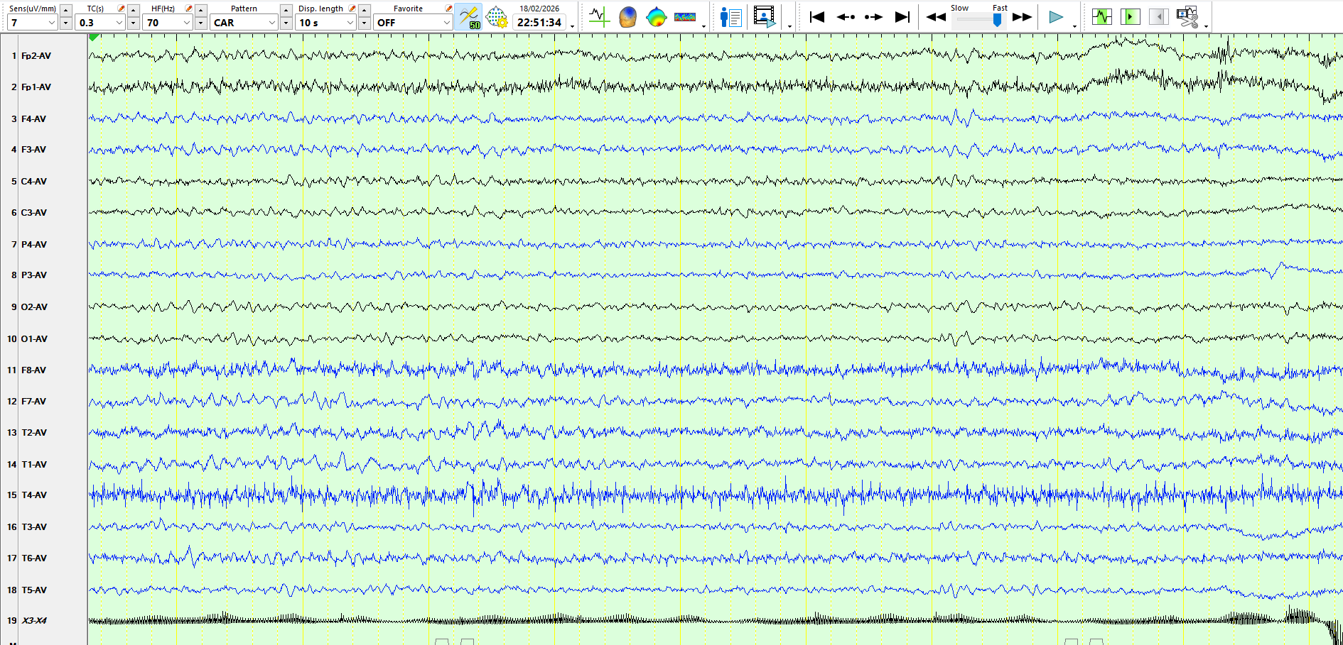
These are three consecutive pages:
Figure 1:
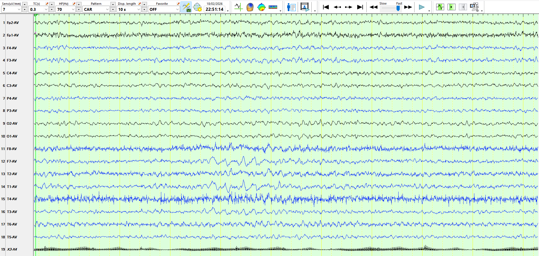
Figure 2:
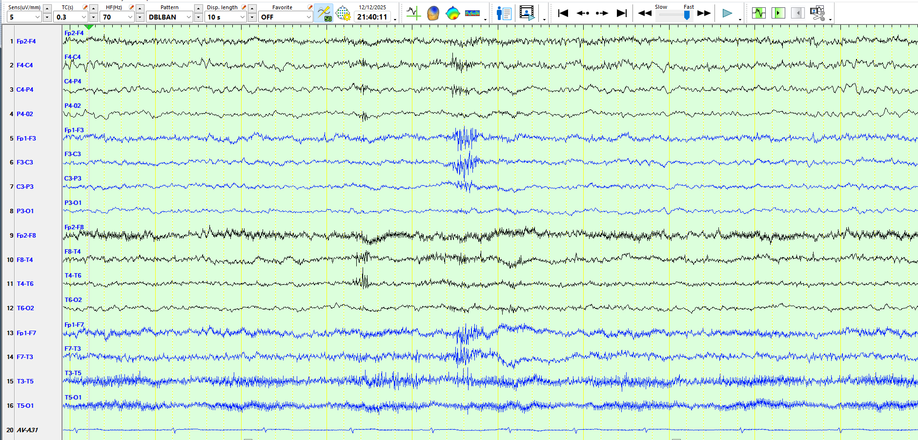
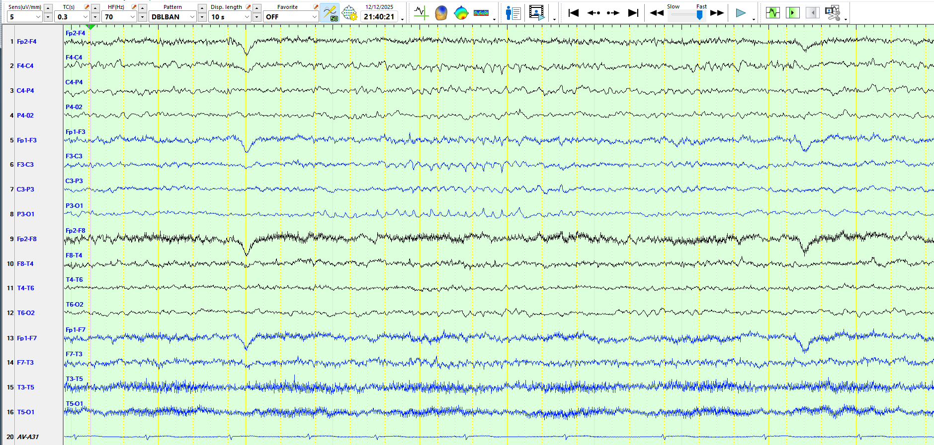
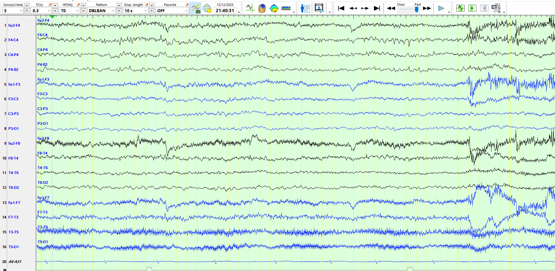
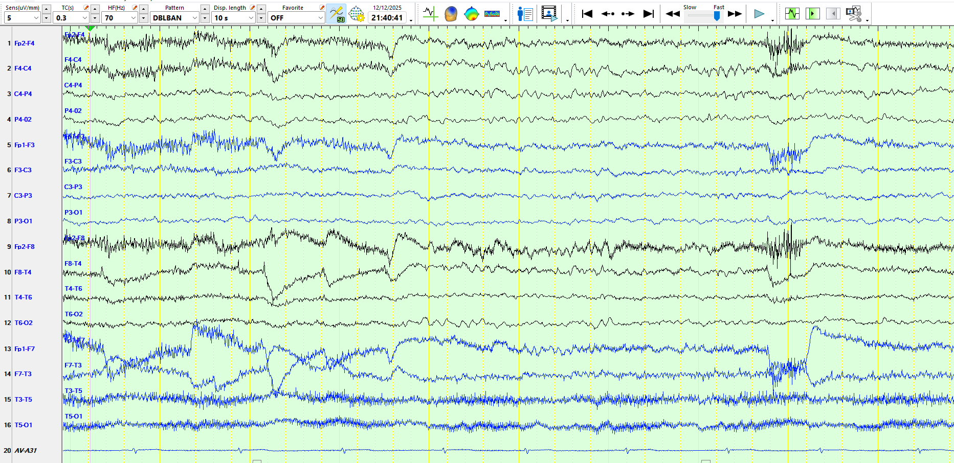
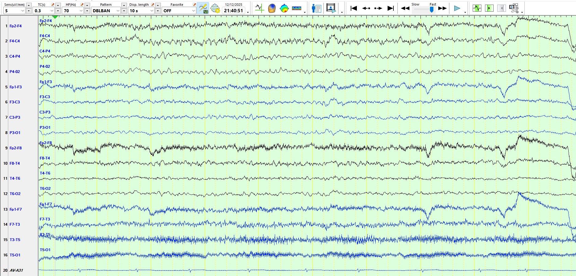
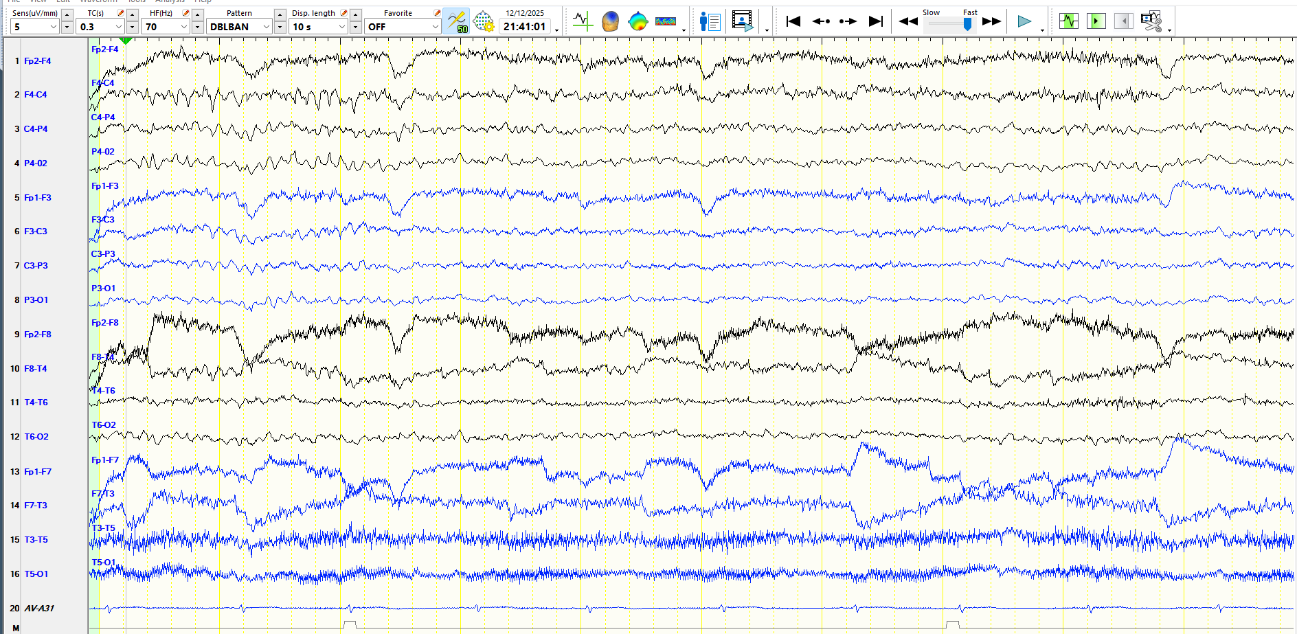
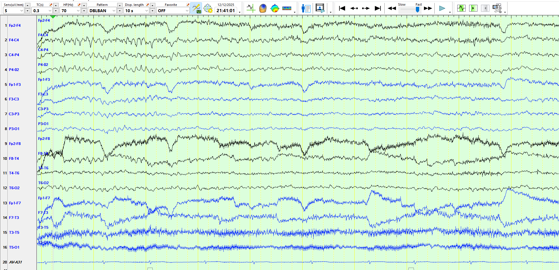
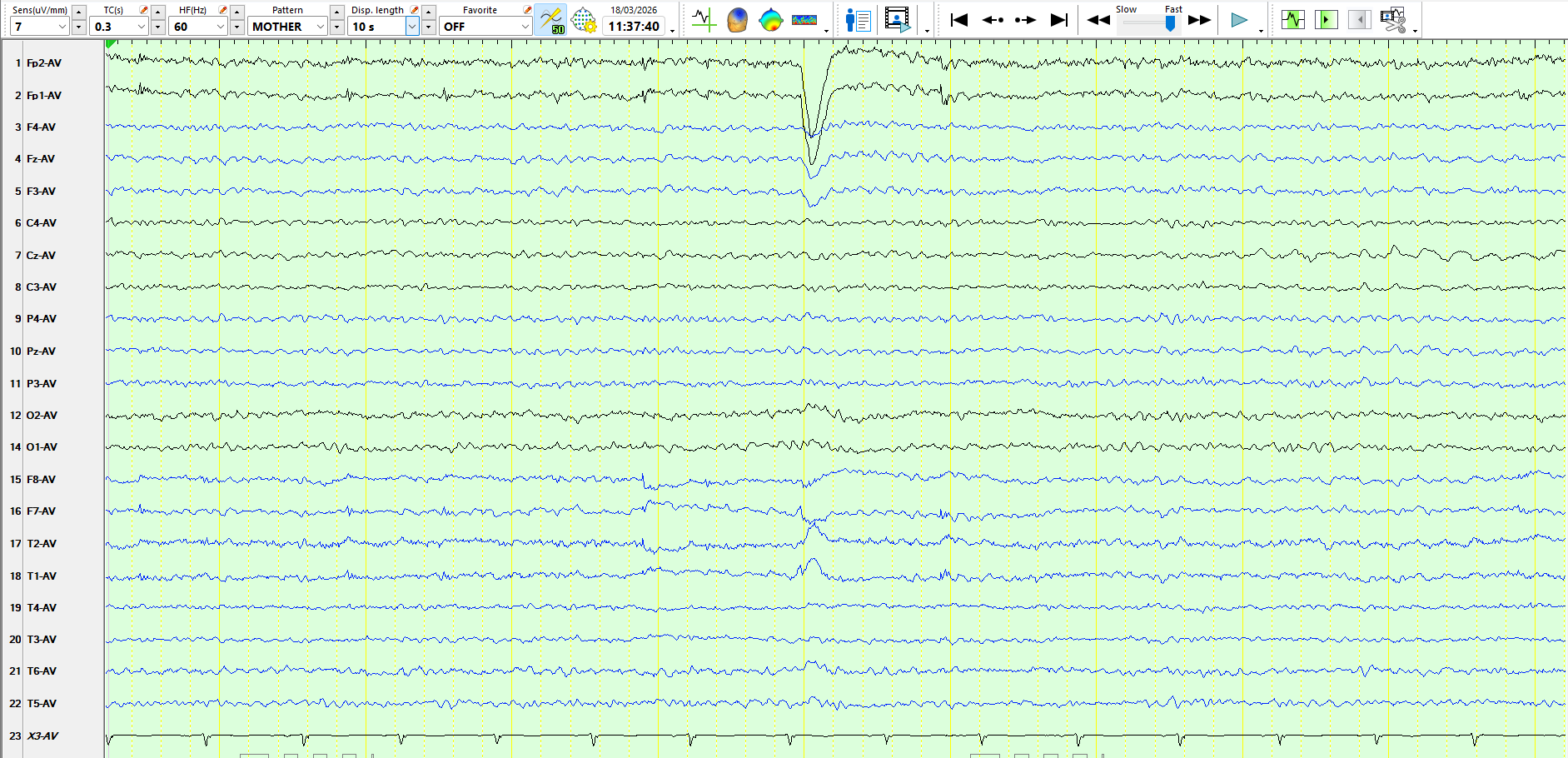
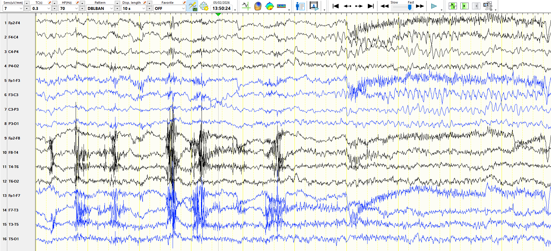
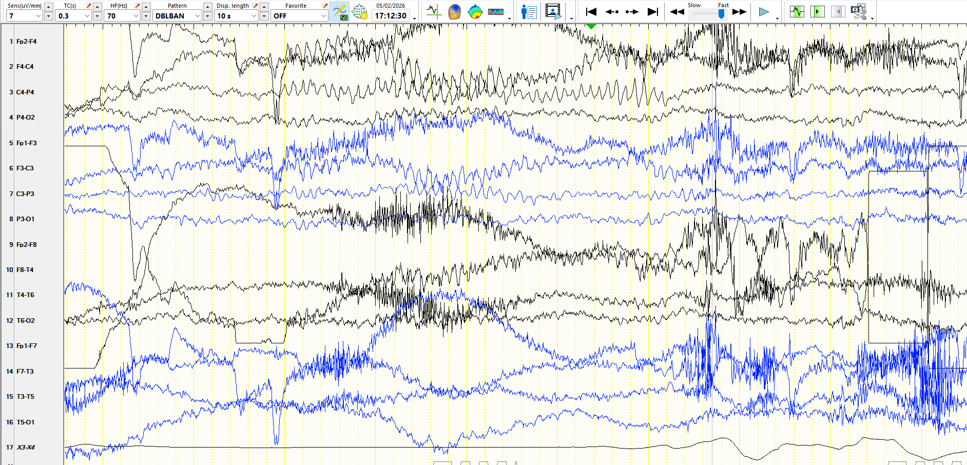
The following represent three consecutive pages:
figure 5
f...
It is important to recognise this, as sooner or later in the same recording, there will likely be isolated waves that resemble spikes or sharp waves.
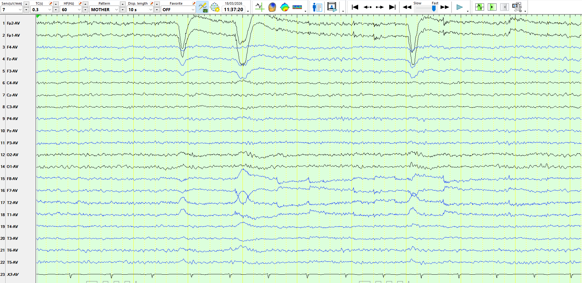
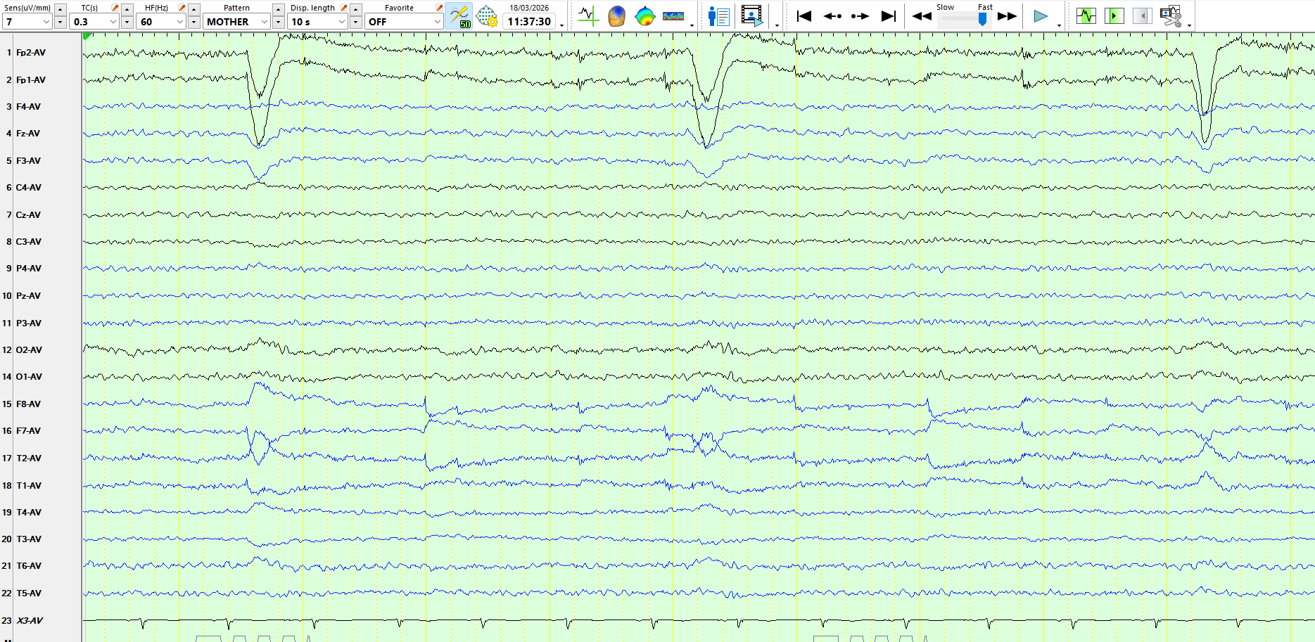
The following is fr...
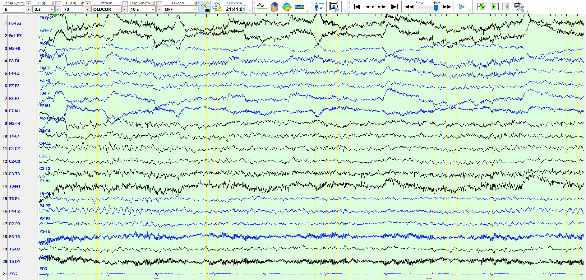
Are these waves on the 2nd page below:
1. A physiological variant
2. Theta and delta waves at M2-F8 (right inferior, anterior temporal region)
3. Theta waves, delta waves, and sharp waves at M2-F8
4. An e...
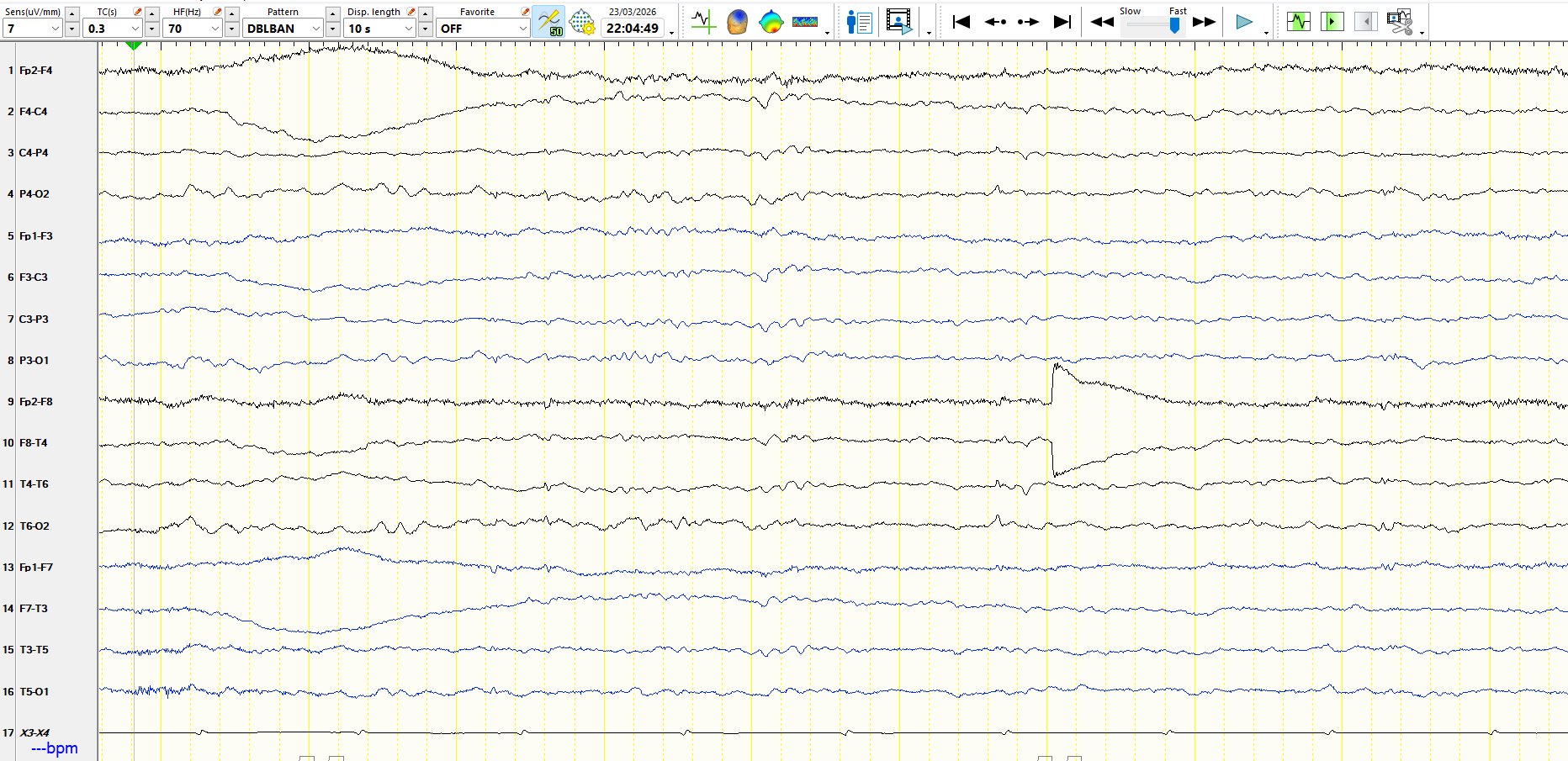
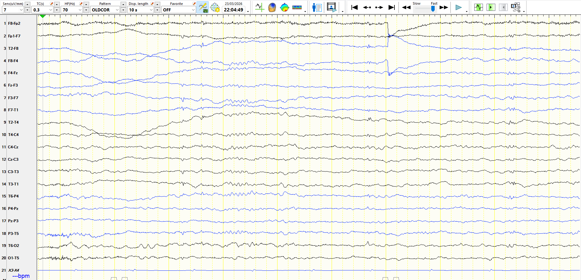
Intellectually normal, traumatic brain injury at the age of 4 months, EEG is normal while awake. The MRI scan is reported to show extensive spongiosis and cavitation in the right frontal lobe. A colle...
Criteria for these have existed for many years, but there have been recent attempts to refine and standardise them, using an evidence-based approach.
Here are some recent attempts:
The appearance of alpha frequencies coincides with eye movements (Eye closure, the Bell's phenomenon, upward movement of the globes), indicative of alpha rhythms. Notice that these are ...