Mid teenage years, "explosive outbursts"
Mar 19, 2026
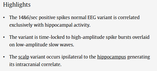
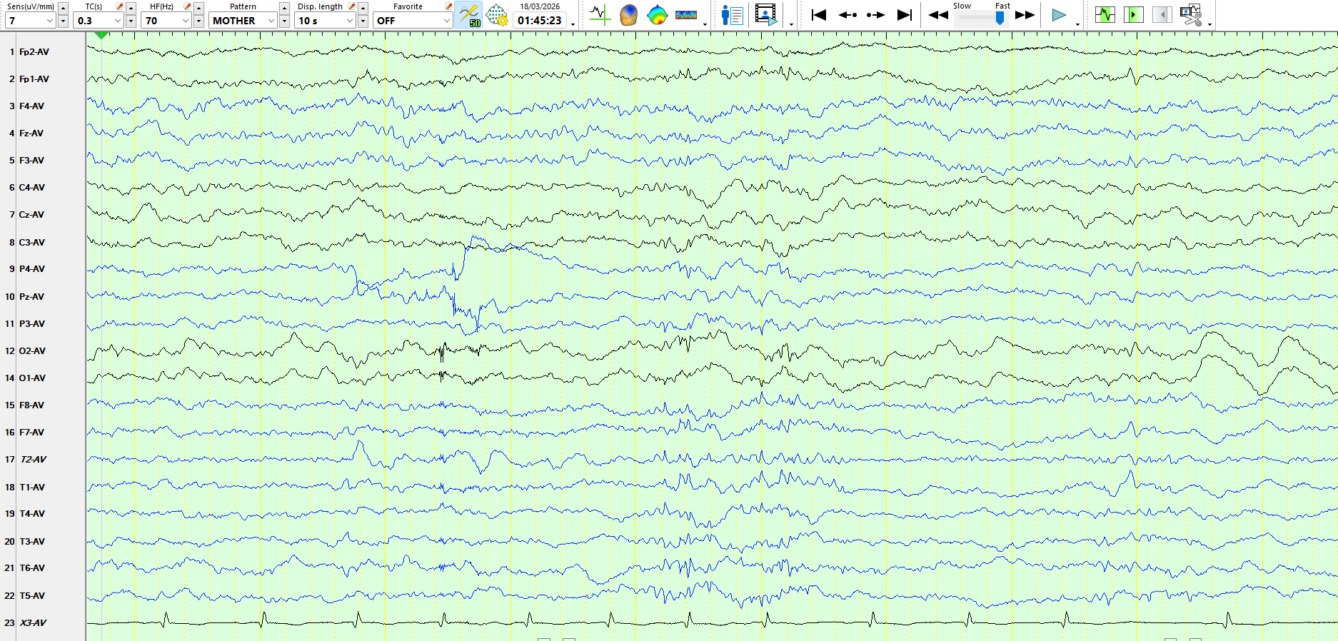
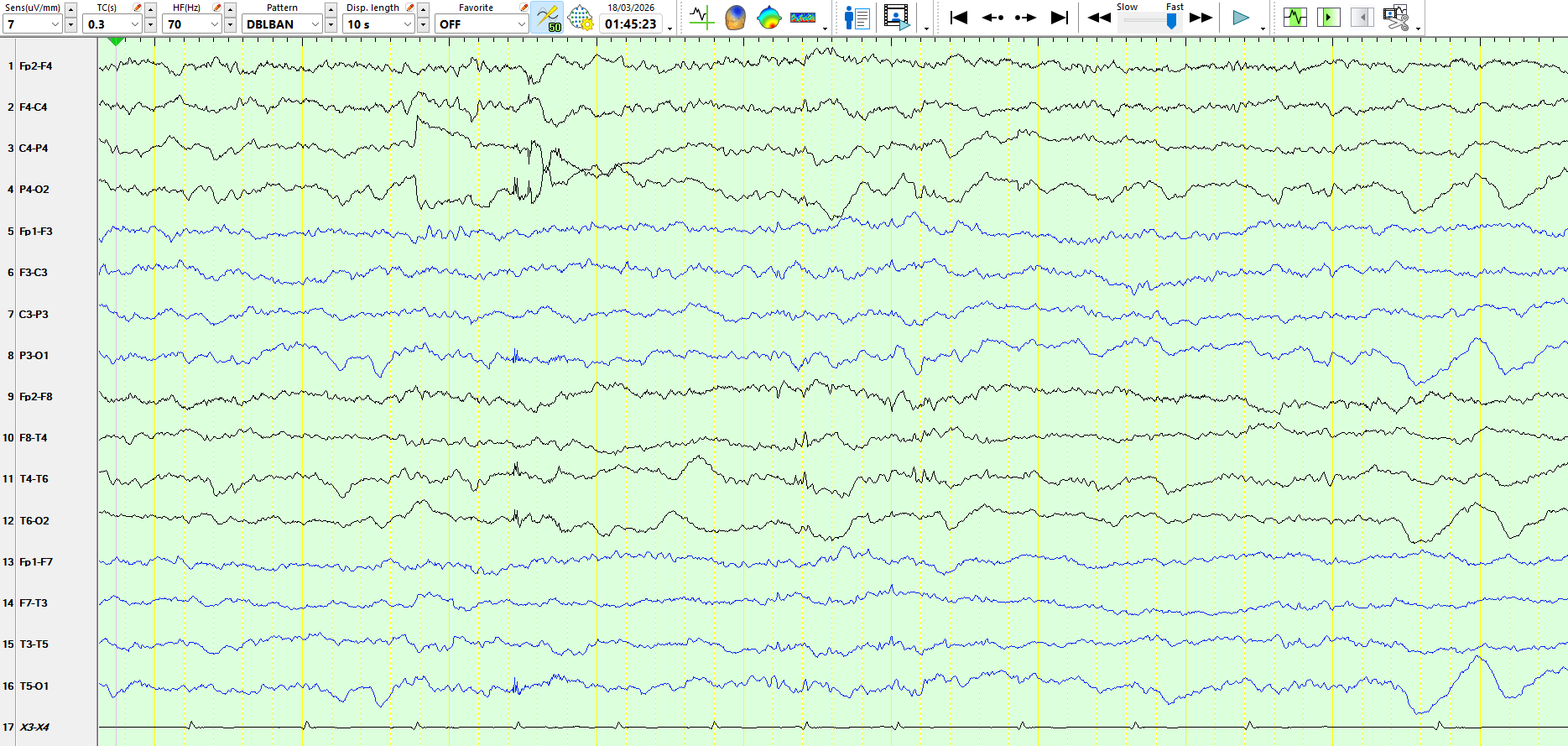
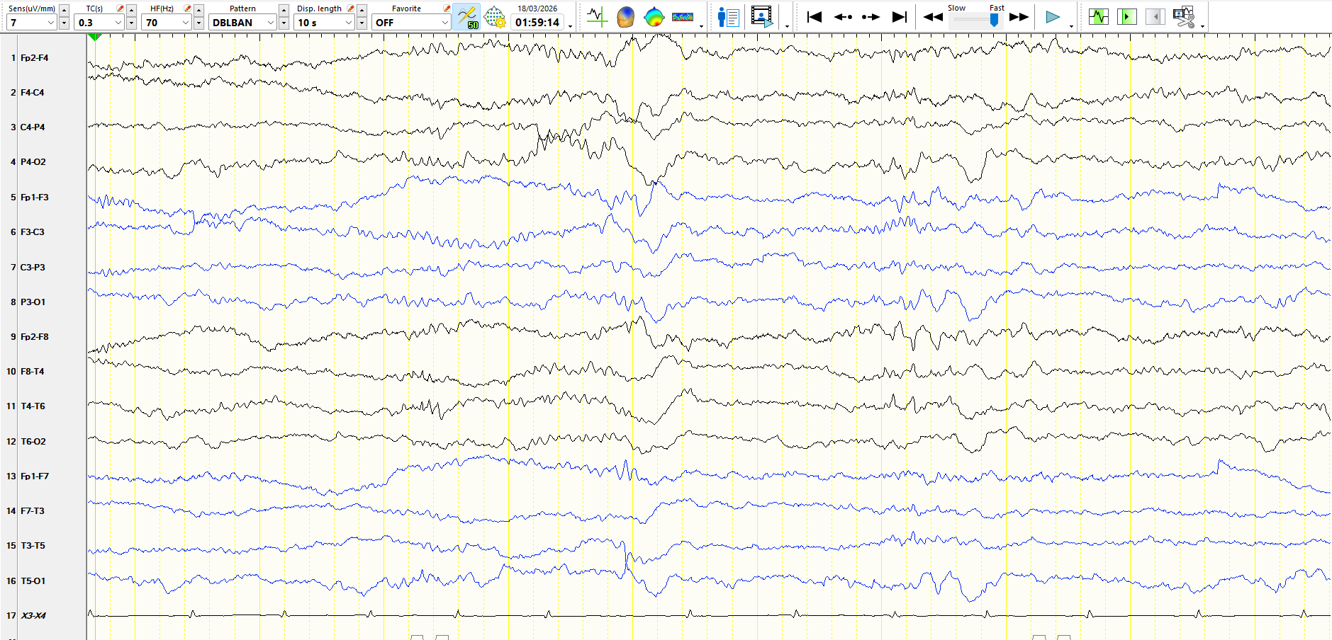
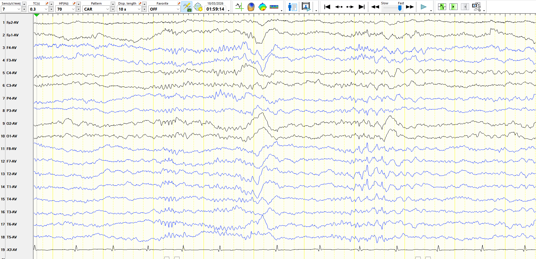
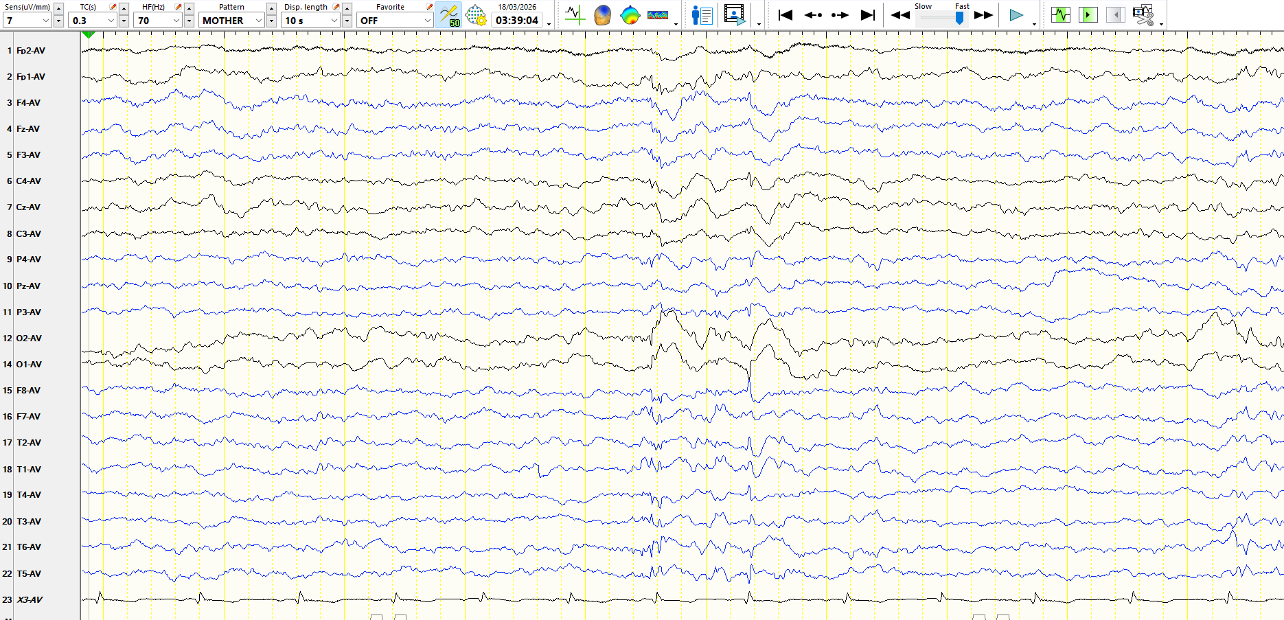
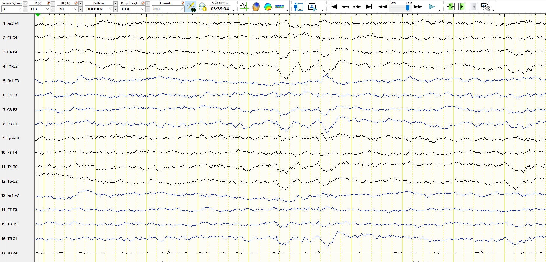
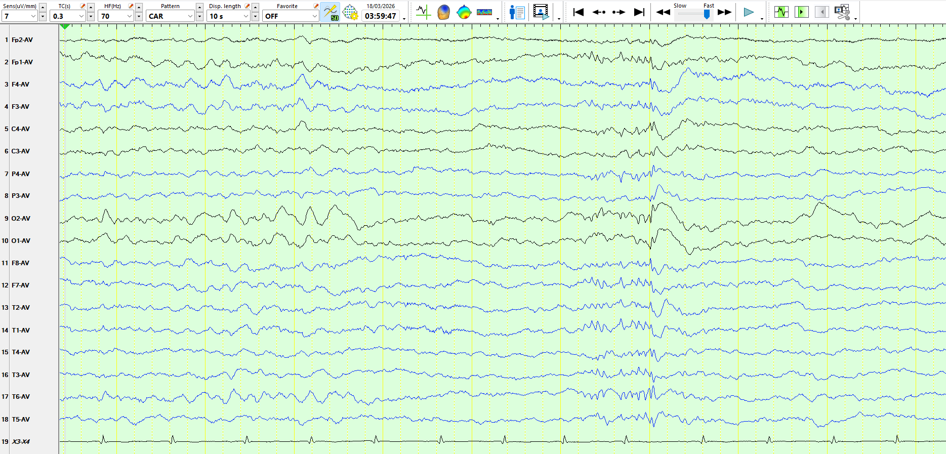
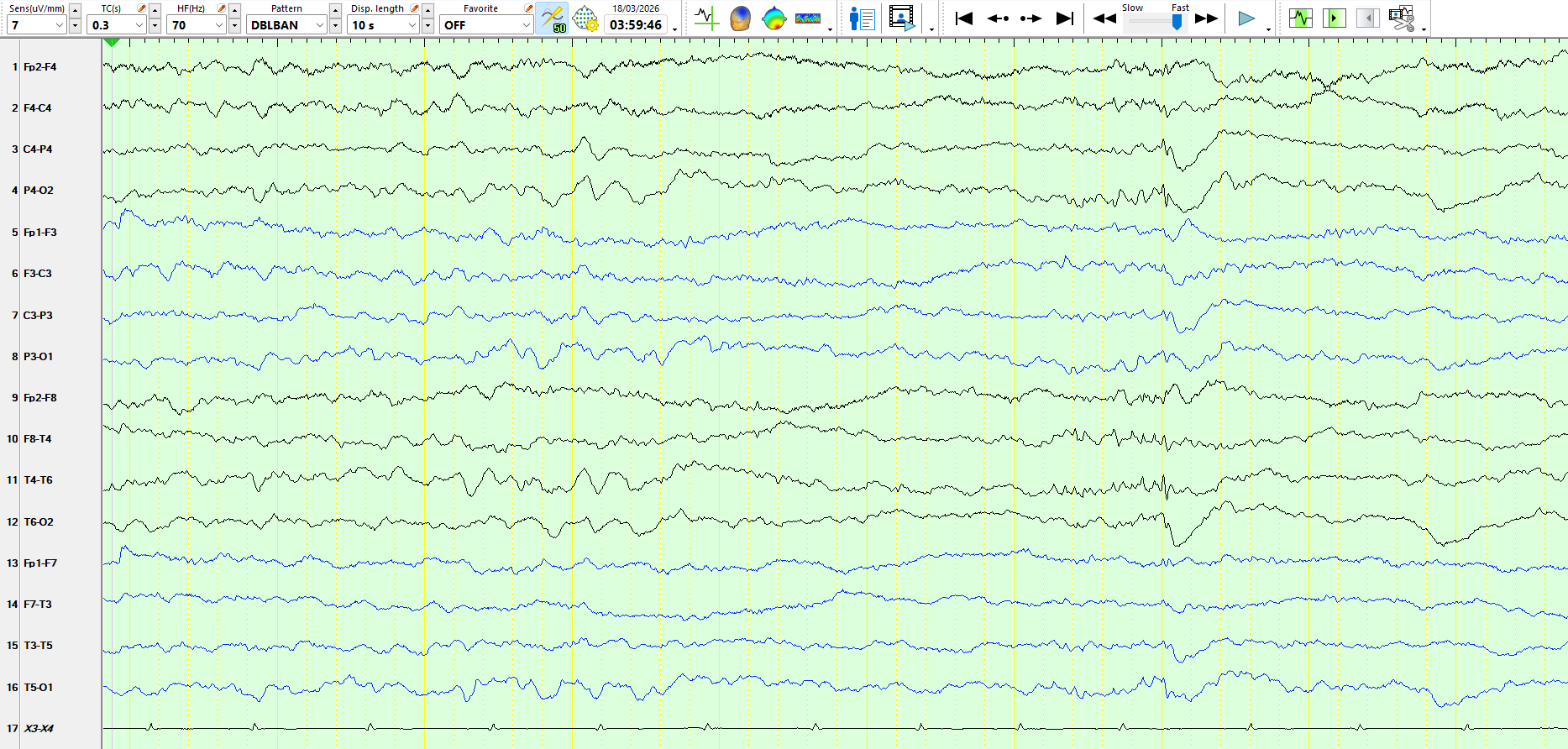
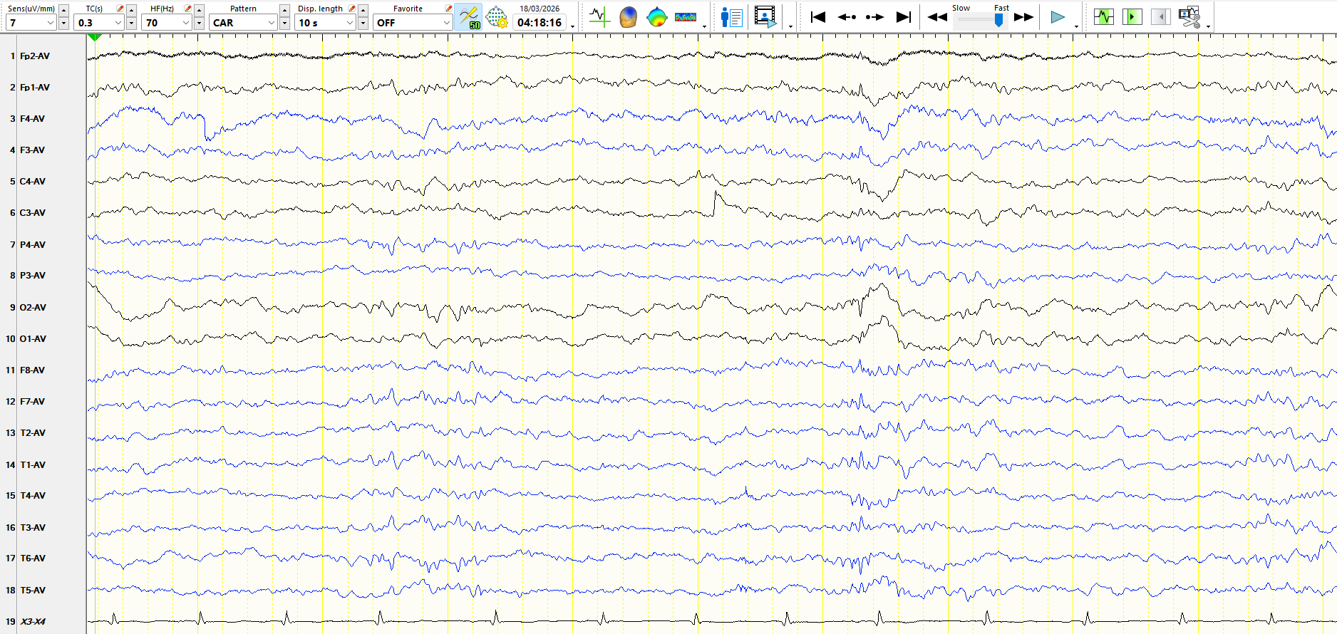
The EEG is normal during wakefulness. Here are a variety of pages from sleep, each represented in different montages. These discharges appeared profusely, typically at least once every 10 seconds during stage I and stage II sleep.
Fig 1
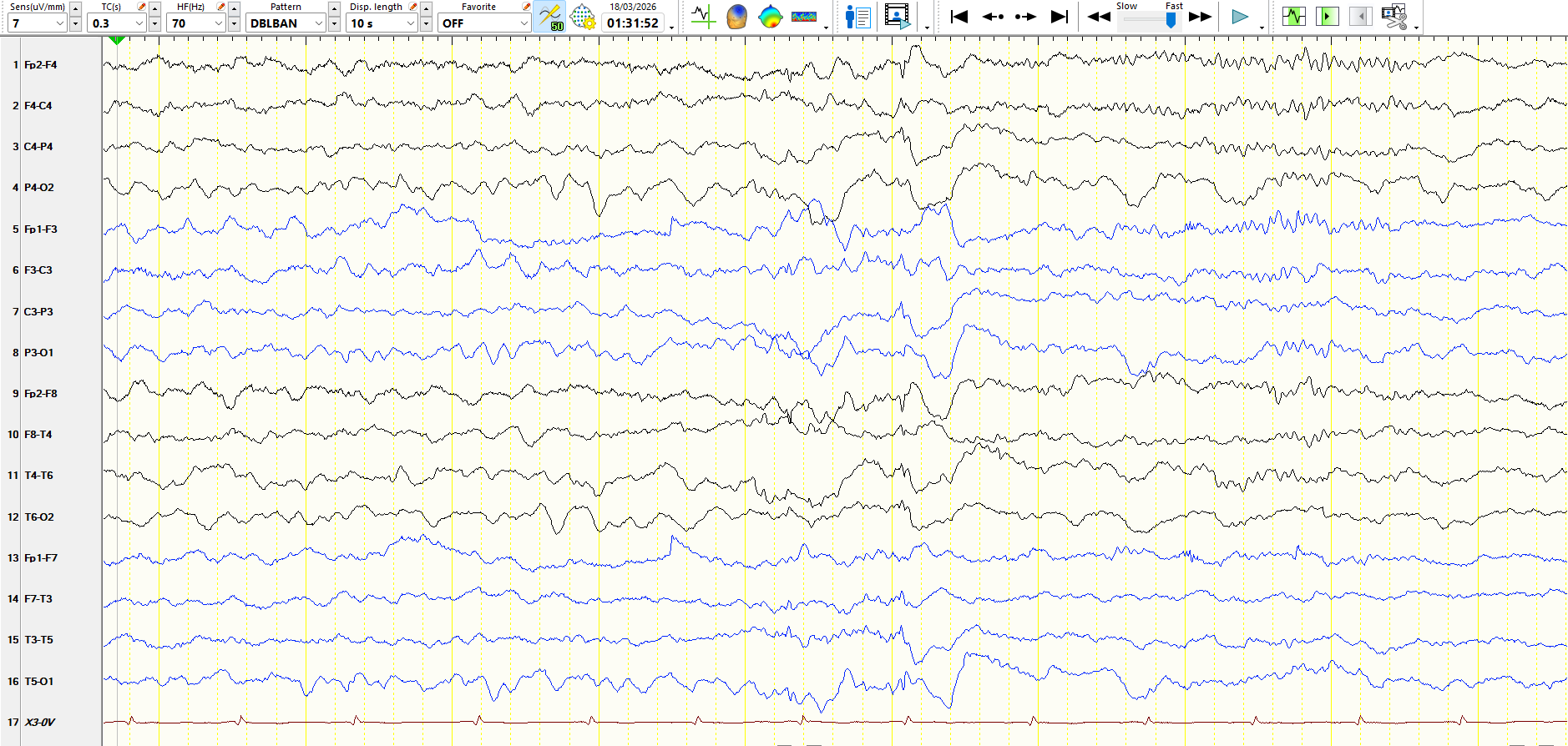
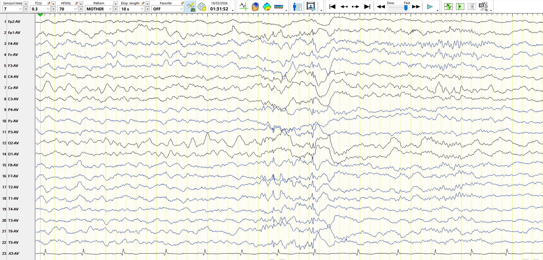
Figure 2
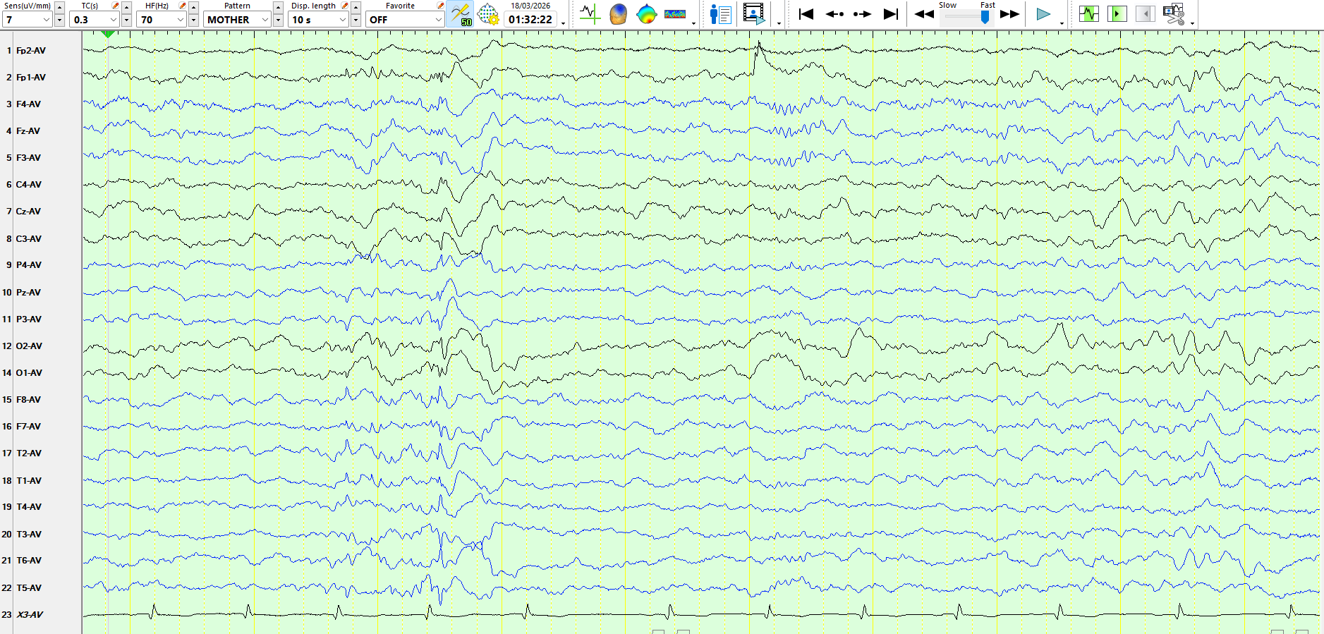
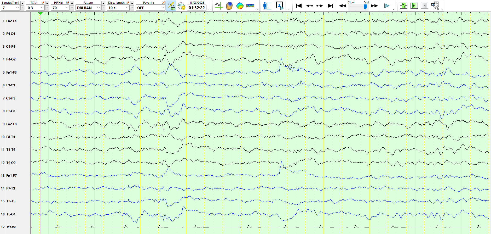
Figure 3
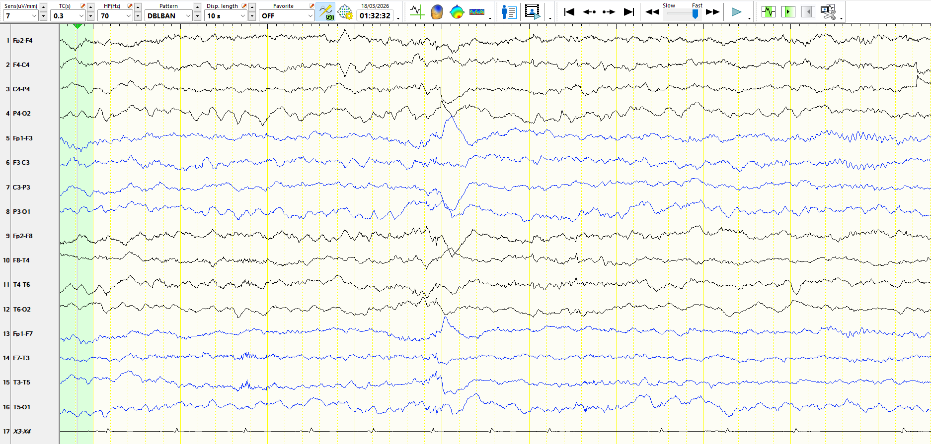
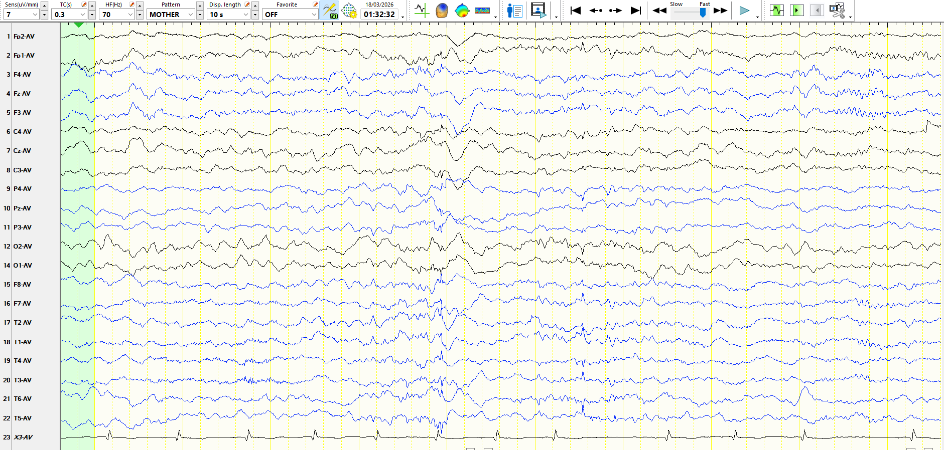
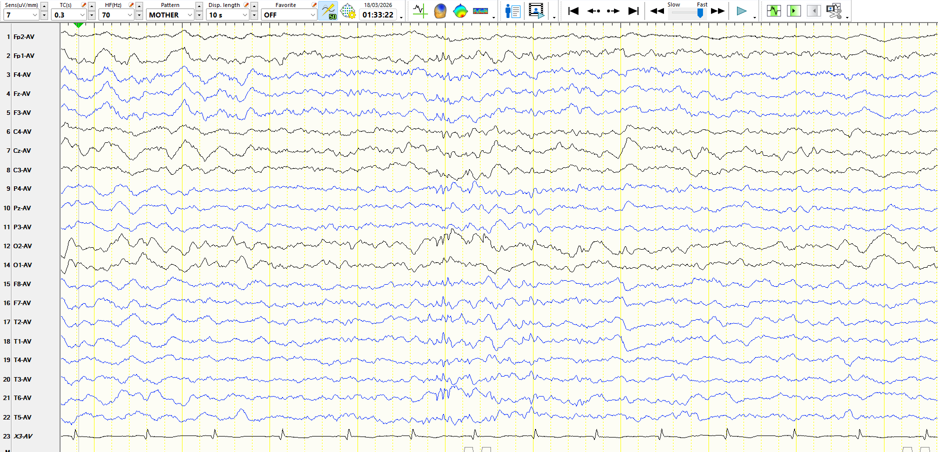
Figure 4
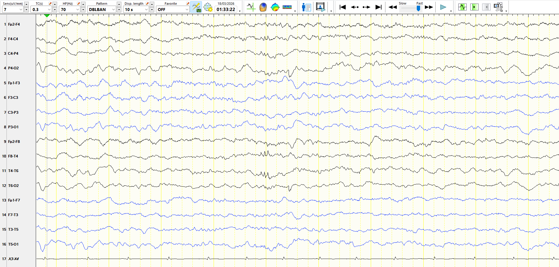
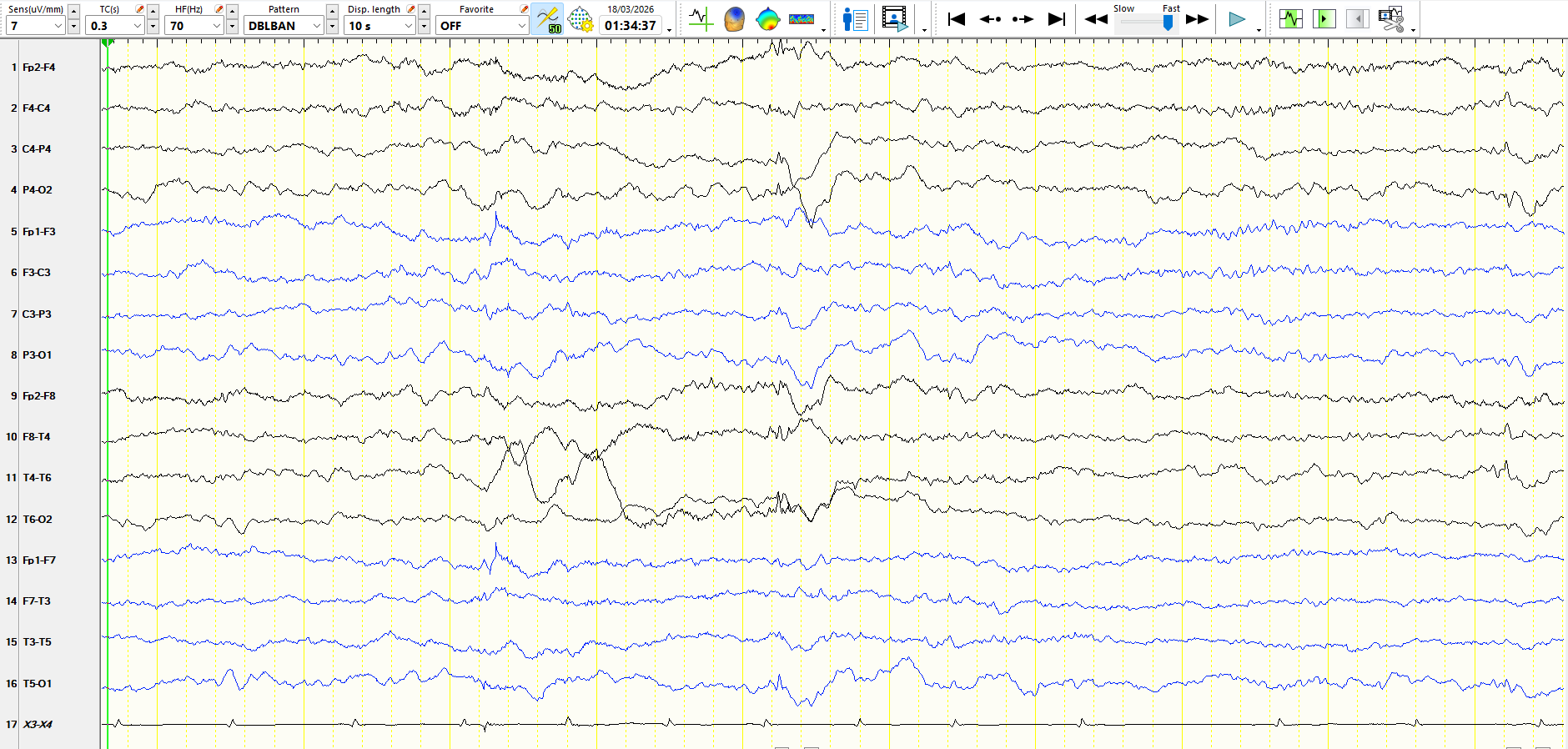
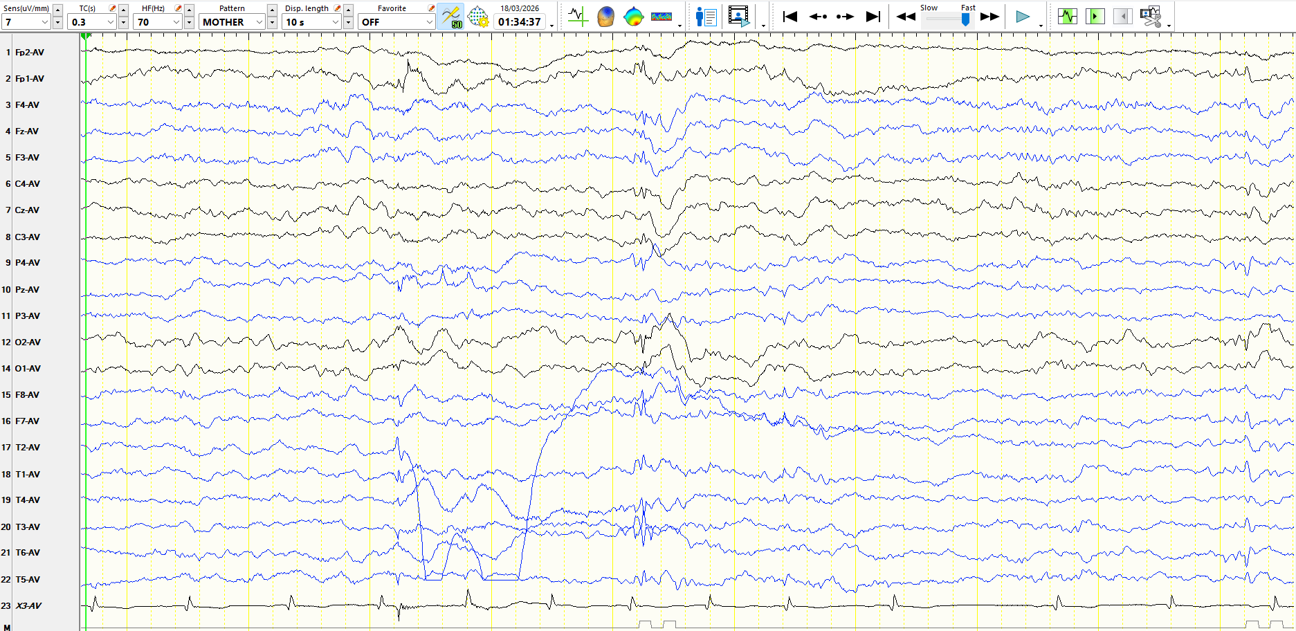
Figure 5:
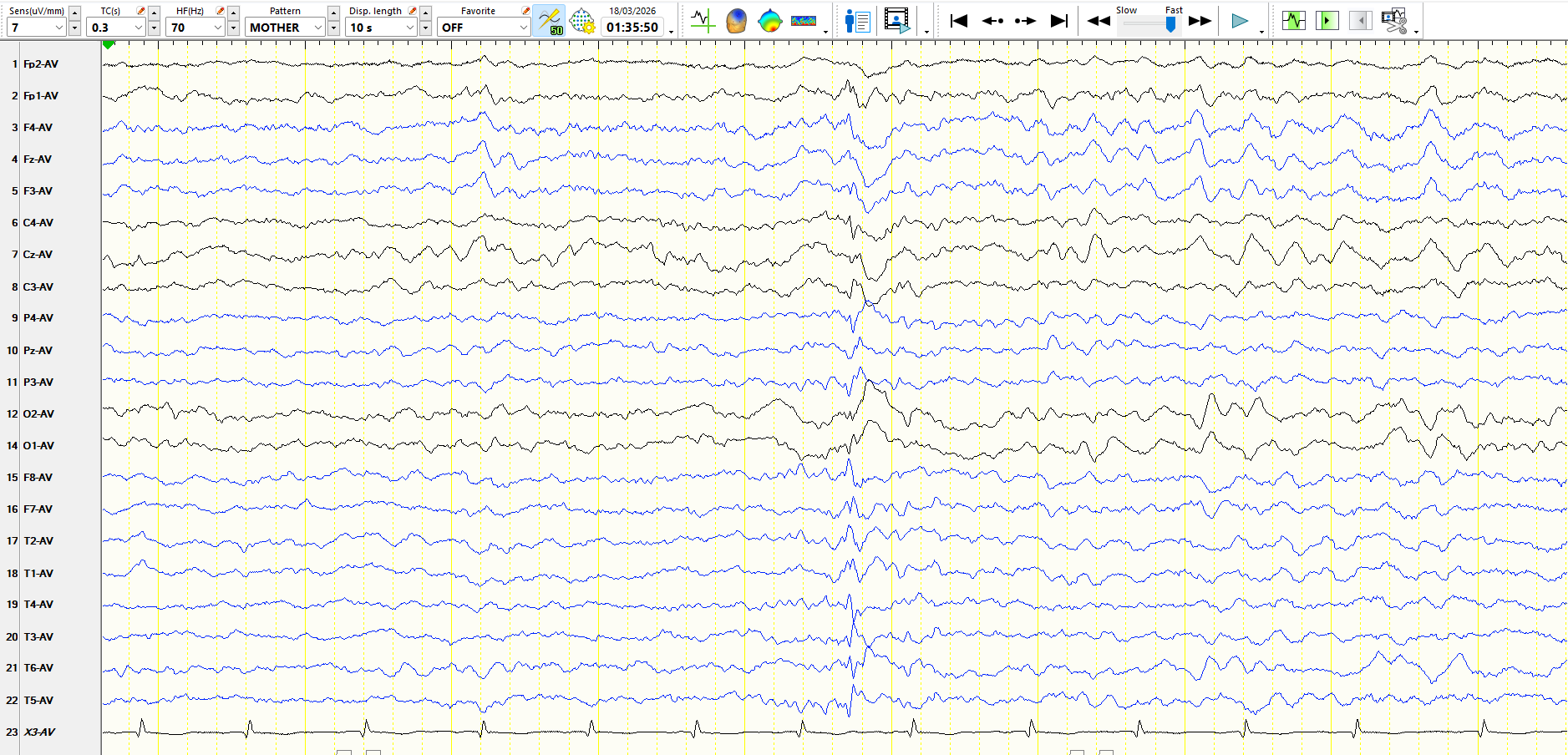
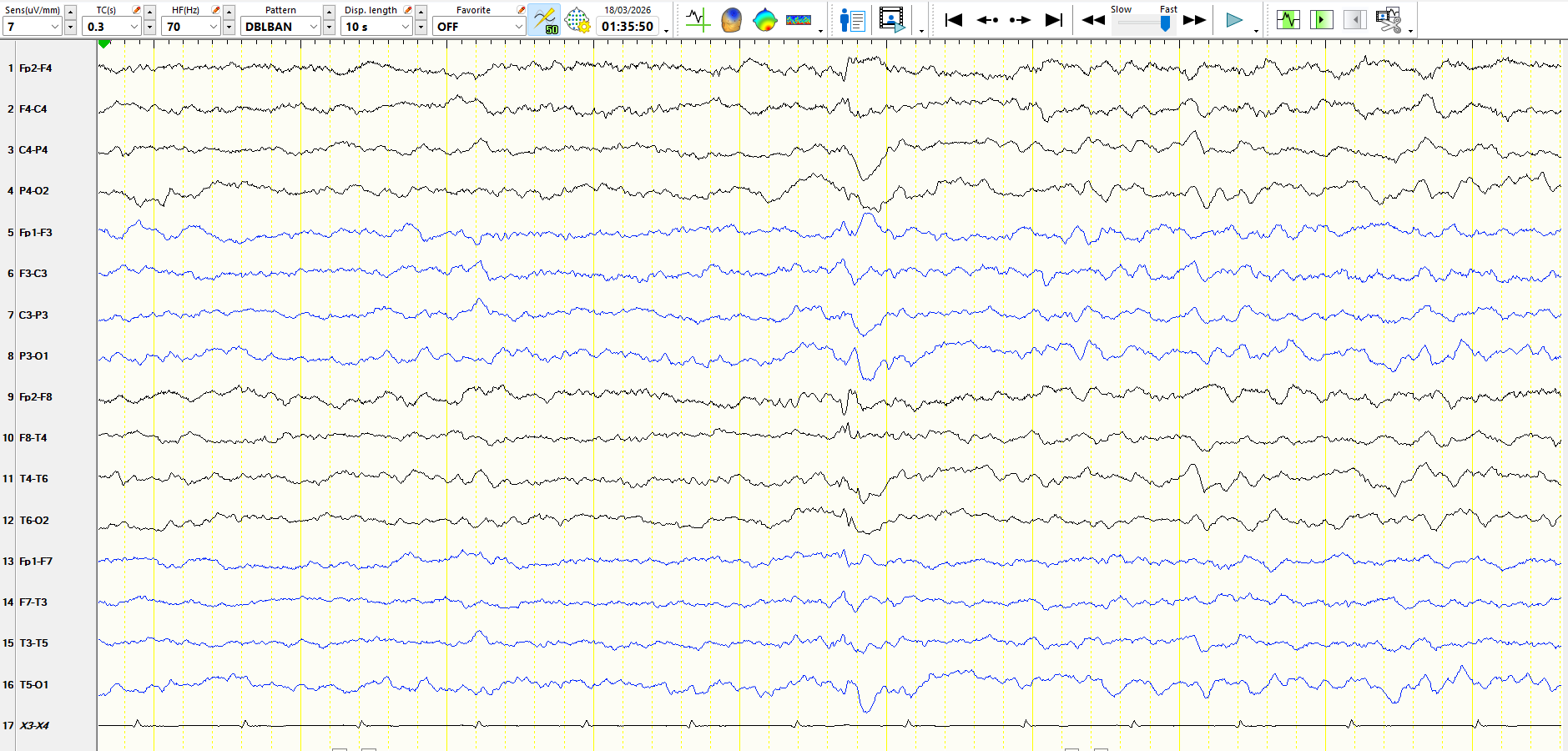
Figure 6
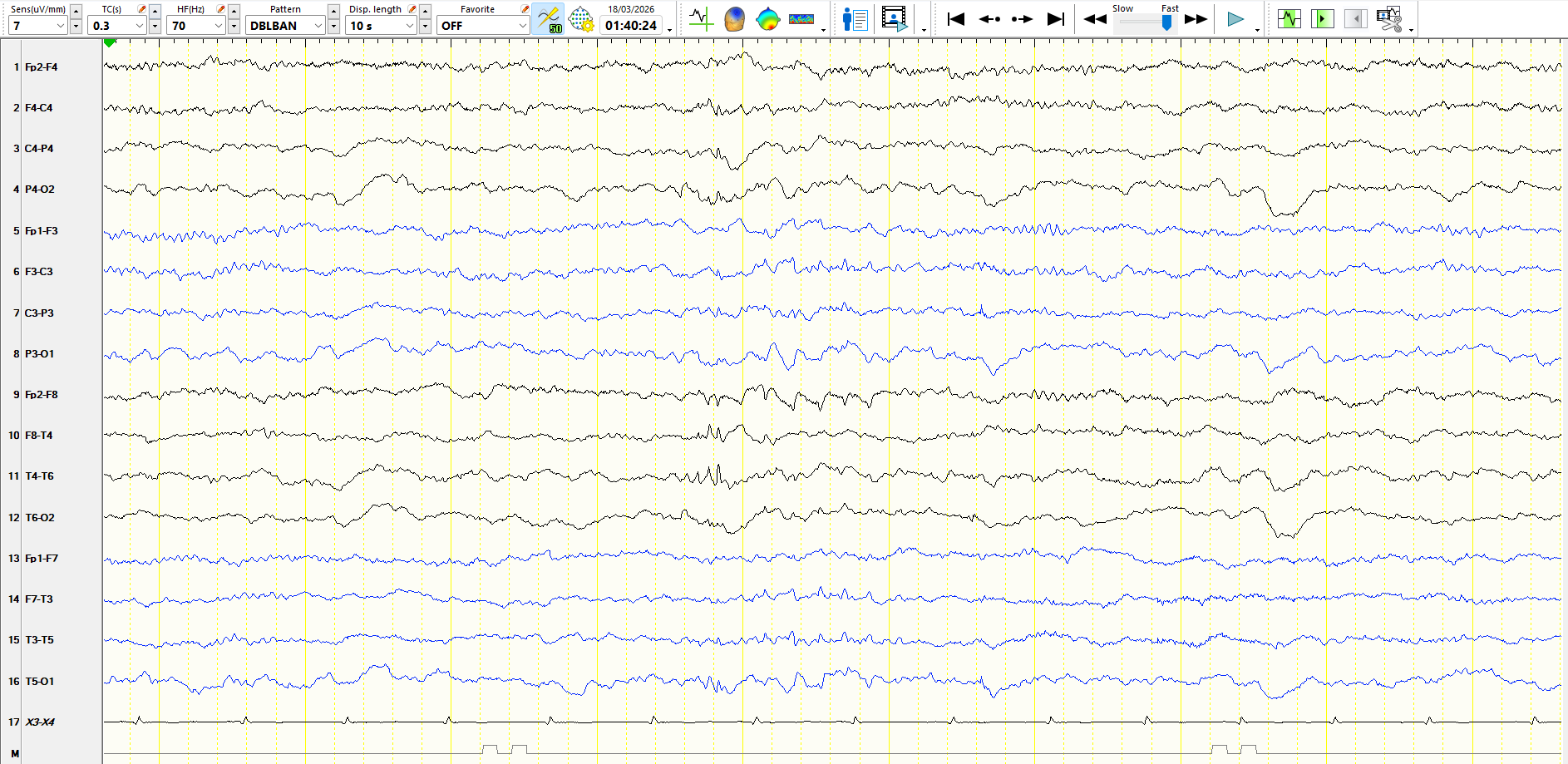
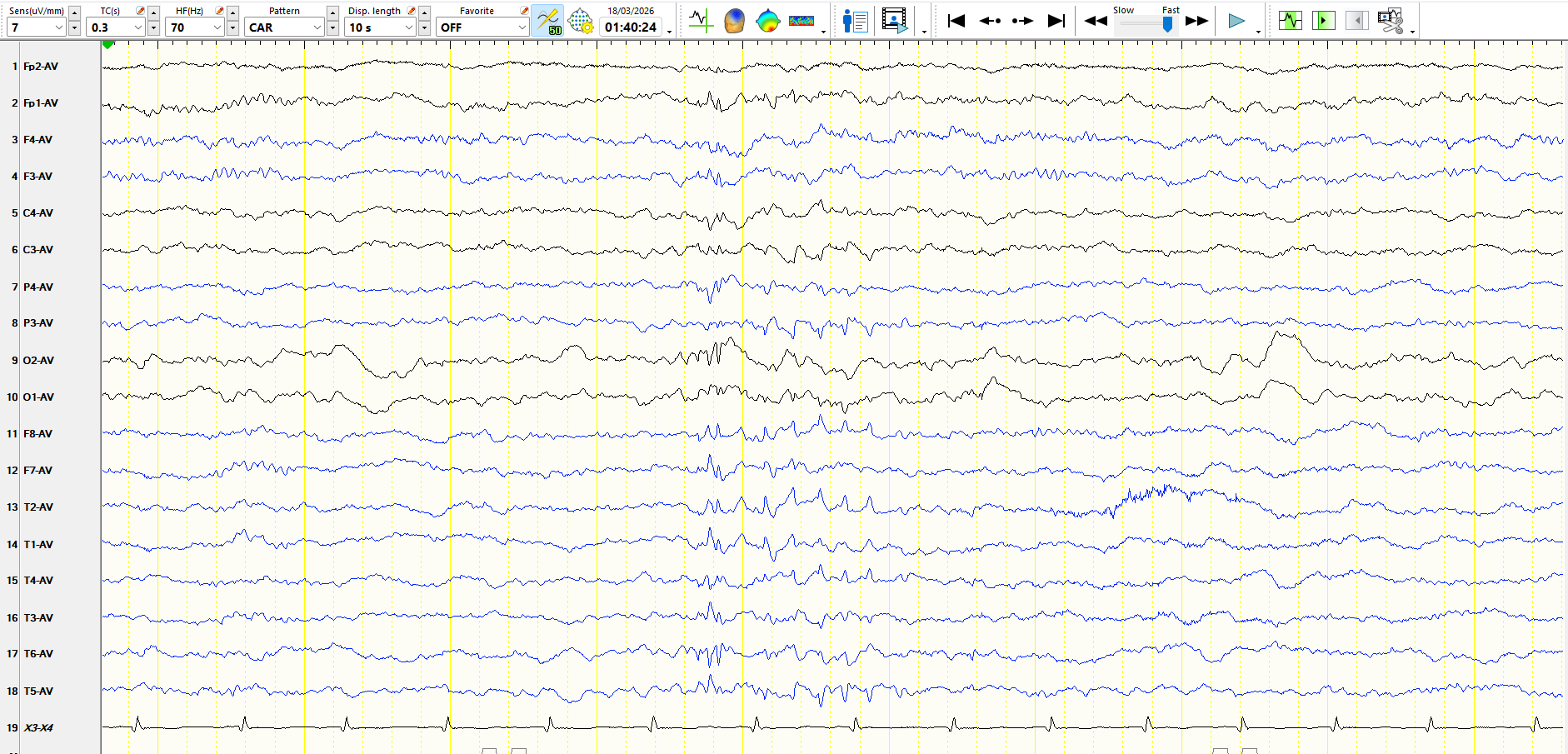
Figure 7
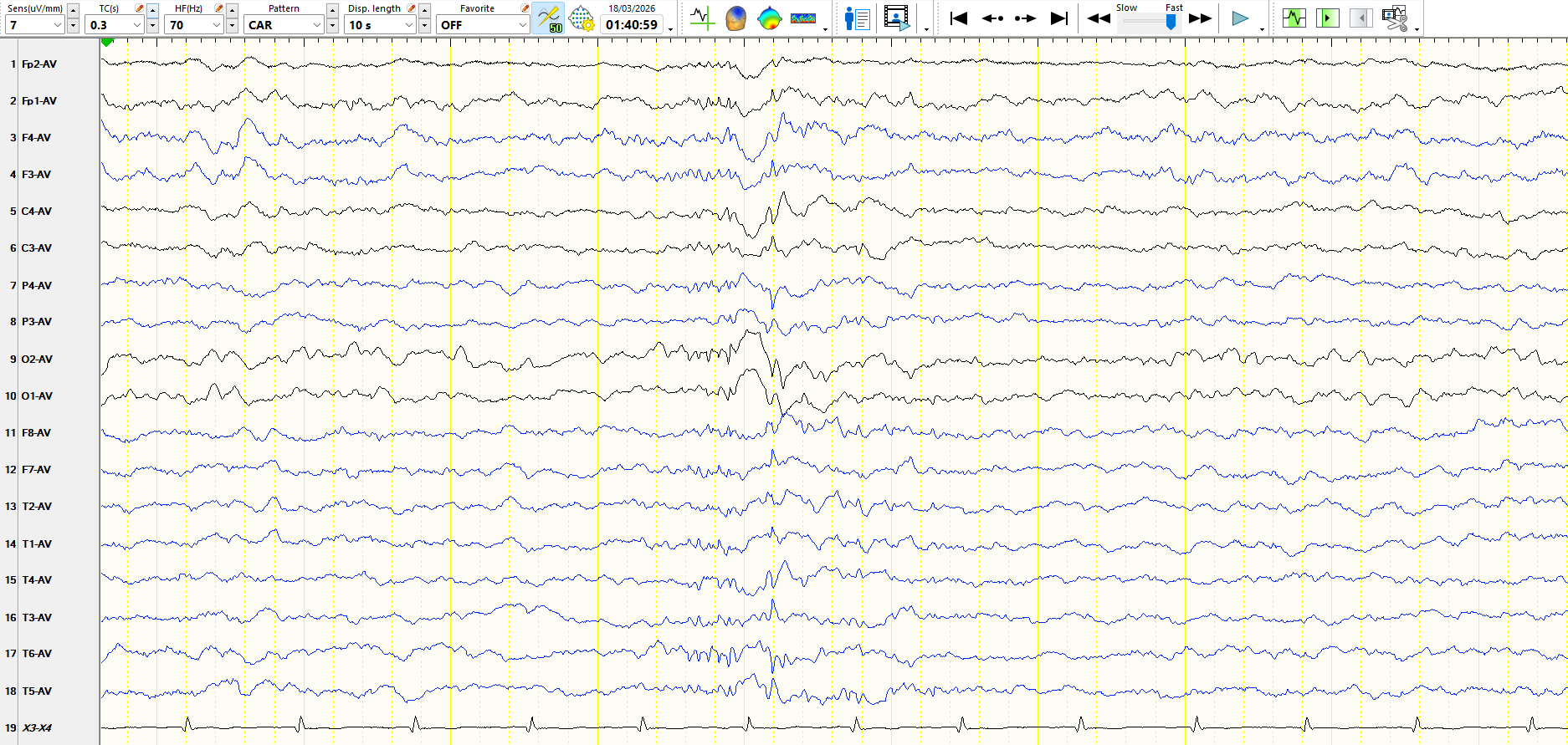
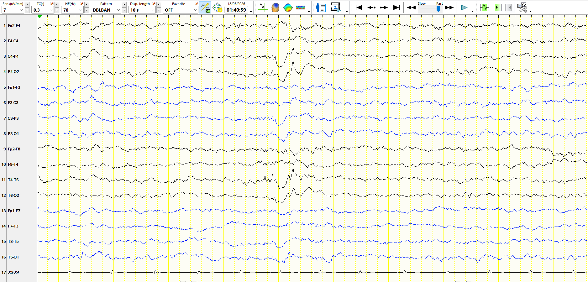
Figure 8
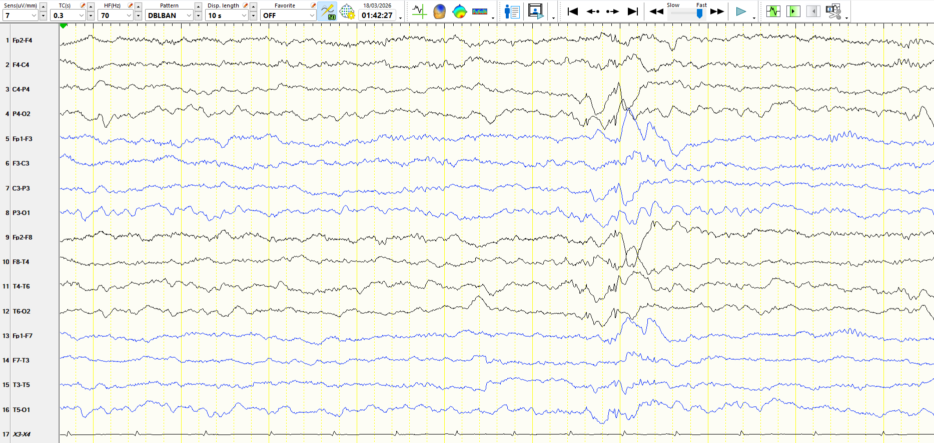
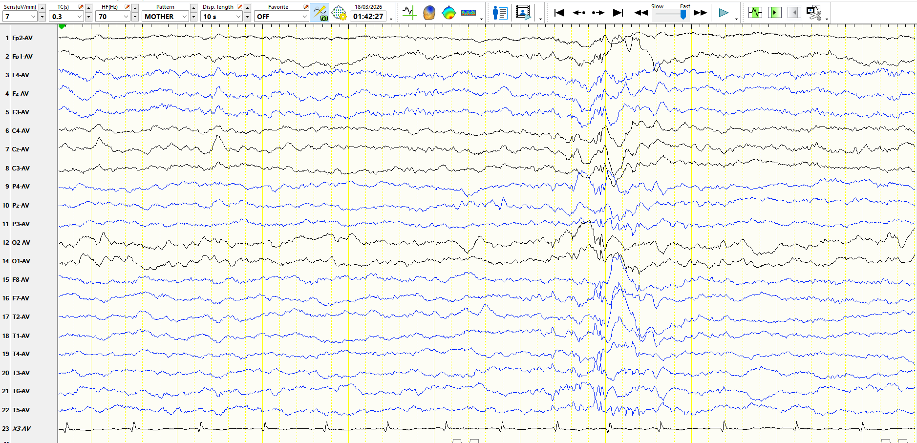
Figure 9
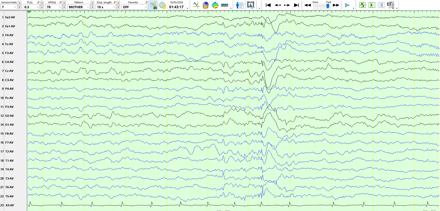
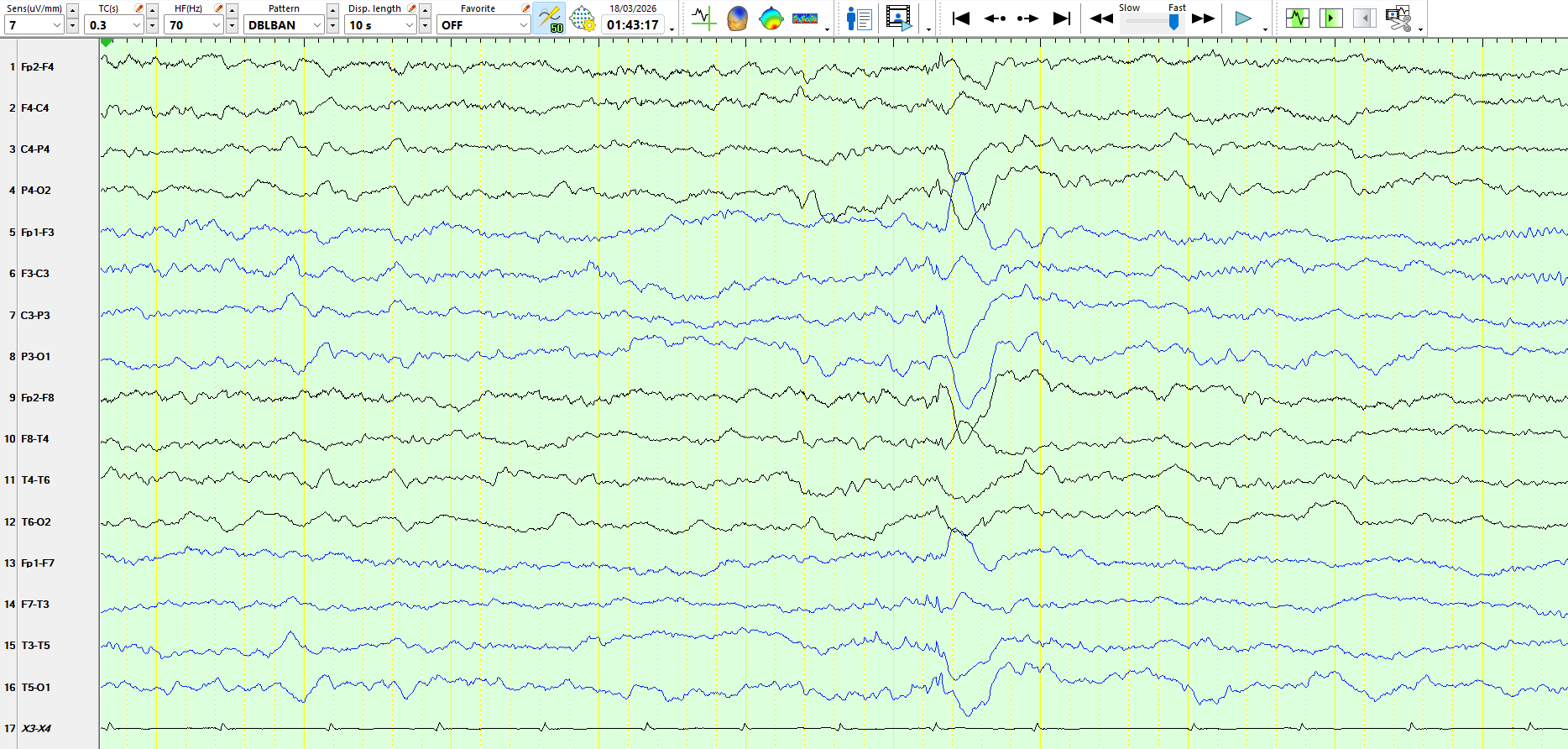
Figure 10
It is debatable whether an EEG should ever have been requested on this patient, but it was and led to a diagnosis of epilepsy. You might call this a compounding of errors.
This is a good example of 14- and 6-Hz positive bursts. The slow waves, preceded by faster frequencies, create the appearance of spike-and-wave. The morphology and frequency of the 14 Hz waves are diagnostic of this benign variant. In some of the pages above ( Figure 6), there are typical 6 Hz discharges. It is not uncommon to see slower waveforms than 6 Hz, and in some of the pages, there are 2-3 Hz slow waves. The key feature is that these slow waves appear at the end of the 14- and 6-Hz positive bursts. You might even be tempted to call these 14- and 6-Hz positive bursts and slow!
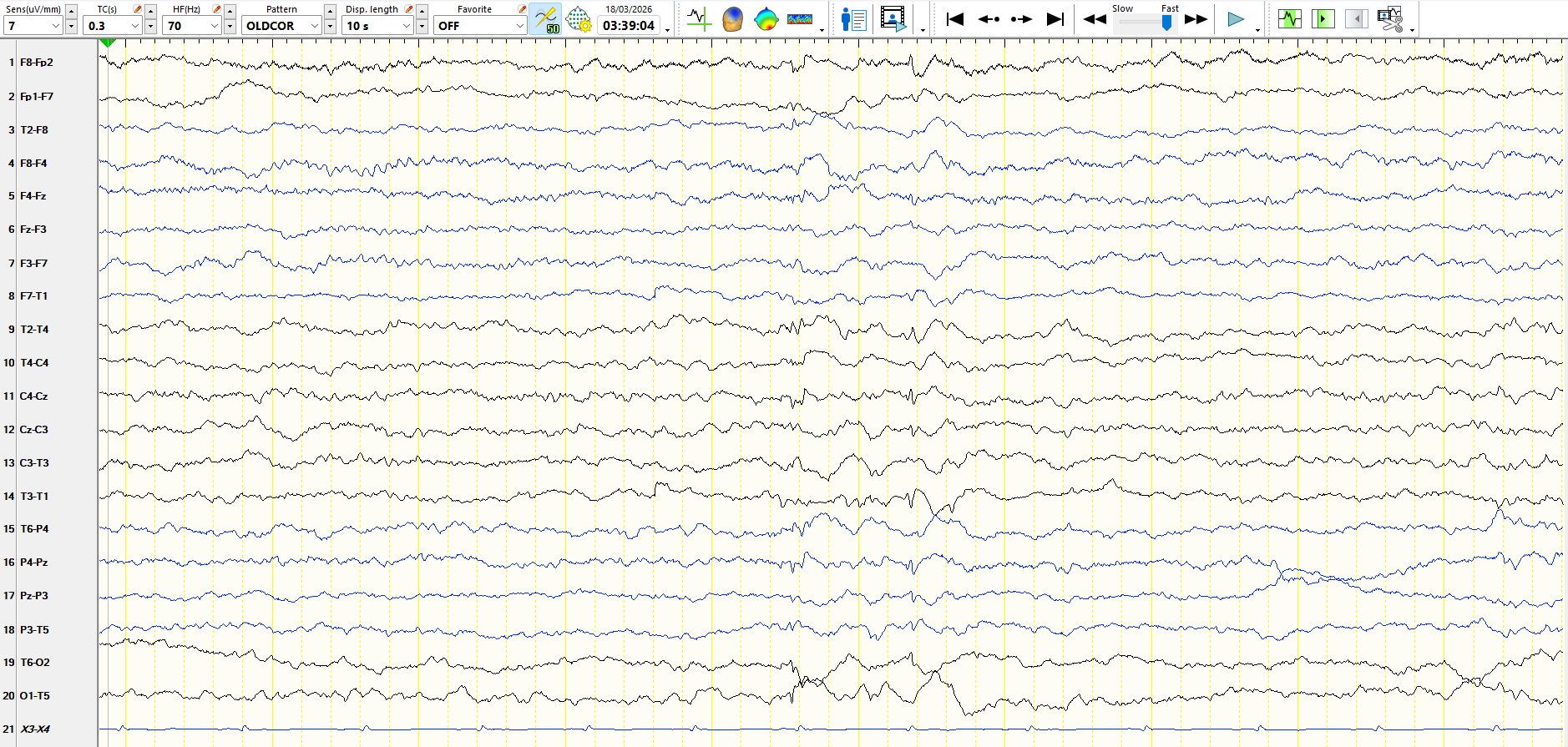
The 6 Hz waves may even appear "focally" on bipolar montages, creating the illusion of temporal sharp waves, as in the following example:
The 14 Hz waves, with their characteristic arciform appearance, can be seen in the 3rd second, while the 6 Hz waves can be seen in the 6th second.
 You may appreciate why these waves are so commonly overinterpreted.

The intracranial correlate of the 14&6/sec positive spikes normal scalp EEG variant - ScienceDirect