
An early Sunday morning call from the emergency unit medical officer working at a nearby hospital concerns the issue of a white cell count of 19 (81% polys & 11% lymphs) and a CRP of 14 in a patient, ...
One can certainly see spikes in one electrode. However, this is an uncommon phenomenon and more often than not a sharply-contoured wave or a spike-and-wave-like discharge is an electrode artifact. Hav...
There are plenty of discharges on an EEG that mimic seizures and many of these are simply normal physiological relevance, either while awake or asleep. Believe it or not, but sometimes "focal" seizure...
At the age of 7 years the patient developed absence seizures, with innumerable daily events characterized by brief episodes of staring and unresponsiveness, without warning and without any post-ictal ...
If this problem vexes you at times, be assured that you are not alone. The distinction between these on morphology alone is often impossible. The former in the title raises the possibility of encephal...
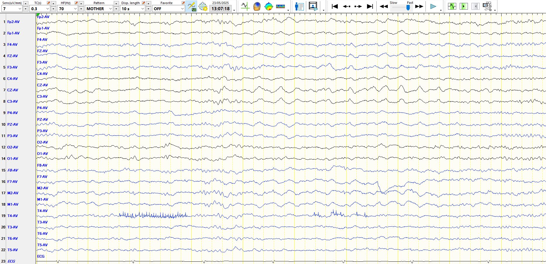
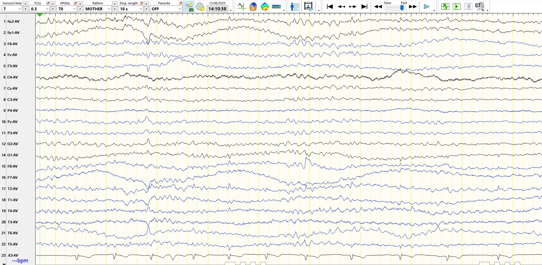
Here are a few more wicket waves. The patient is 52 years old and likely has left temporal lobe epilepsy, with a normal MRI scan.
In the above, the patient is asleep. Notice the low amplitude i...
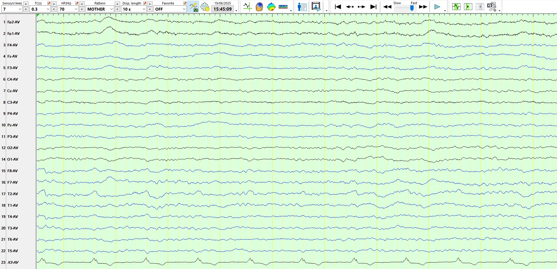
Here are some examples of wicket waves in the occipital and temporal regions from a patient who is 33 years old:
It is always a good idea to pay attention to background rhythms whenever you look a...
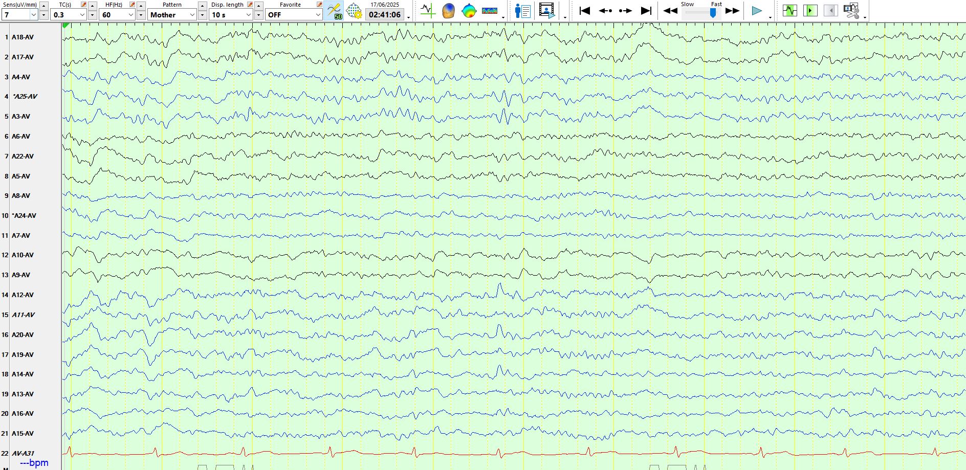
This is a good example of electropositive wicket waves. Try to invert the waves in your mind; if are able to do so, you might notice the resemblance of these waves to wicket waves posted previously.
...A lesion on the MRI scan. What do you make of these theta waves on the first page? Seizure while working on a yacht; his career depends on this.
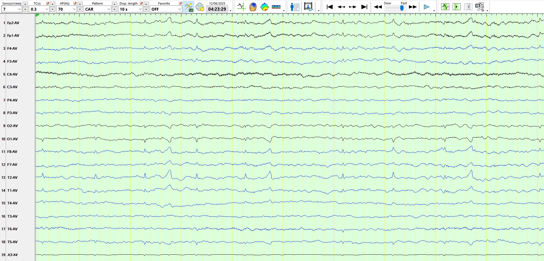
An isolated wicket wave (not synchr...
4-year history. Once or twice a year, "I would lose peripheral vision" (bilaterally), "objects would look crystallized" and then in some "I could not read, the words appeared jumbled" and others notic...
