
Triphasic wave vs sharp and slow wave
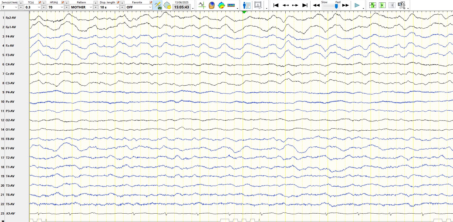
Jun 18, 2025If this problem vexes you at times, be assured that you are not alone. The distinction between these on morphology alone is often impossible. The former in the title raises the possibility of encephalopathies, especially those of metabolic cause, and prion disease, while the latter in the title predicts the diagnosis of epileptic seizures. Here is an example of the latter, 65 years old, with a history of epilepsy since the age of 12 and symptoms predictive of left frontal opercular seizures. He has a history of a propensity to clusters of seizures and status epilepticus. This is his 20-minute EEG 8 days after an admission via the emergency unit. He is up and about, talking, interactive and yet executive functioning is not completely normal. Notably, his language function appears to regress slightly at times during the course of the day, when he has word-finding difficulties during interaction with the nurses and me.
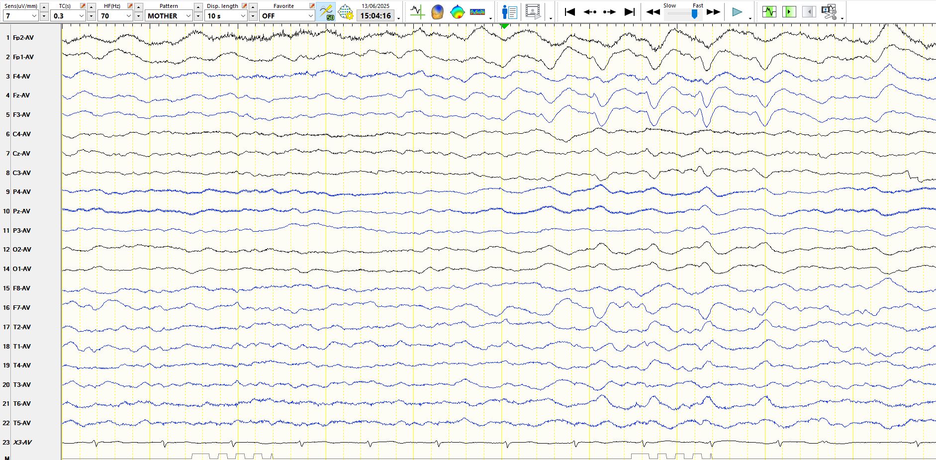
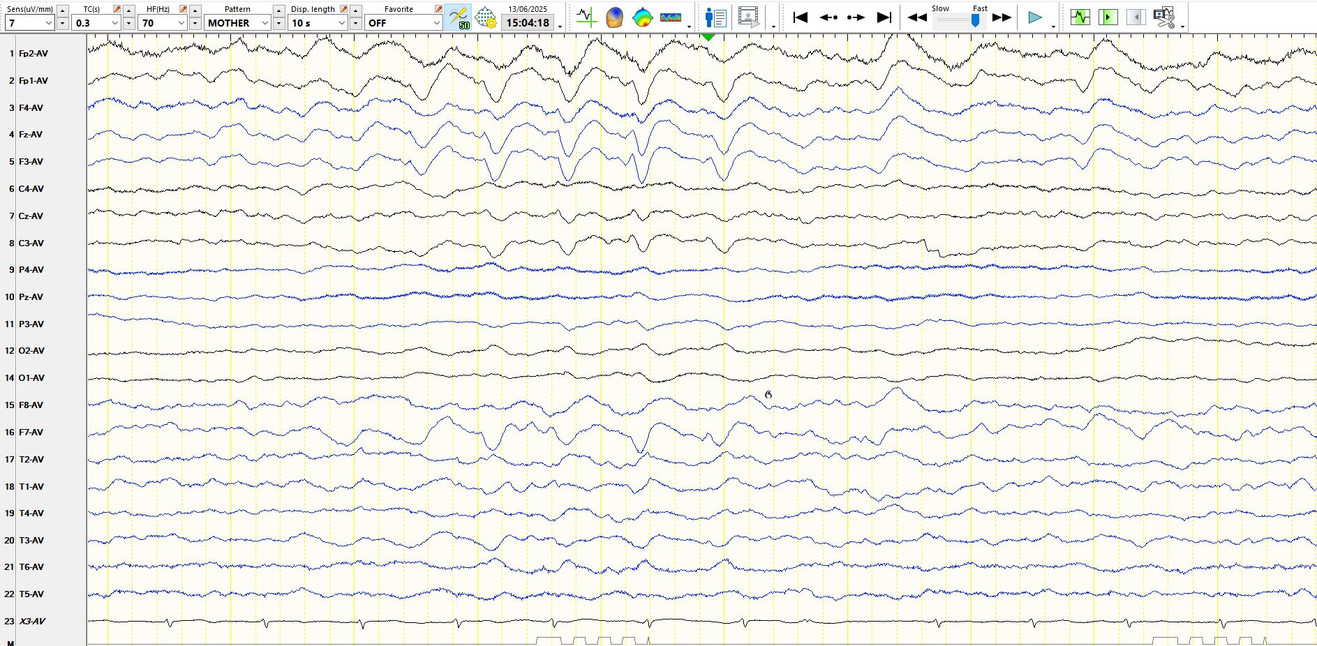
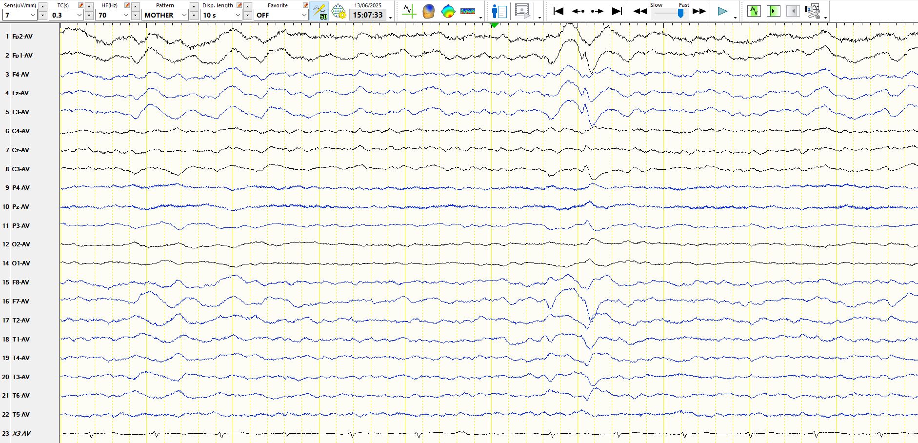
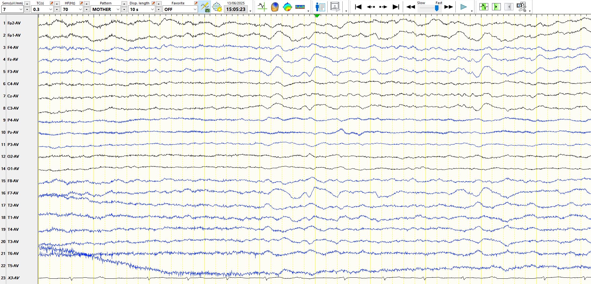
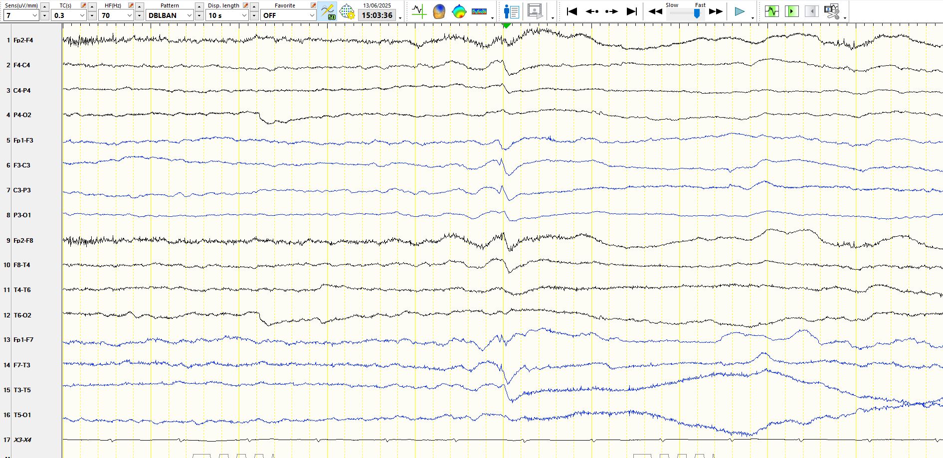
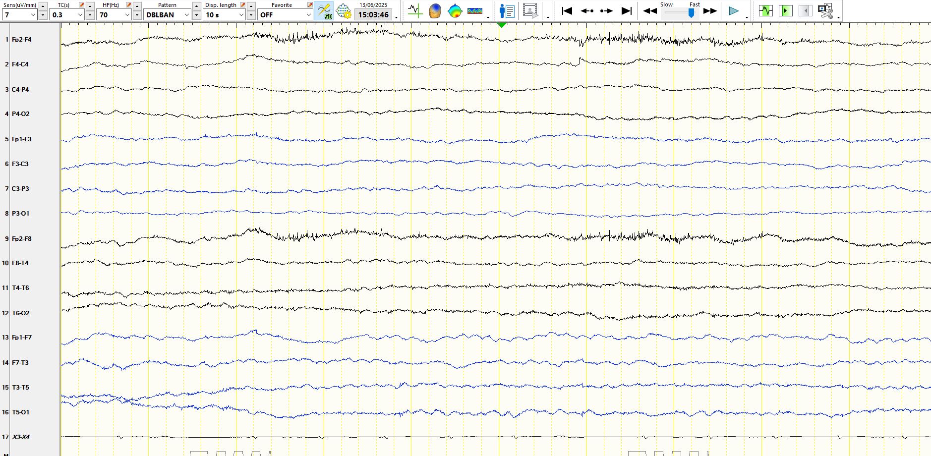
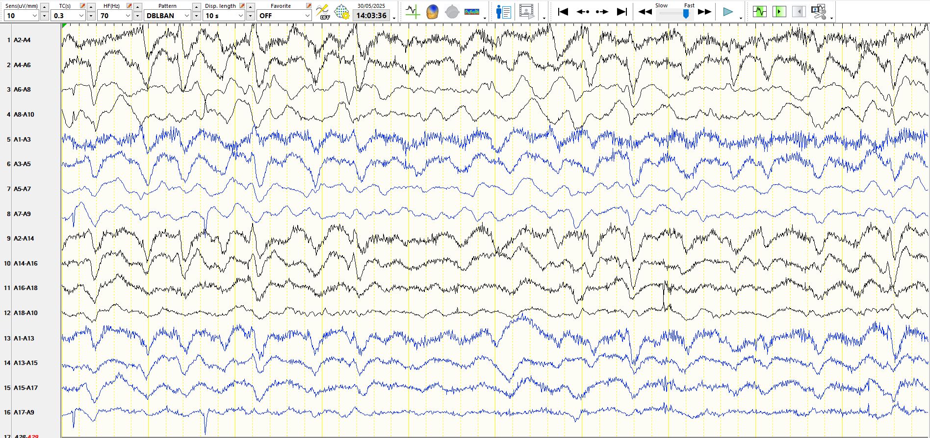
The following 4 are consecutive pages:
A.
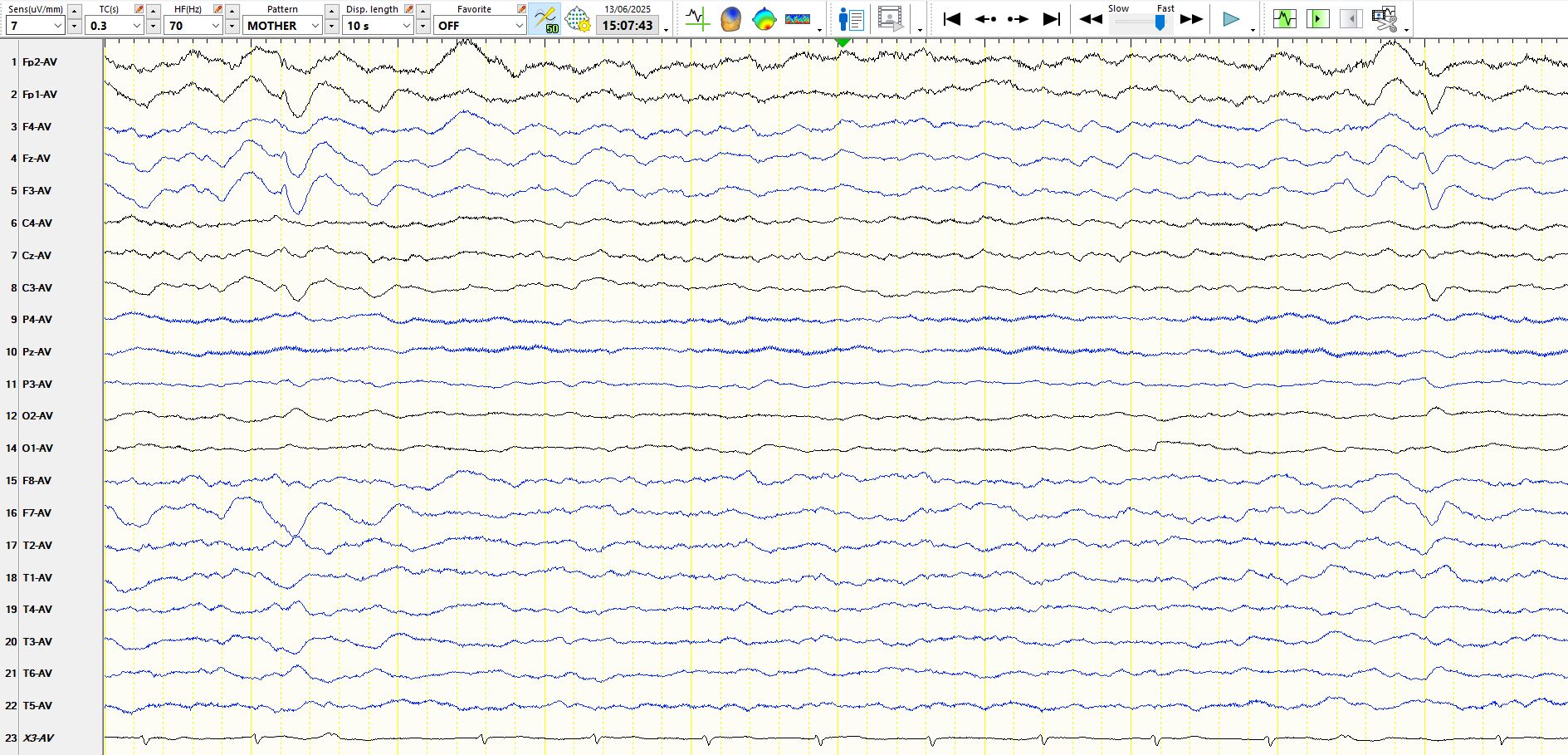
B.
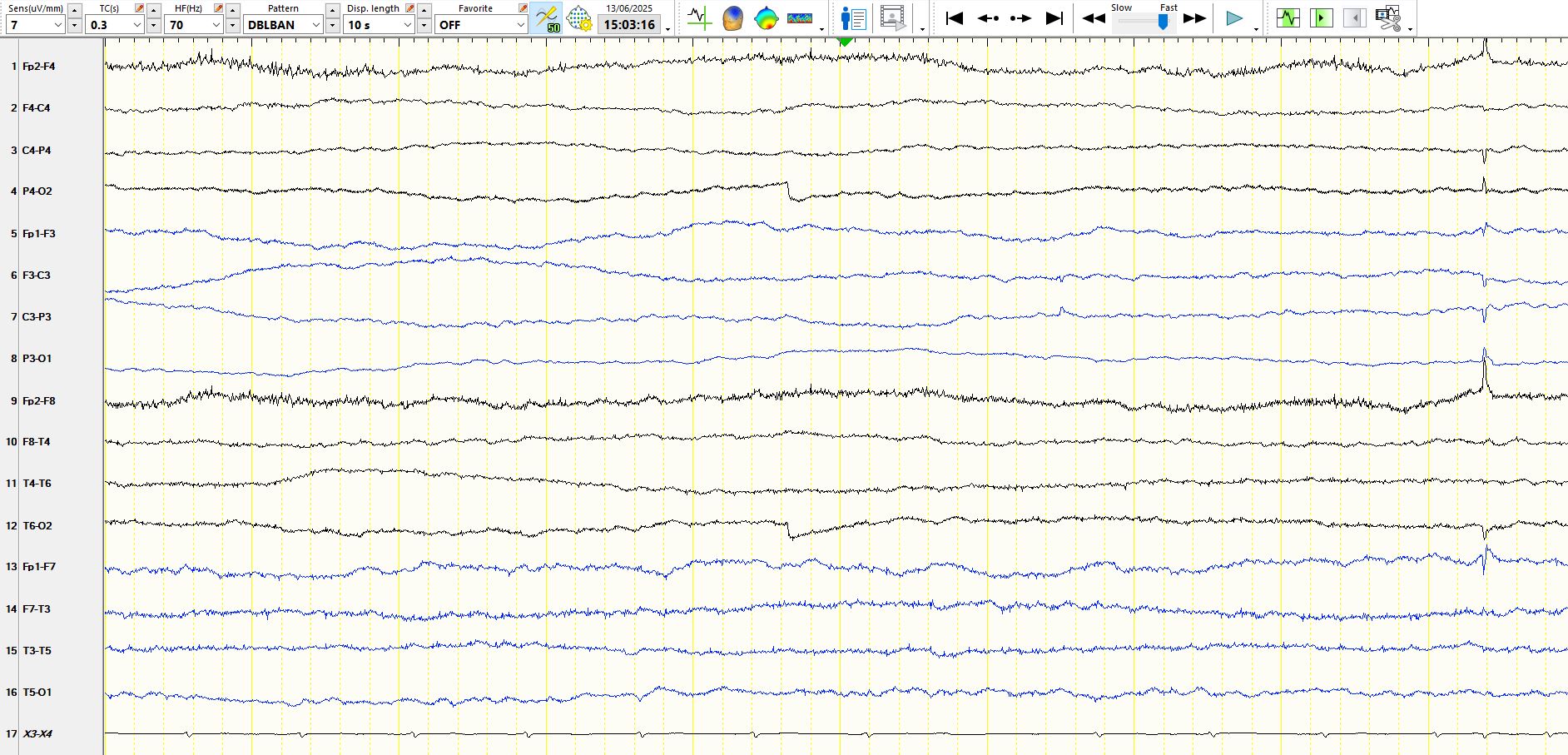
C.
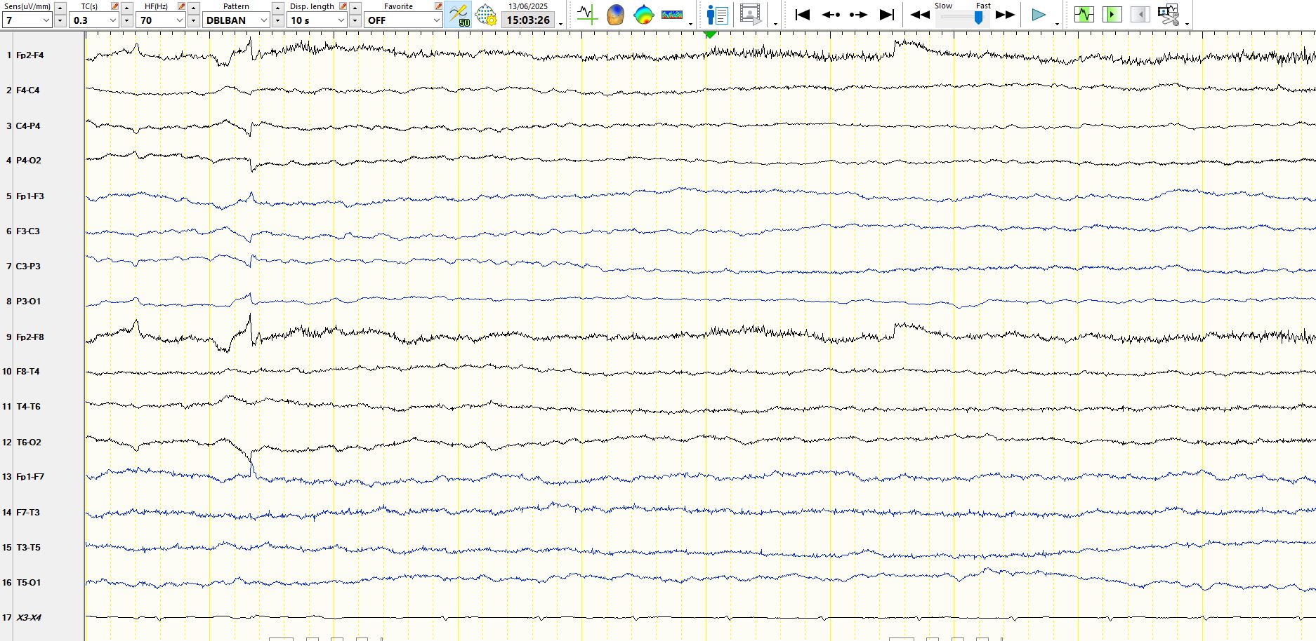
D.
This is a repeat of one of the above pages
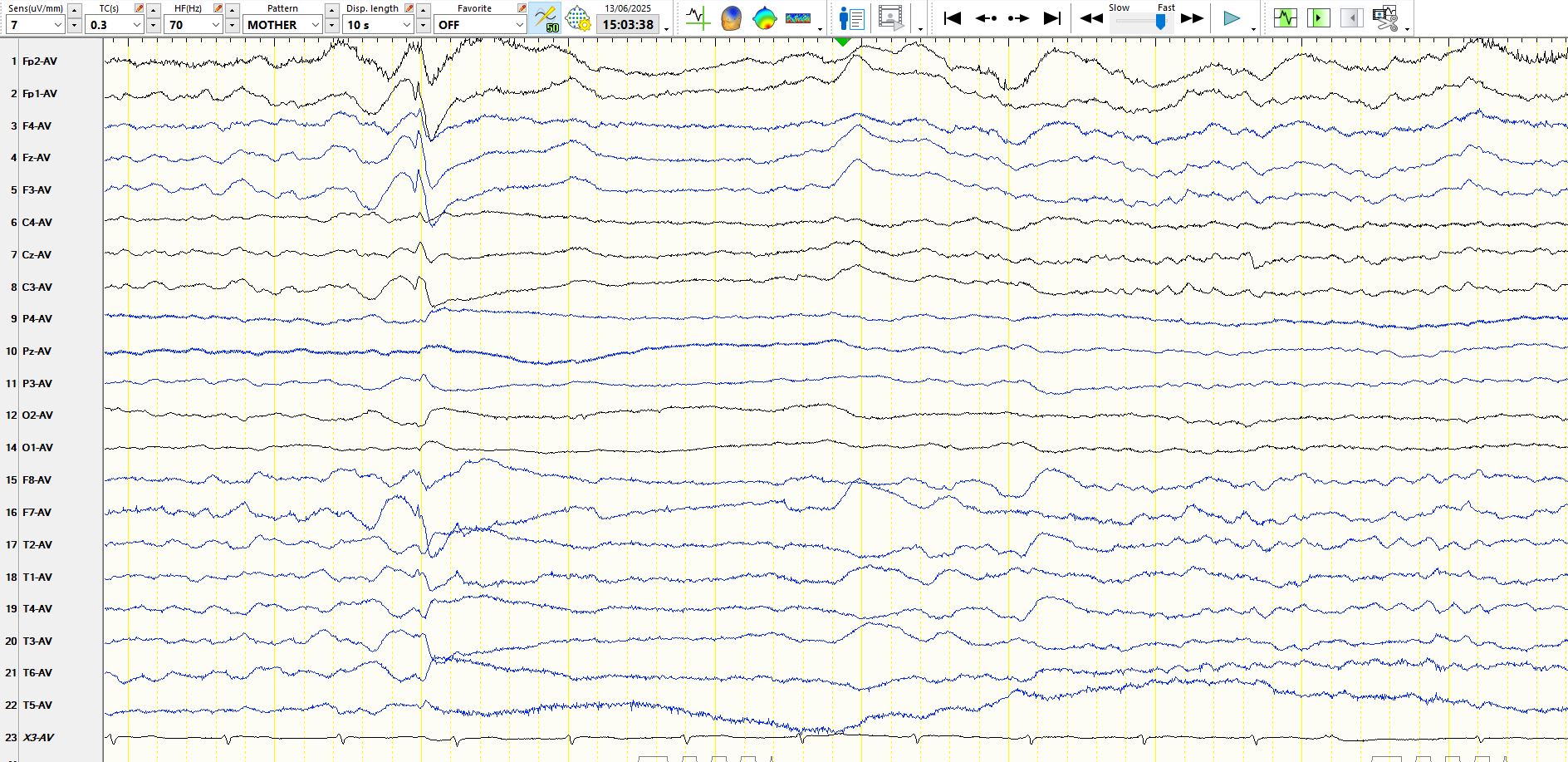
Believe it or not, but the above is a seizure, not noticed or remarked upon by the technologist. There is generalised desynchronization for approximately 3 seconds, followed by delta waves, before the EEG recovers. This almost certainly was a focal seizure and the presence of a spike at its commencement suggests that the seizure was primarily generated with in the left frontal lobe. The desynchronization likely would have had very low amplitude widely synchronous beta frequencies as its electrographic correlate on intracranial recordings. There may also have been a small region of a disproportionately high amplitude beta frequencies in the left frontal region on intracranial recording. These are the betting probabilities, based on experience. The remarkable feature is that desynchronization like this can be the manifestation of focal seizures. The 3rd EEG from the top in the sequence of 4 consecutive images (C) is the same subclinical seizure.
Compare this to the following EEG from a patient with Rett Syndrome, 7y
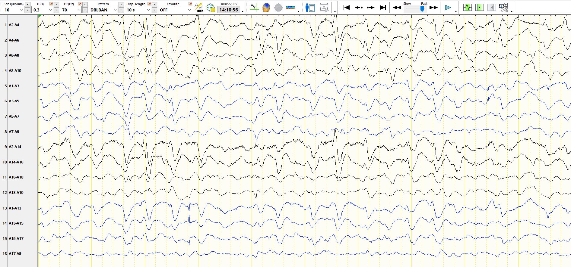
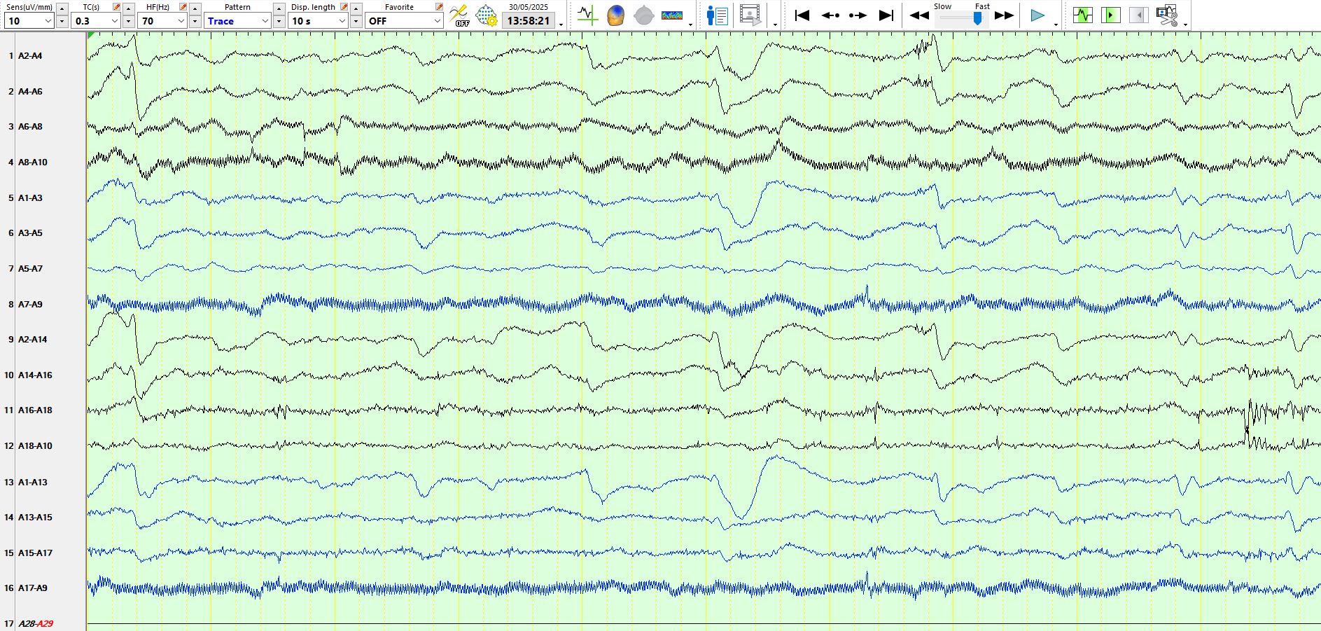
Some of the waves have a "triphasic" morphology, while others are unambiguously spikes and sharp waves. The patient has epilepsy.
Rett syndrome and the electroencephalogram - PubMed
Electroencephalographic findings in Rett syndrome - PubMed
PS, it should really be Rett Syndrome