19y, seizures?
Jun 13, 2025A lesion on the MRI scan. What do you make of these theta waves on the first page? Seizure while working on a yacht; his career depends on this.
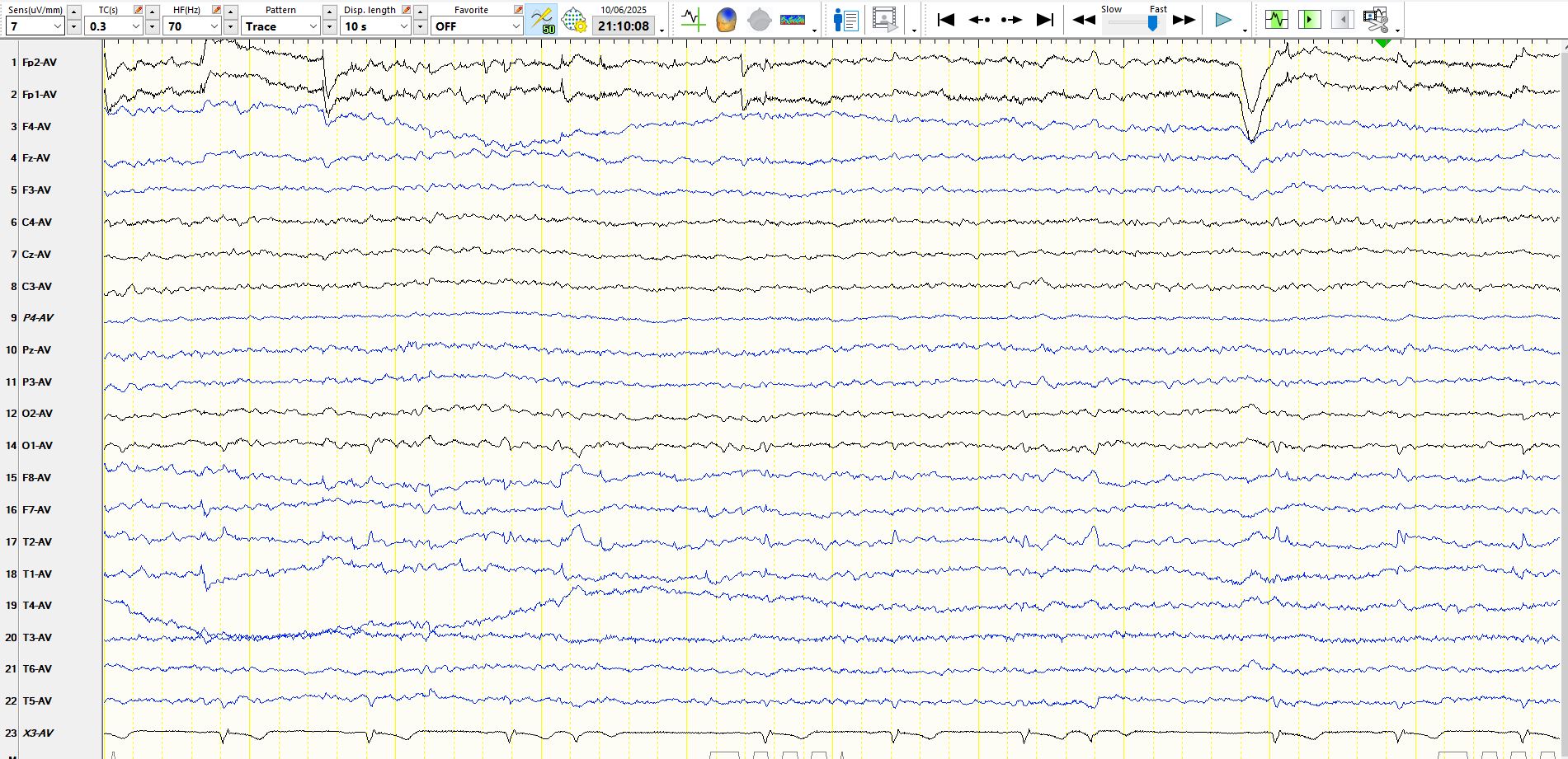
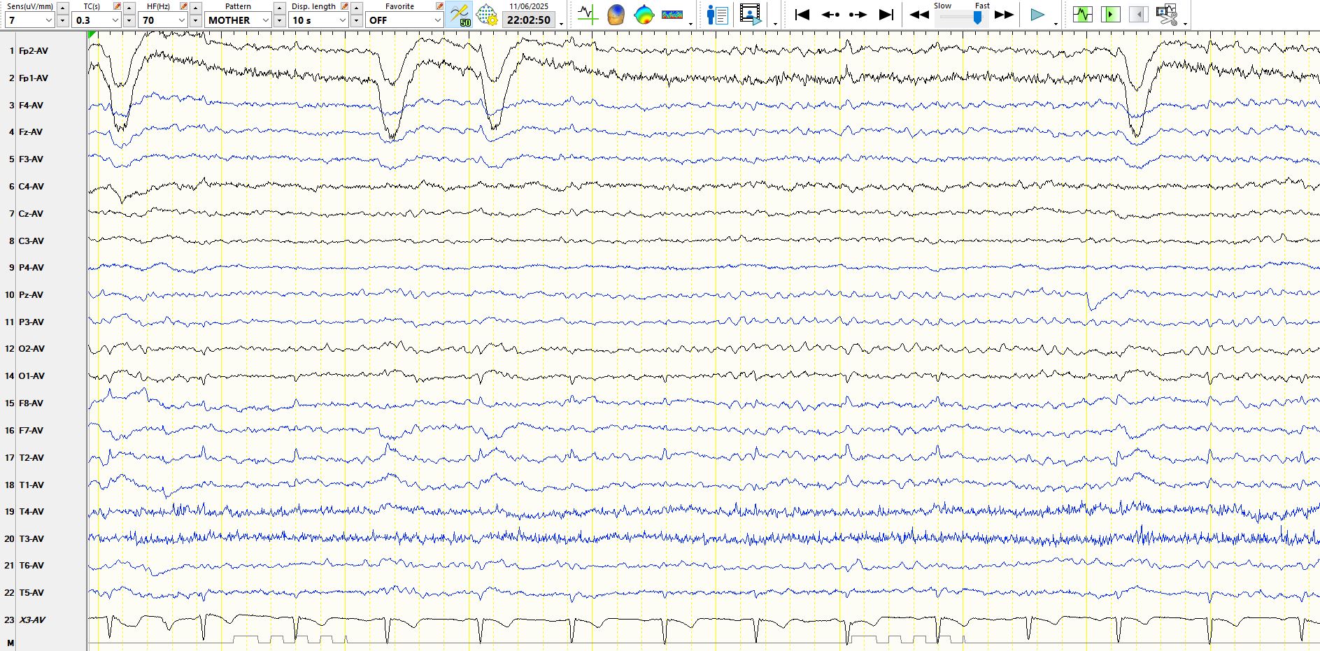
An isolated wicket wave (not synchronous with the ECG).
This is the same wicket wave as on the previous page, albeit starting a few seconds earlier (time can be seen at the top of each page)
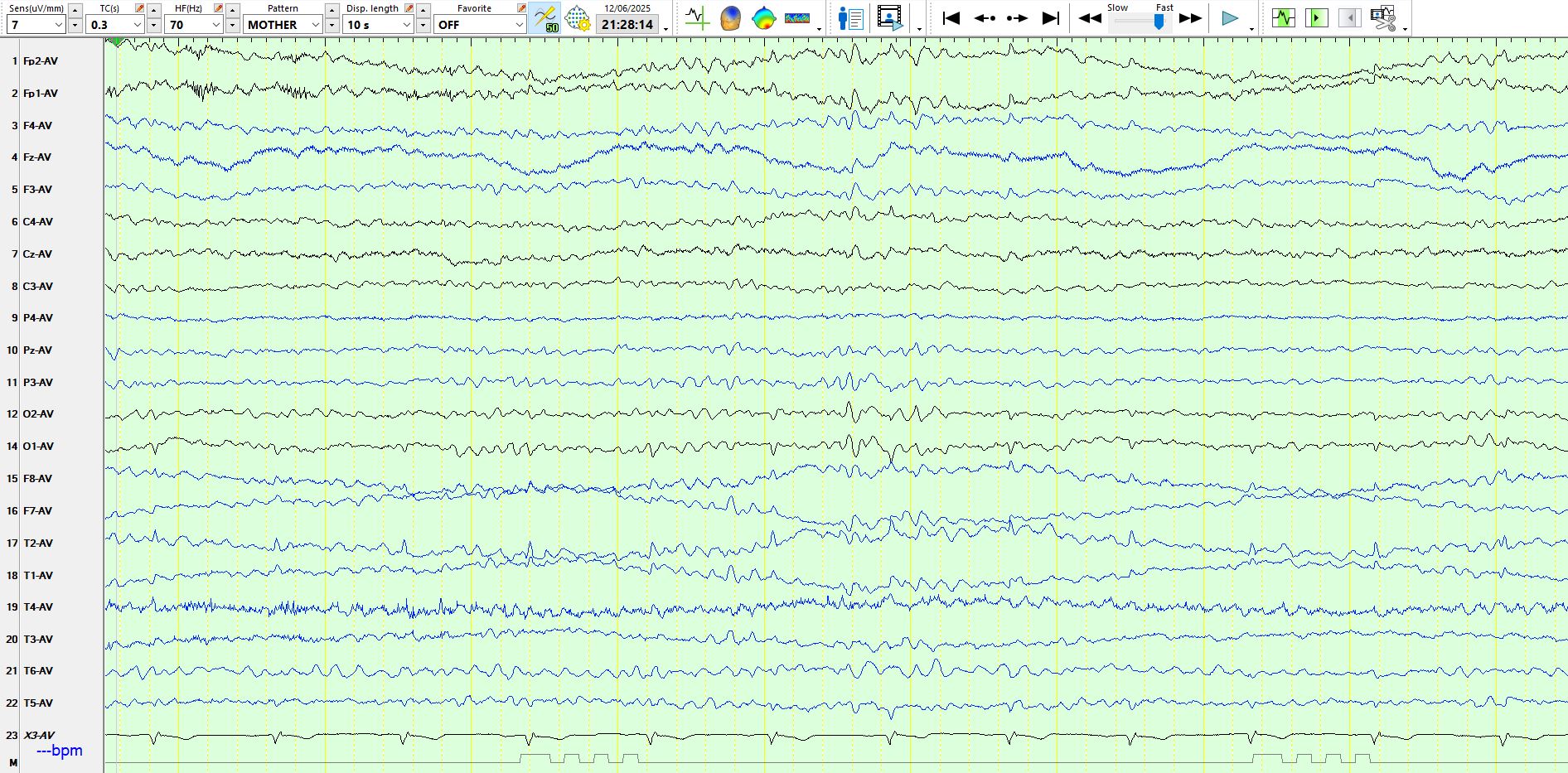
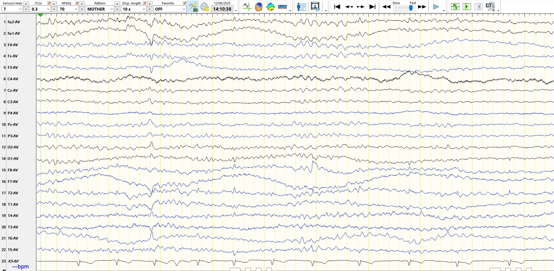
The above page is normal (drowsy) and precedes the page below
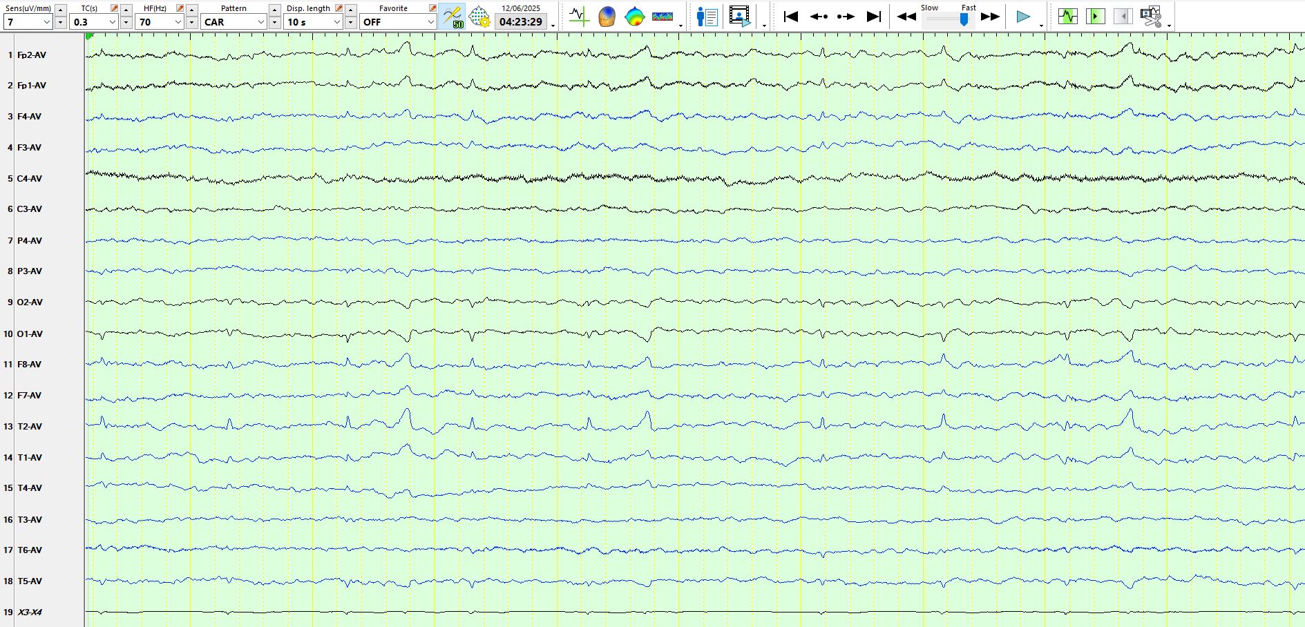
This is another good example of a wicket wave, which happens to be synchronous with the ECG beat). Compare the waveform to those in previous posts Red Alert 2
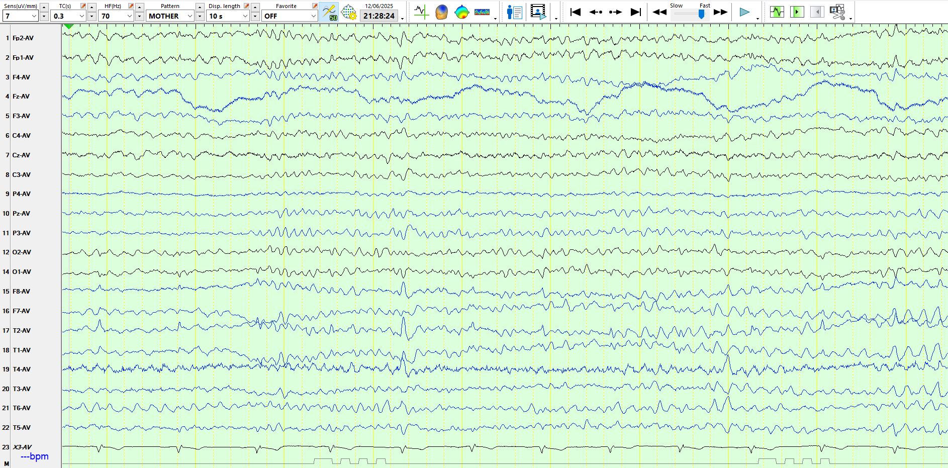
The page below follows previous page
The above demonstrates evidence of drowsiness, including the appearance of progressively slowing theta frequencies in the temporal regions, especially the left
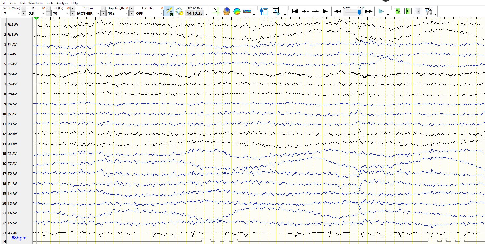
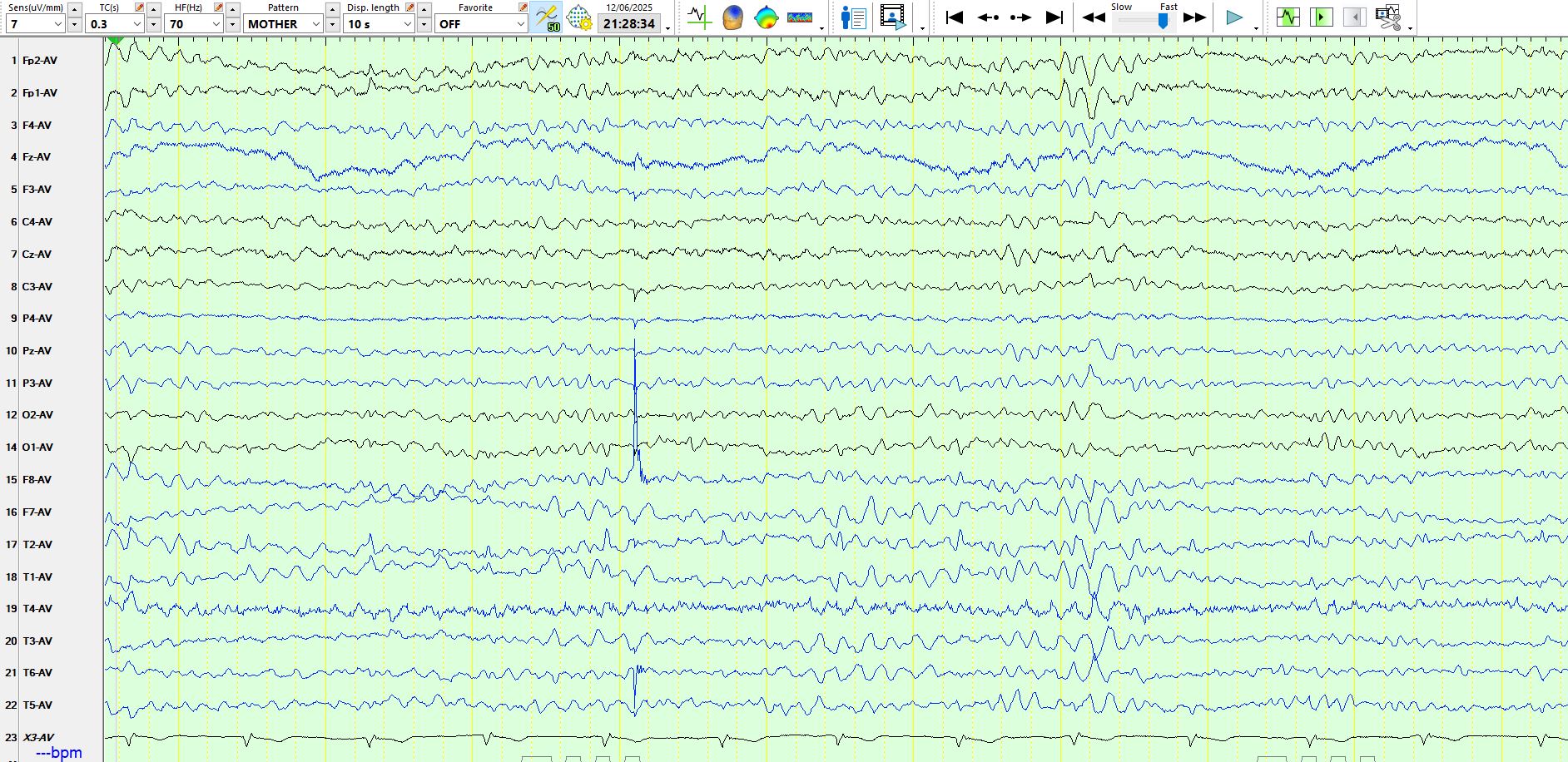
Having looked at all of the images above, go back and study the ECG trace. The top page is a good example of what you might call "pseudo-theta" (not an existing term). This of course relates to wide- complex extra-systolic beats, if my limited cardiological skills are correct. THE EEG is entirely normal.
The MRI demonstrates a juxta-cortical, linear lesion in the white matter of the left post central gyrus extending medially to above the insular cortex. There is no mass effect and no enhancement. The nature of the lesion is unclear and does not relate to any of his symptomatology. His loss of consciousness almost certainly was related to a very large binge on alcohol, with very high blood alcohol levels. There is no history of any right facial or any other sensory symptomatology, nor of any episodic or persistent language dysfunction. There is no history of previous cranial trauma. His perinatal and developmental history is unremarkable.
 \
\
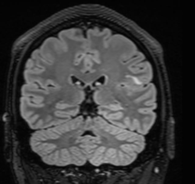
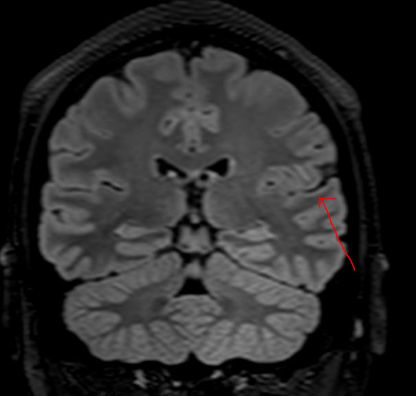
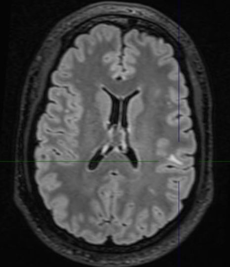
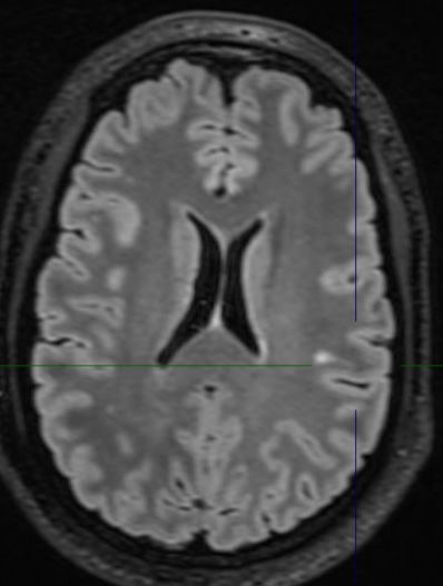
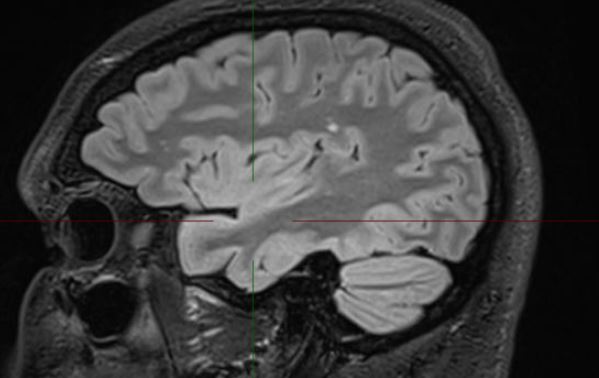
In addition to the above lesion there are a few other punctate white matter lesions. It is presumed that these are old and have been discovered coincidentally as they do not relate to any past or recent symptomatology and are therefore "asymptomatic". I discussed the case with experienced neuroradiologist who reports that the lesions appear "innocuous". The case also makes the point that diagnostic tests may result in a lot of harm; in this instance the patient was effectively "medically deported" within 24 hours of the scan having been done in a highly developed country. If the EEG is misinterpreted, the patient's problems will only be compounded. His EEG was normal for 72 hours; the rationale for performing it for this duration will be discussed at a later stage
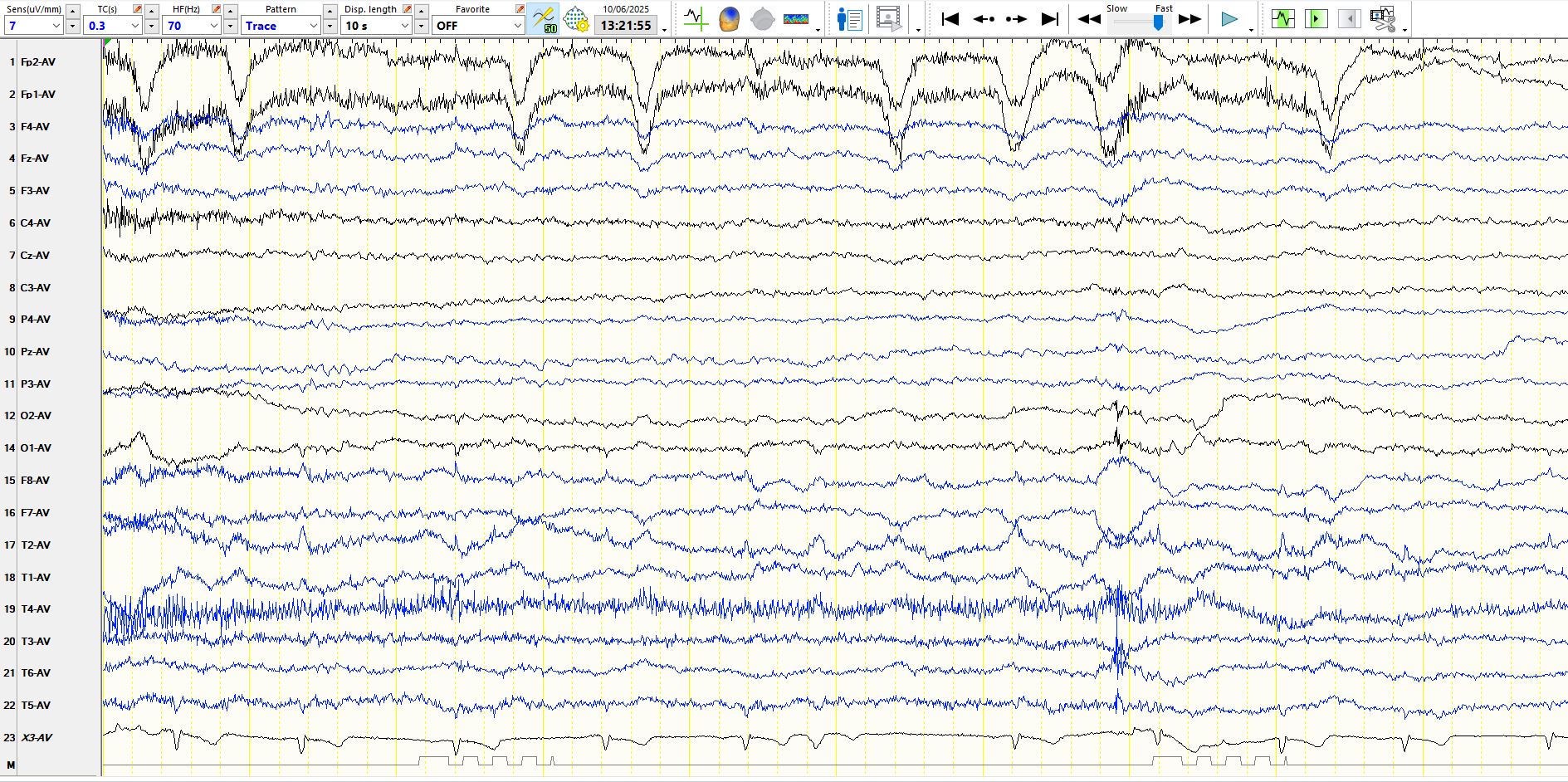
Notice the broad-complex, premature extra systolic beat in the ECG rhythm trace in the images above and below, with a compensatory pause and the effect of these waves on the EEG trace