More cricket
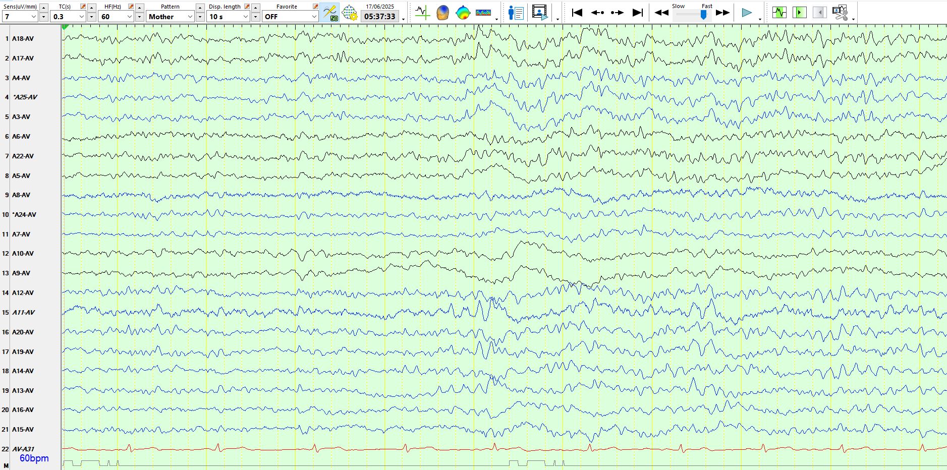
Jun 17, 2025Here are a few more wicket waves. The patient is 52 years old and likely has left temporal lobe epilepsy, with a normal MRI scan.
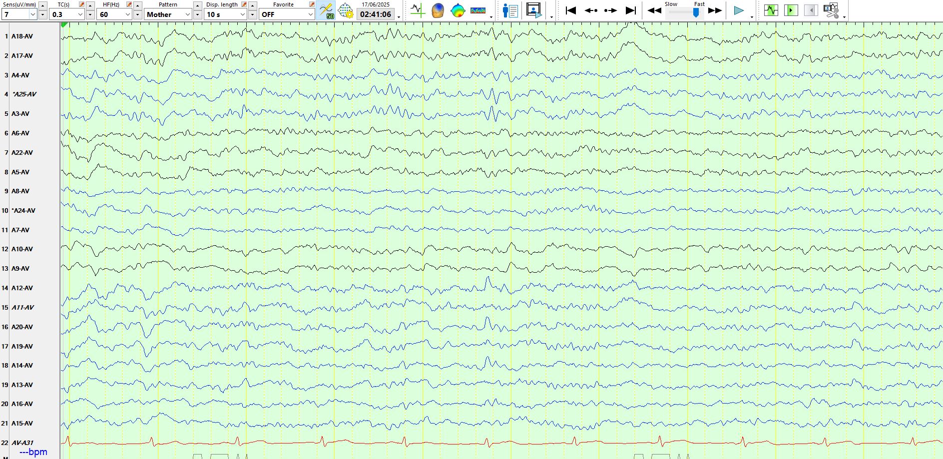
In the above, the patient is asleep. Notice the low amplitude isolated wicket wave above the 6th ECG beat.
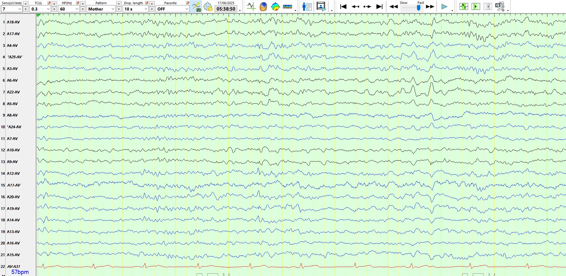
Sleep, above.
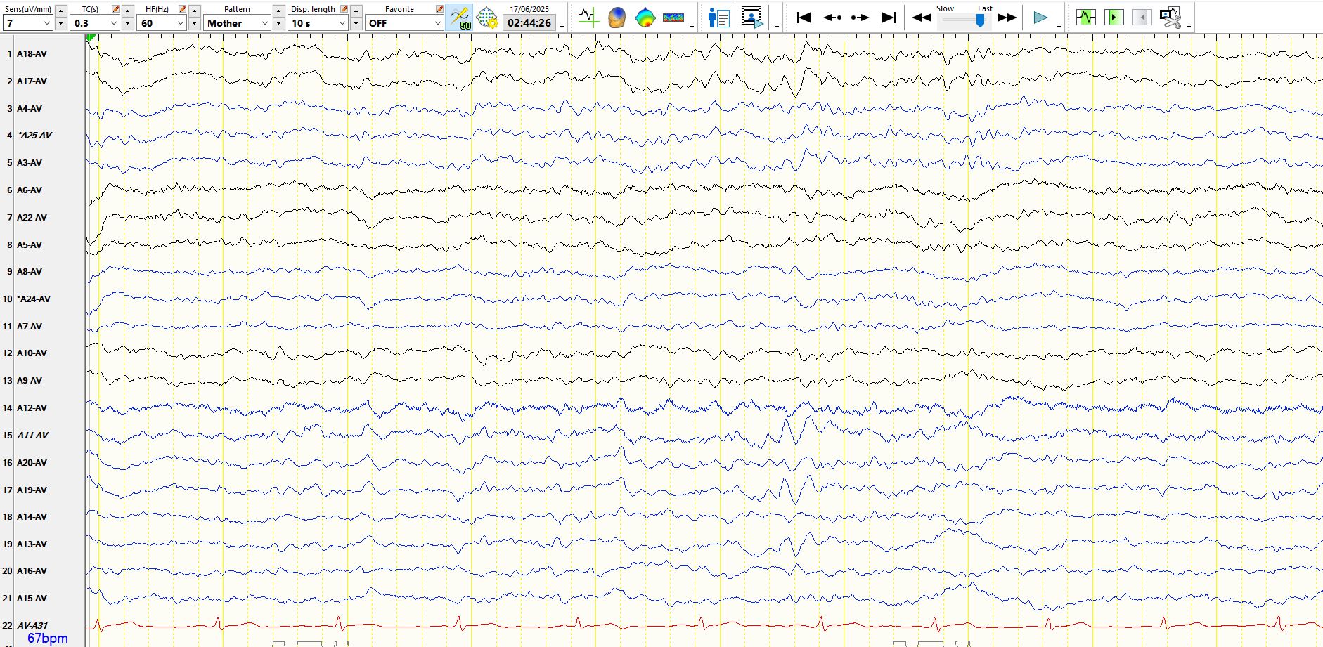
In the above example, there are a sequence of wicket waves between the 4th and 5th ECG beats, and again between 6th and 7th ECG beats.
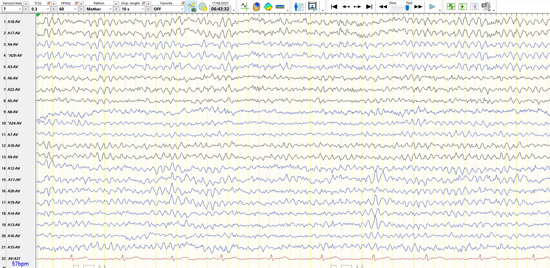
Sleep, above.
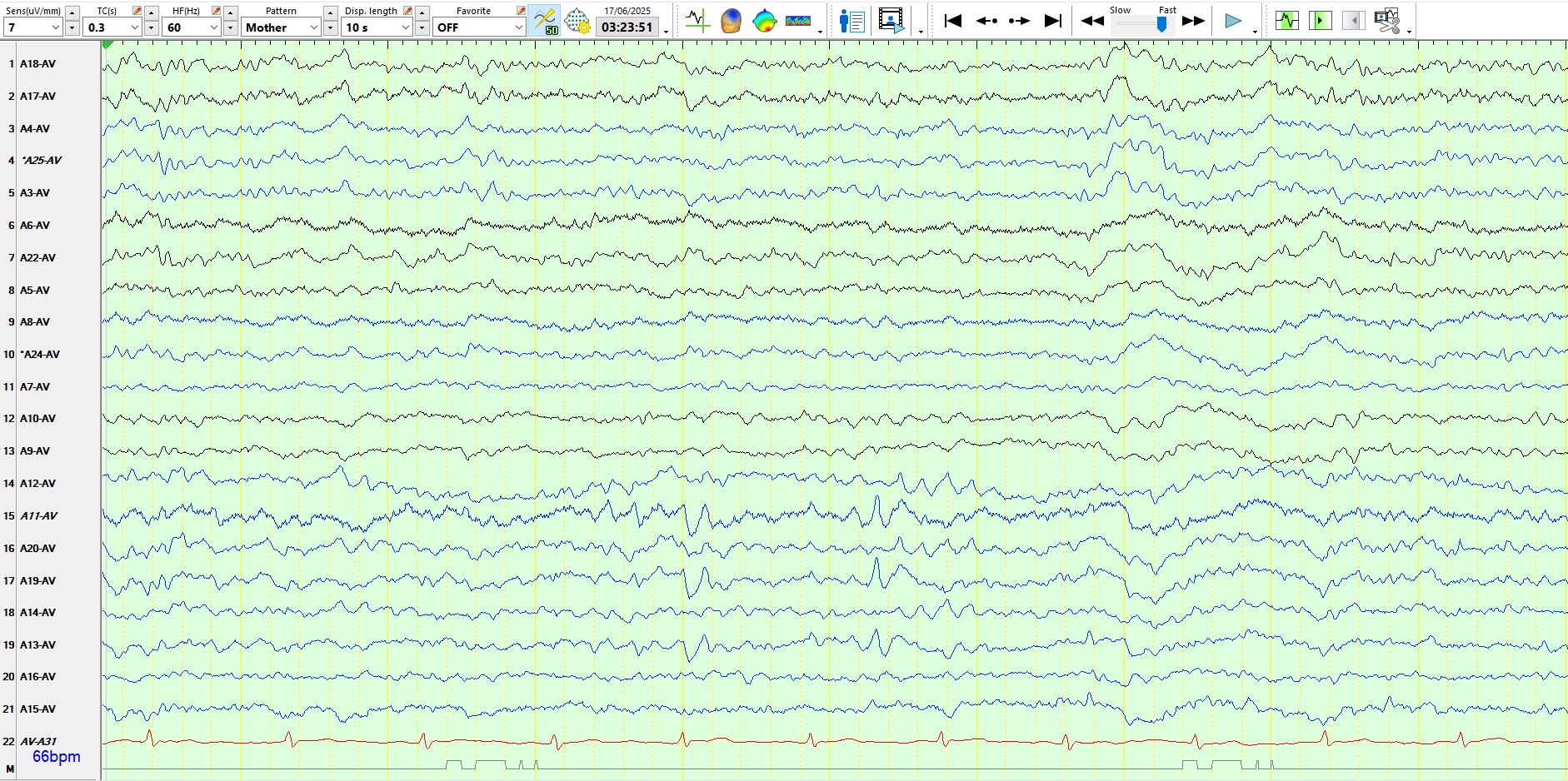
Low amplitude isolated wicket wave above, just after the 5th ECG beat.
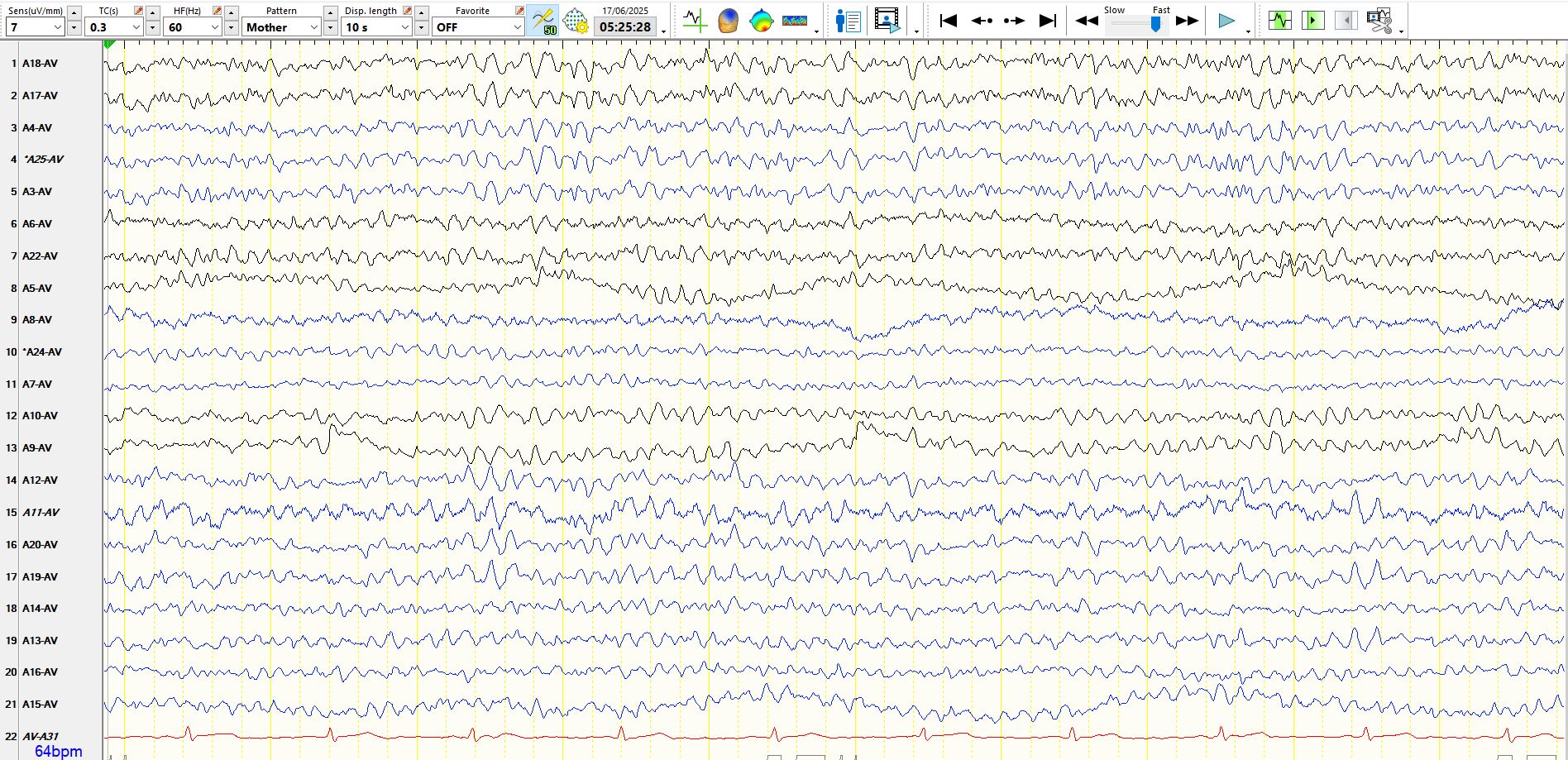
And here are your sighters:
Look above the 7th second