
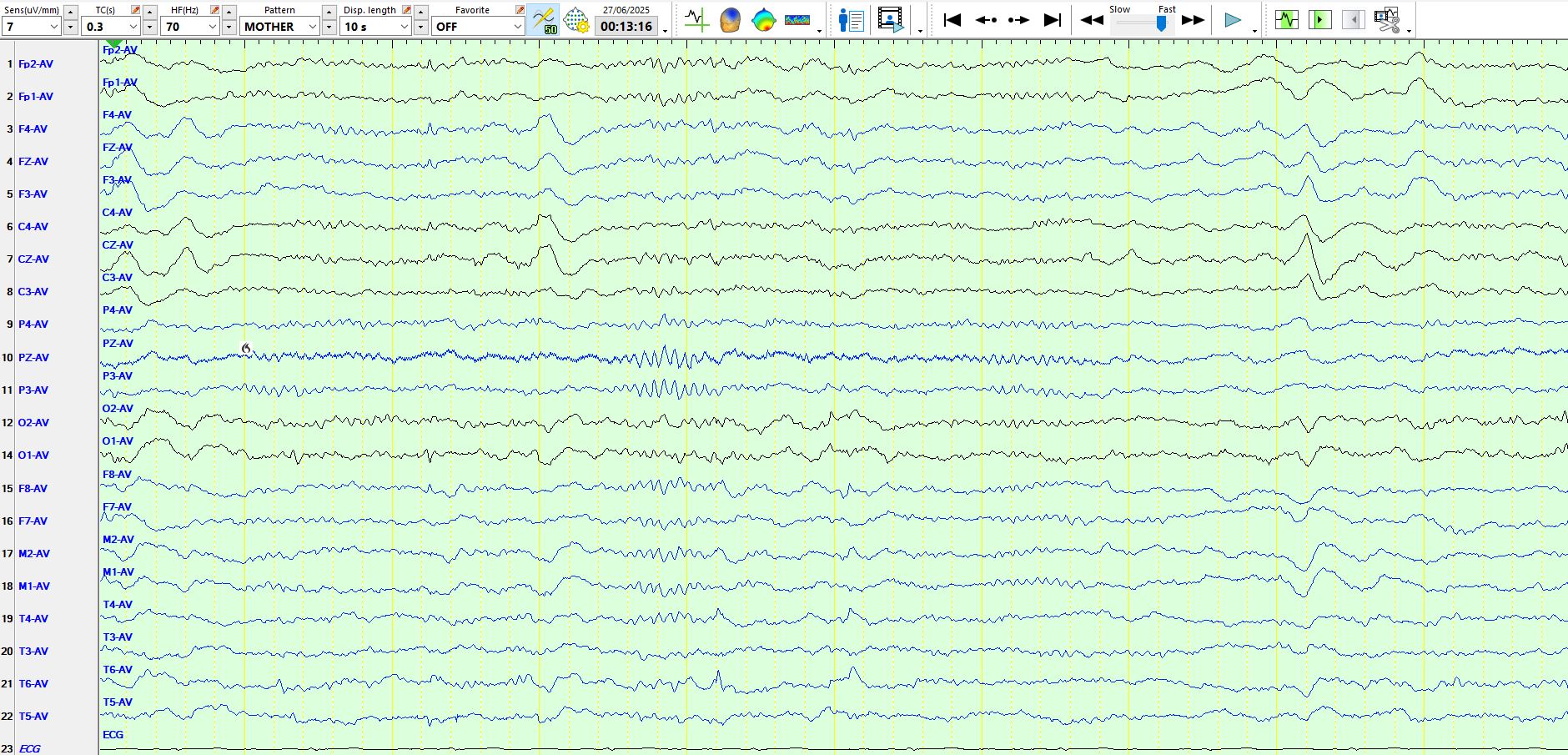
Background rhythms vary quite a lot from one person to the next.
If you count carefully, you will find 13 Hz rhythms primarily over the occipital regions of the above patient. These are re...
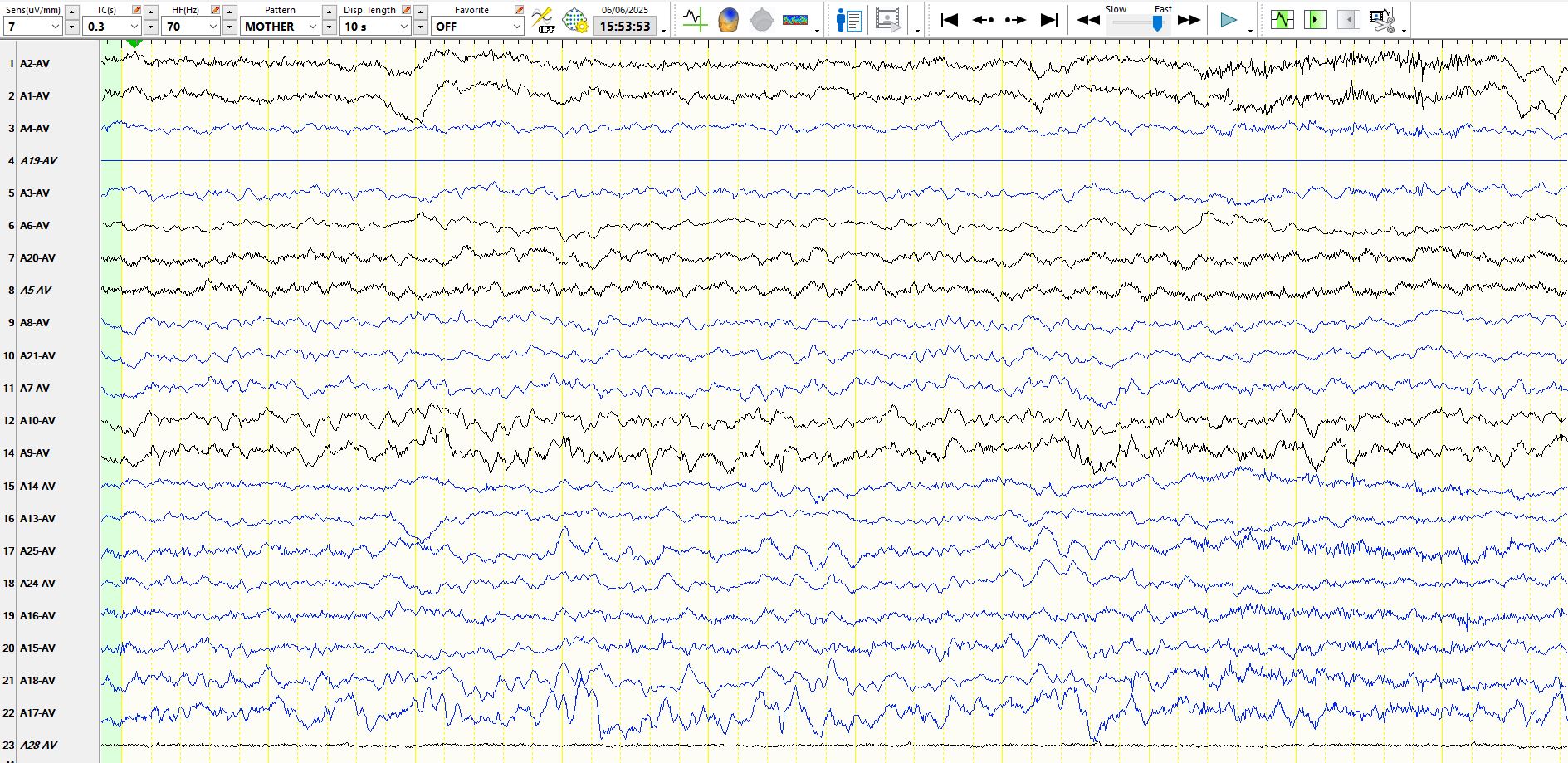
Consider the following page;
Although the discharges resemble spike-and-wave, if you look backwards or forwards in the top 2 channels, you will recognise similar sharply contoured waves. Thes...
A suggestion? The null hypothesis for people working in the field of epilepsy should be that "there is no epilepsy". The doctor is therefore required to disprove the null hypothesis. Yes, even for peo...
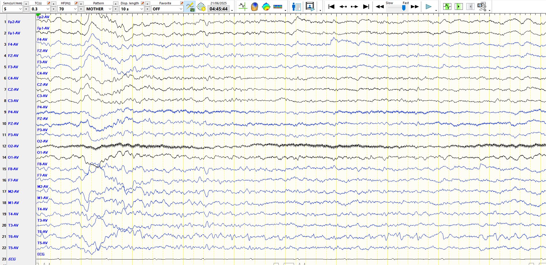
As is typical of these, the following example is better seen on the referential montage than on the bipolar montage:
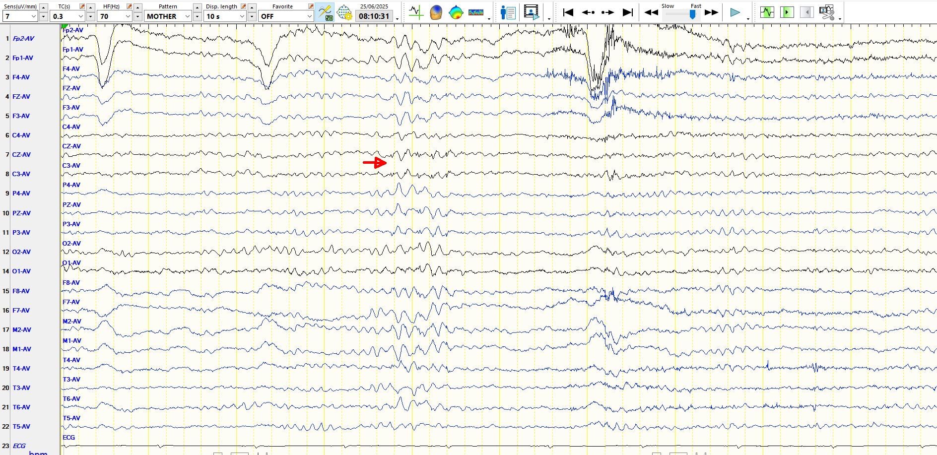

To reiterate, as a general rule, avoid calling low amplitude sp...
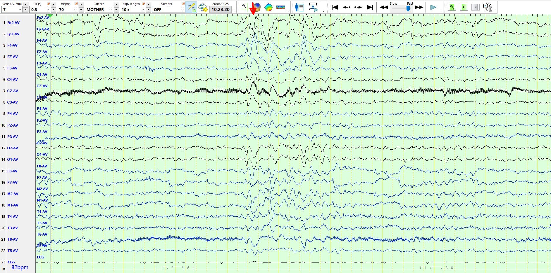
Is the arrowed wave?
1. An electrostatic artefact in sleep
2. A generalised spike, primarily electro positive over the posterior head regions, during sleep
3. A very sharply contoured vertex wave
...Years ago, I taught myself the following trick to guard against calling delta waves in the temporal regions and elsewhere spike-and-wave. If you see something resembling the following and wonder wheth...
Have a look at the following few pages:
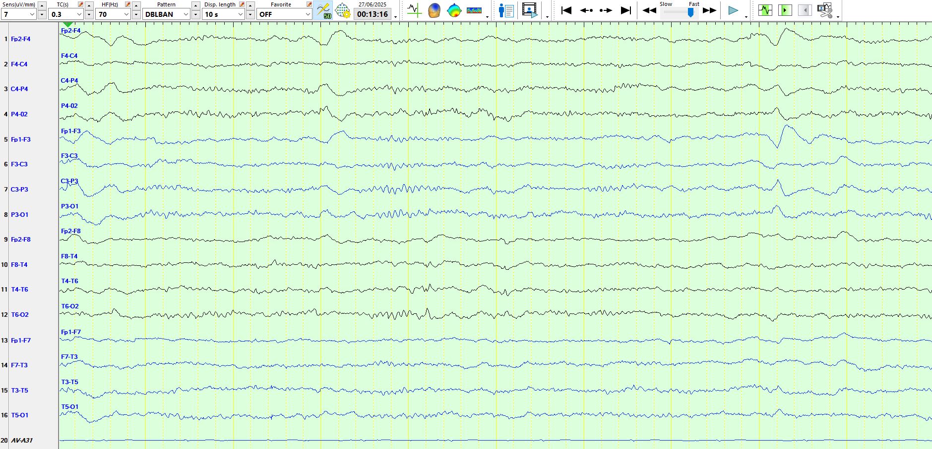
The following page is the bipolar representation of the above page:
In the above, the wave is seen at T6-T4 and is electro-negative, exc...
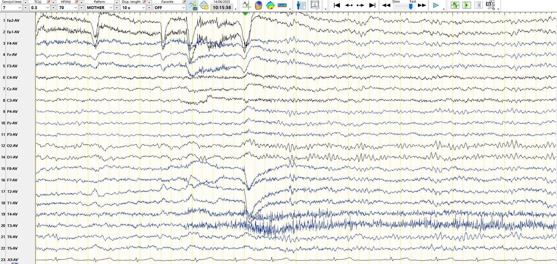
What do you make of the following discharge that appears on the following 2 sequential pages? Try and answer the question for yourself, before reading the comments right at the bottom.

...
The following two images of the same page, viewed on different montages, illustrate the problem discussed in the previous post, emphasising rule 2.
in the image above, while the patient ...
This can be tough. Really tough. Rule of thumb? Don't call them spikes if they appear in 1 electrode only! There are exceptions. But be very careful. Even worse, artefacts can appear in adjacent elect...
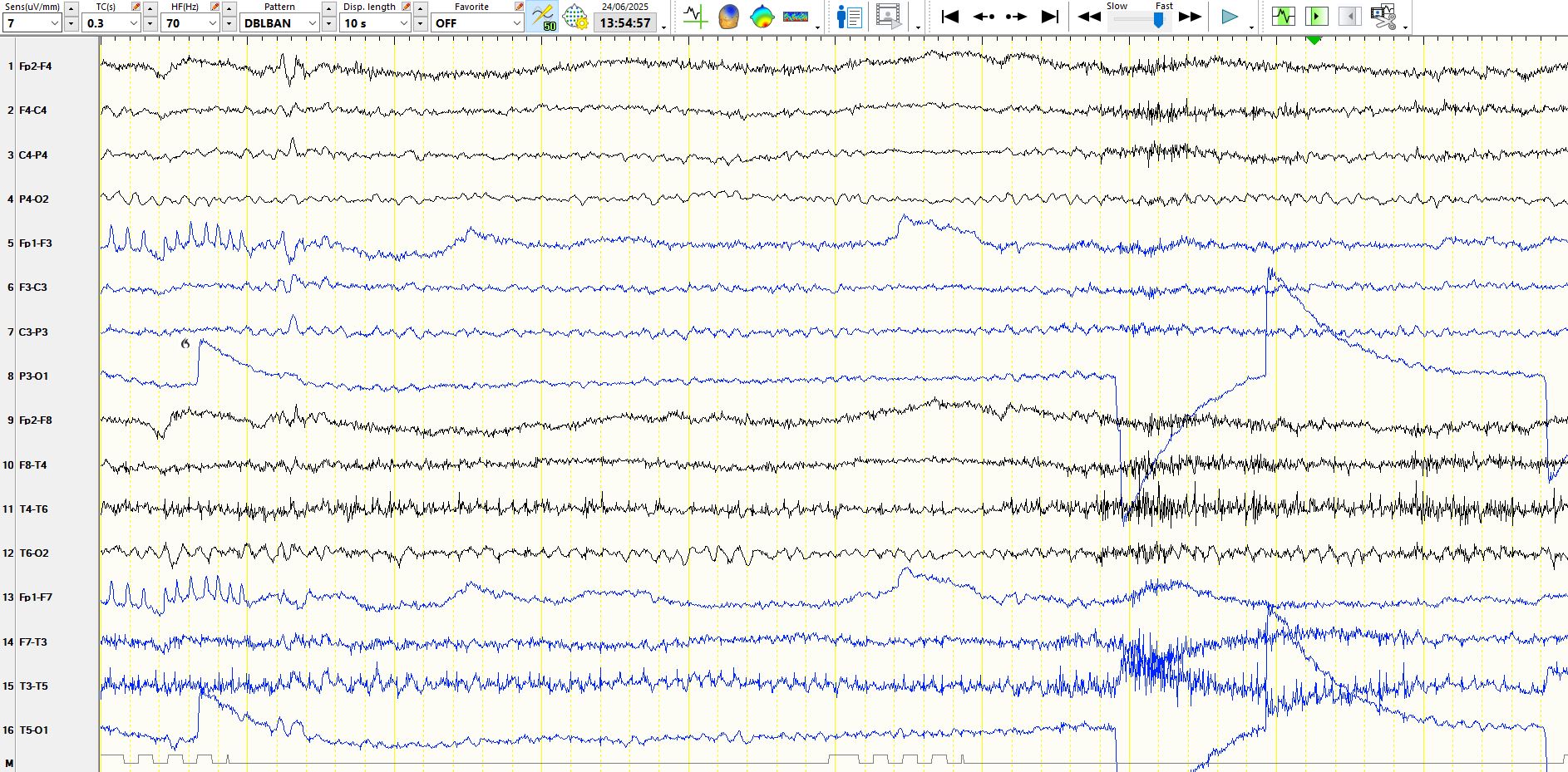
It may sometimes be hard to tell cortical abnormalities from artefact. Have a look at this:
Referential montage
In the above, T5 (A17) appears artefactual. Hence, one may assume all abnormal...
The patient is five years old. These are best seen on referential montages, where they may be confused with a train of sharp waves or even electrographic seizures; hence they are regarded as "pseudo-e...
