
Artefact vs theta, delta and spikes
Jun 23, 2025It may sometimes be hard to tell cortical abnormalities from artefact. Have a look at this:
Referential montage
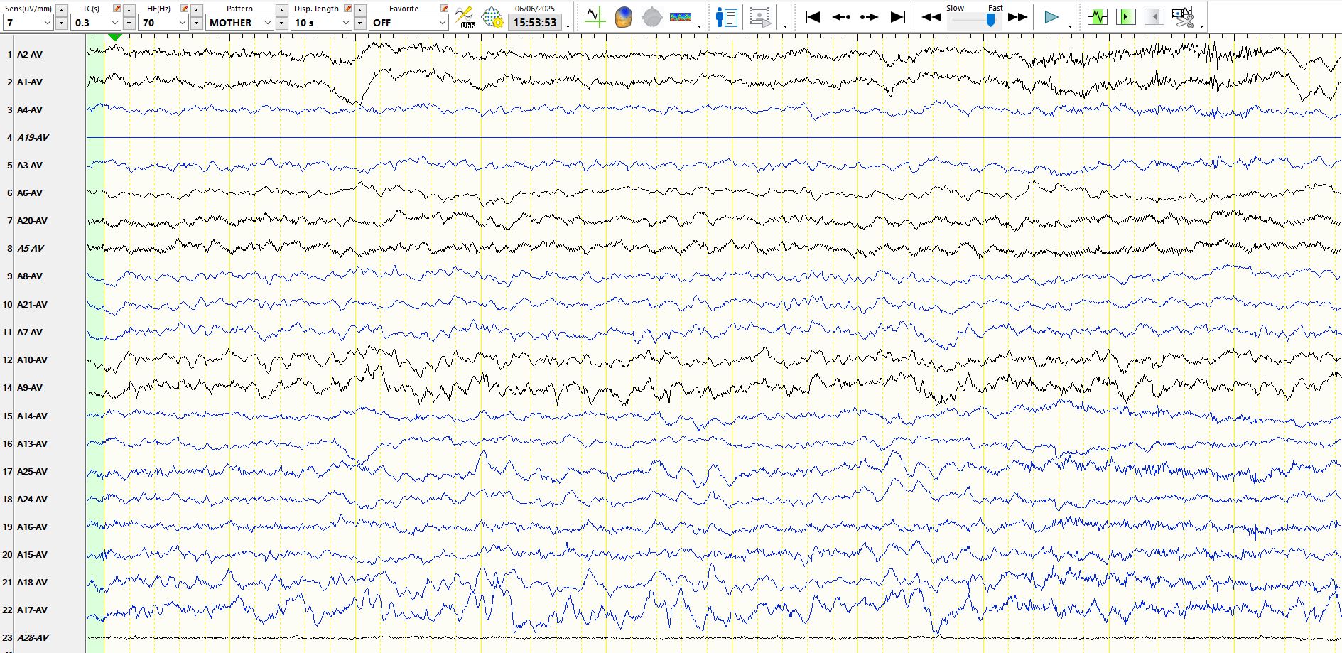
In the above, T5 (A17) appears artefactual. Hence, one may assume all abnormal looking waves in at T5 are artefacts, including the delta-looking wave at T5 (at 3 seconds, 3rd darker yellow line)), as it is not seen at O1 (A9). About 500ms later there is an apiculate wave at O1, but not at T5. Skip on to 4.5s (halfway between the 4th and 5th solid yellow lines) and there is an apiculate wave at T5, but woah, it is also seen at O1-P3. That looks like a biological field! But the artefact raises doubt (which is a good reason to hold fire and an even better reason for the technologist to try and avoid or minimise artefacts). Now, take a look at the isolated delta wave in T5 again and compare it to the waves below.
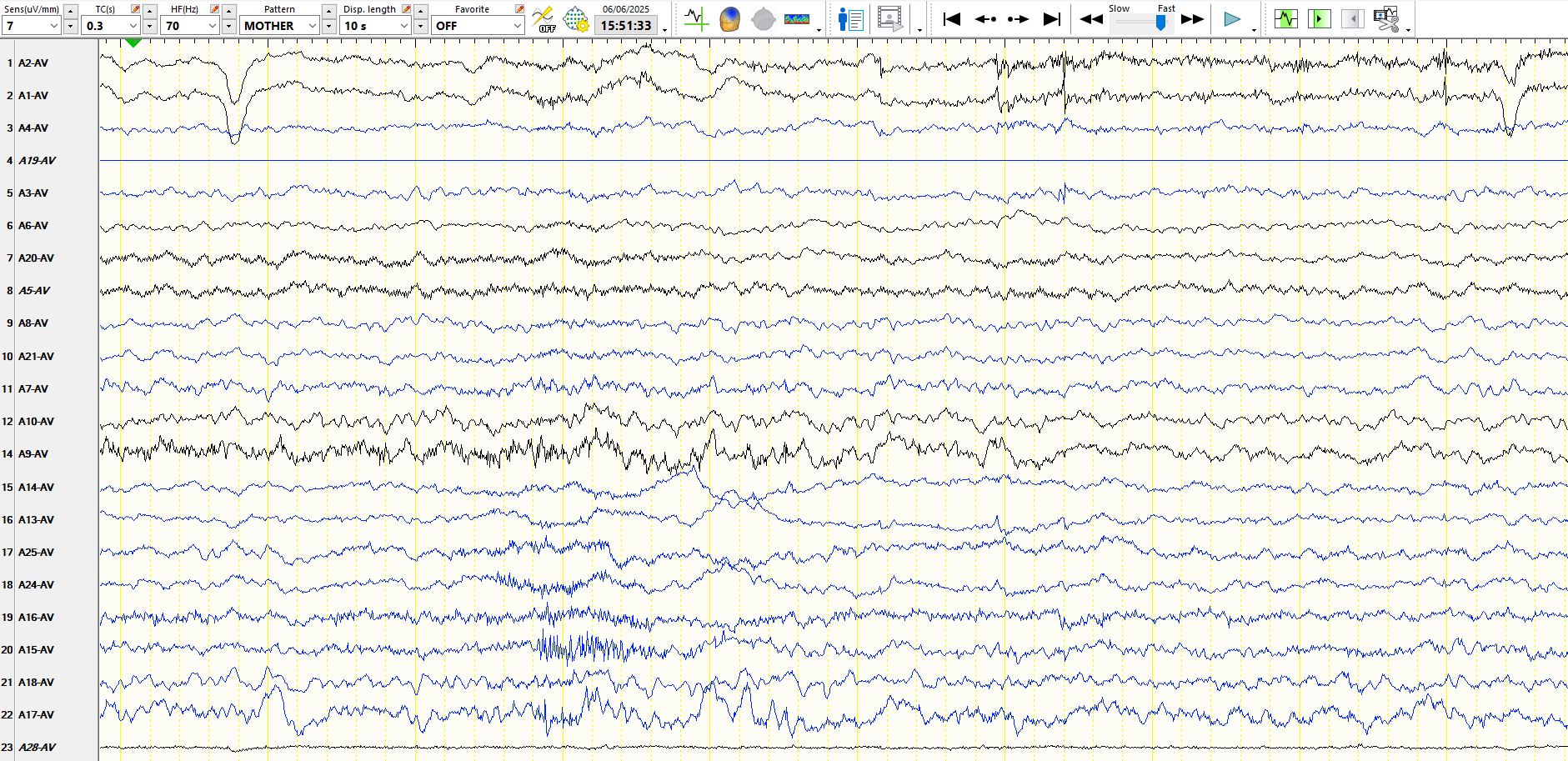
In the above, the slow waves appear synchronously at T5-O1, therefore are not artefact.
In the above, at 5s there is a sequence of somewhat unusual looking spike and wave at T5-O1-O2. The field, morphology and absence of artefact are compelling for spikes.
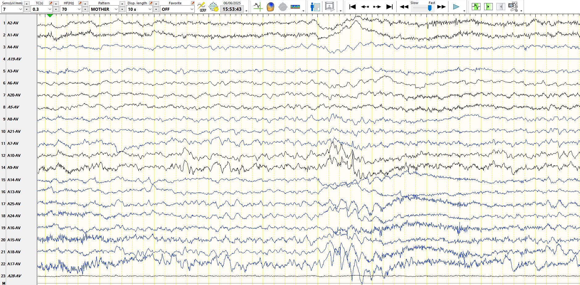
The above is arguably an electrographic seizure as it lasts about 5 seconds at O1-T5, although it does not evolve. At the minimum, these are sequential spike and wave discharges. The patient is asleep and nobody noticed anything. The patient has Angelman's. The paediatric neurologist and the parents are concerned about subclinical seizures. Oh, there are also spike and wave discharges at T6-O2-T4 (18, 25) at 1, 2 and 3s, and at O2-P4 (10, 8) at about 3s.
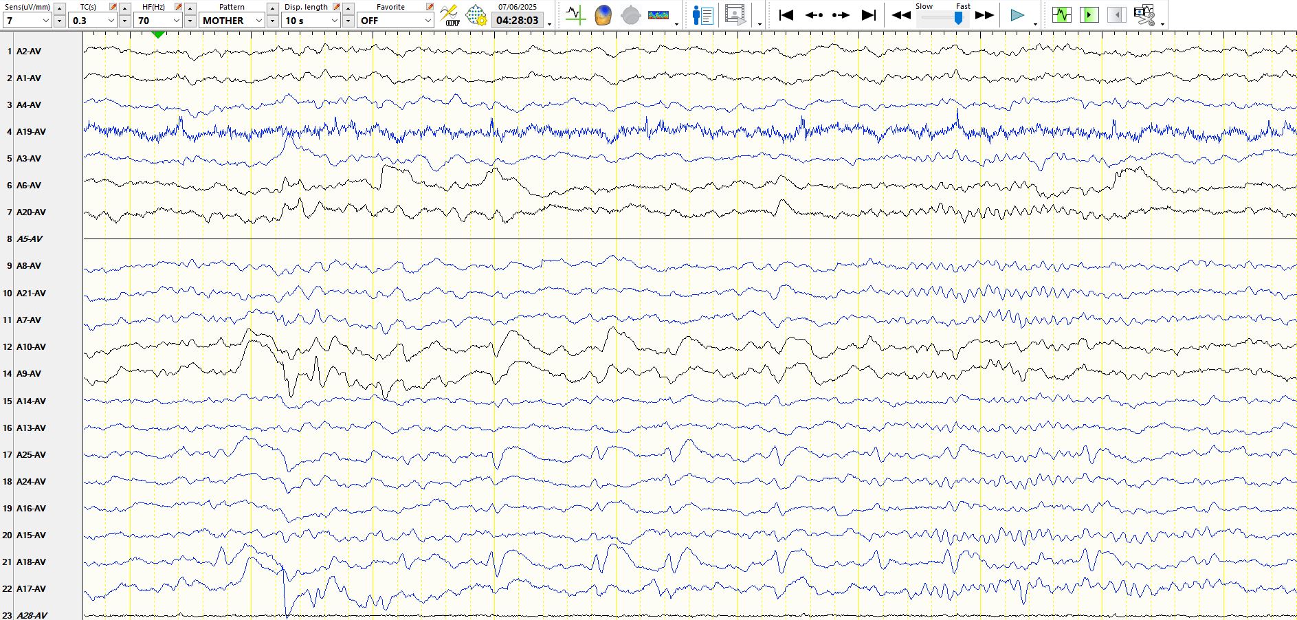
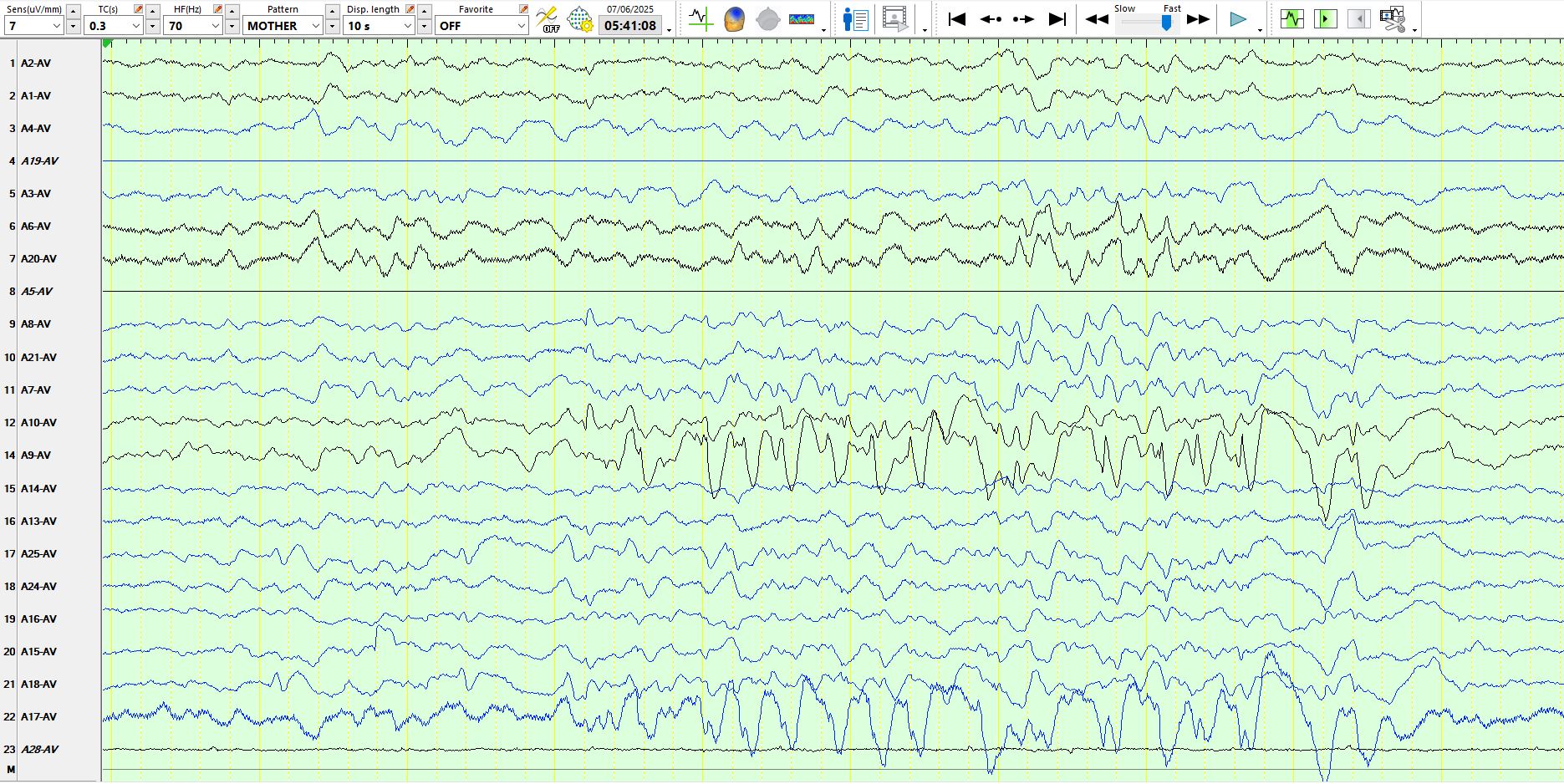
No doubt about the right posterior temporal (T6) spikes