Adult, episodic events 3-4 times per year, possibly representing epileptic seizures
Mar 07, 2026What do these waves during wakefulness and sleep represent?
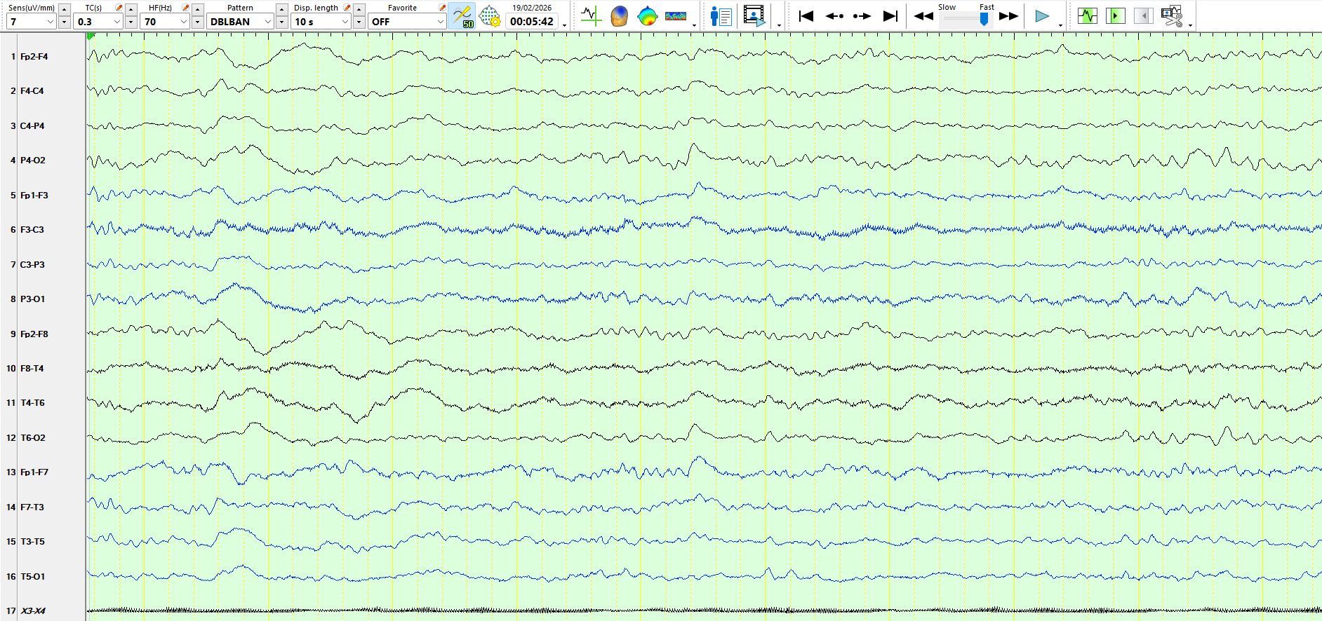
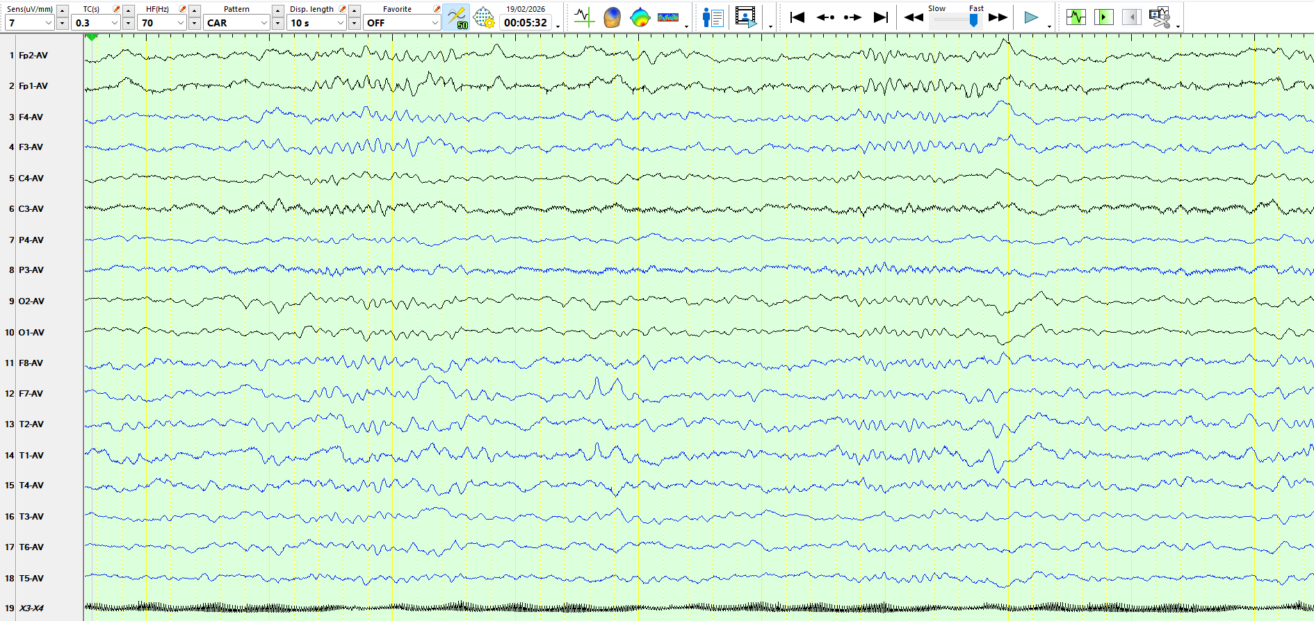
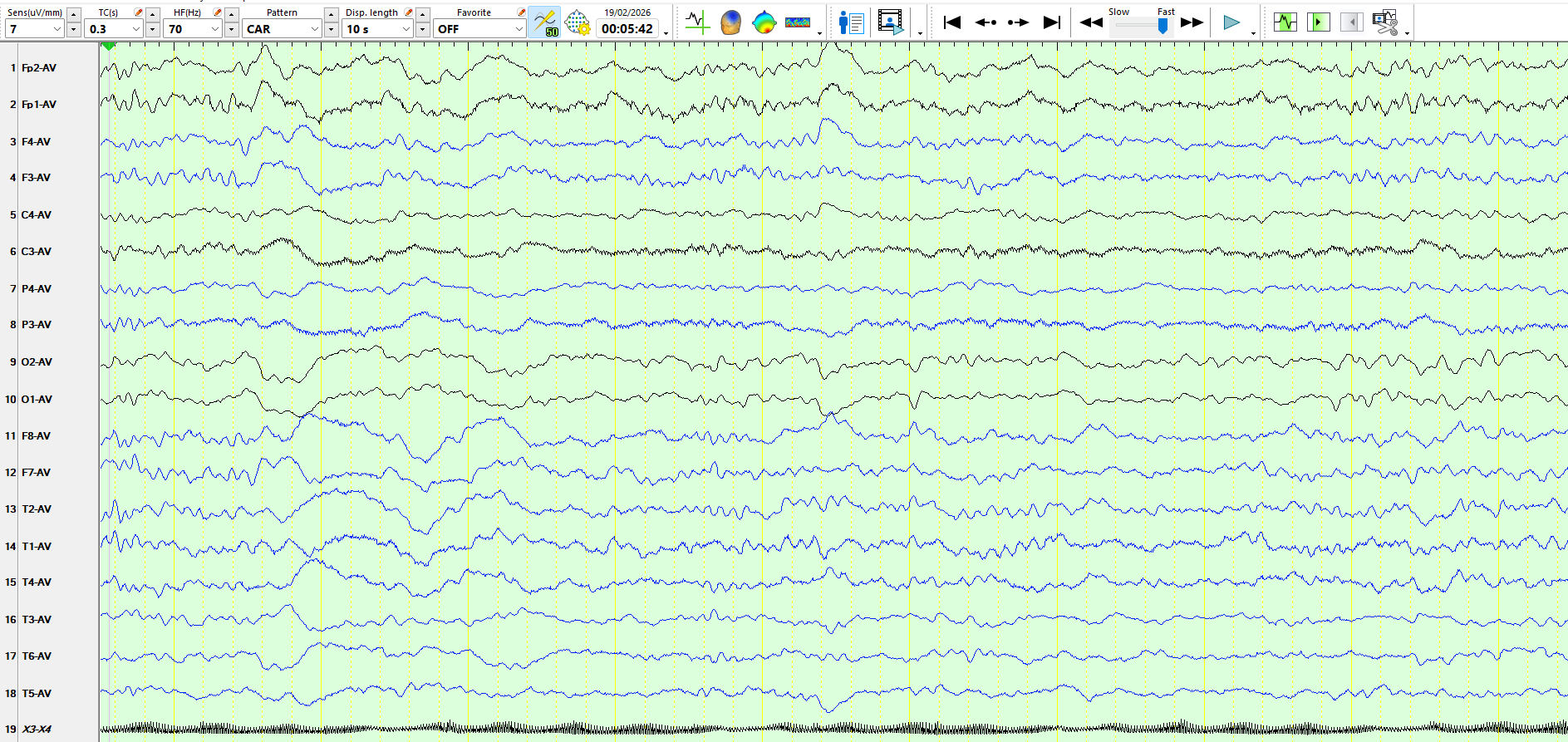
These are three consecutive pages:
Figure 1:
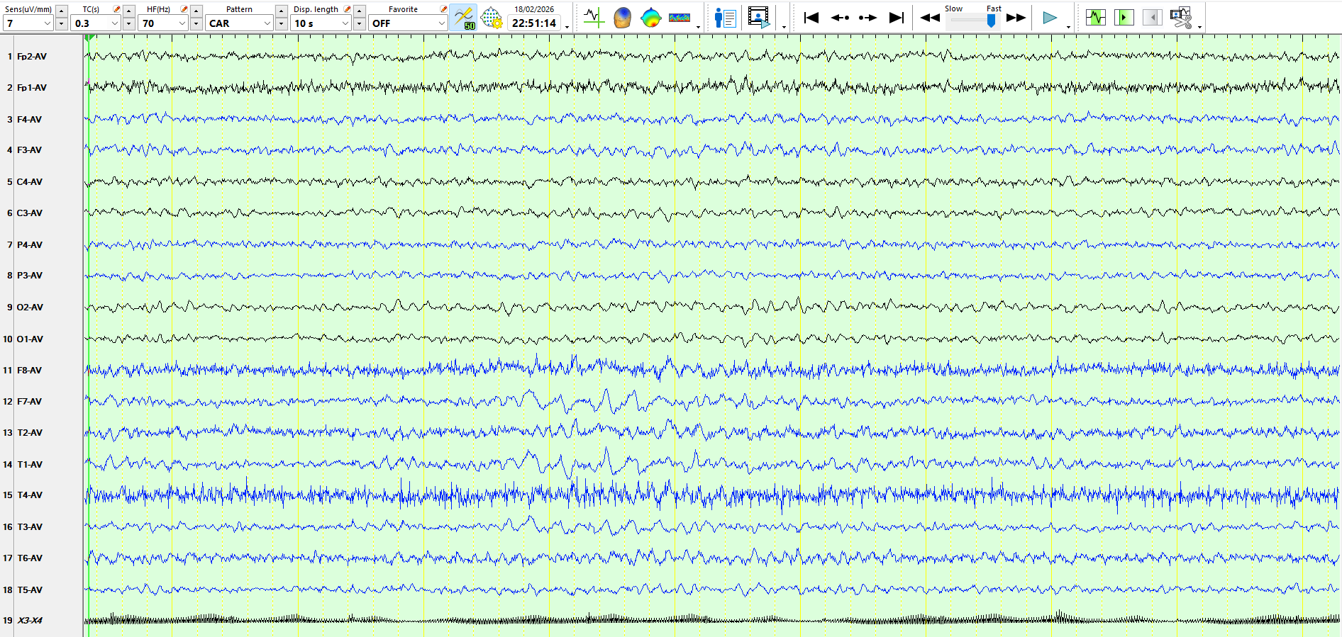
Figure 2:
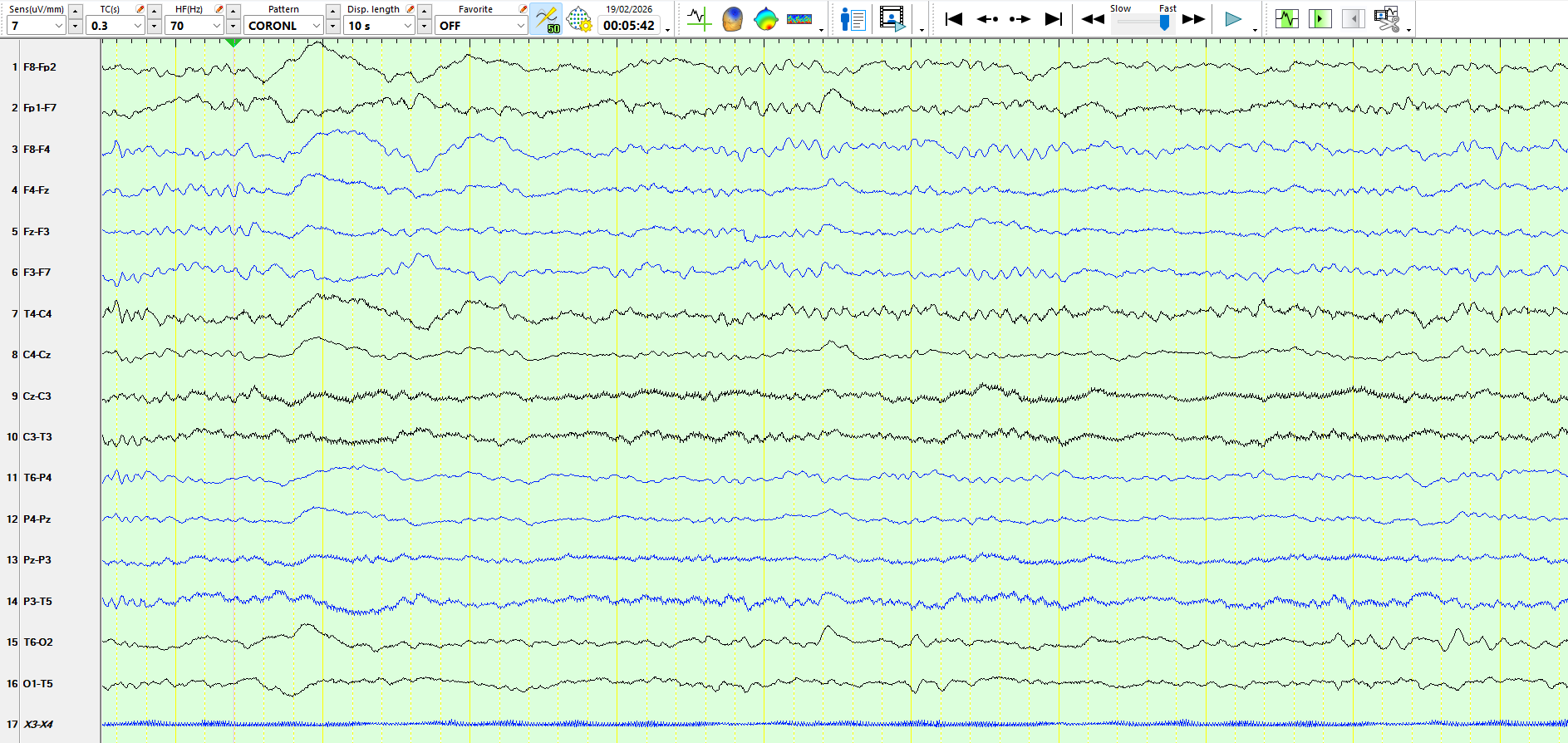
The following represent three consecutive pages:
figure 5
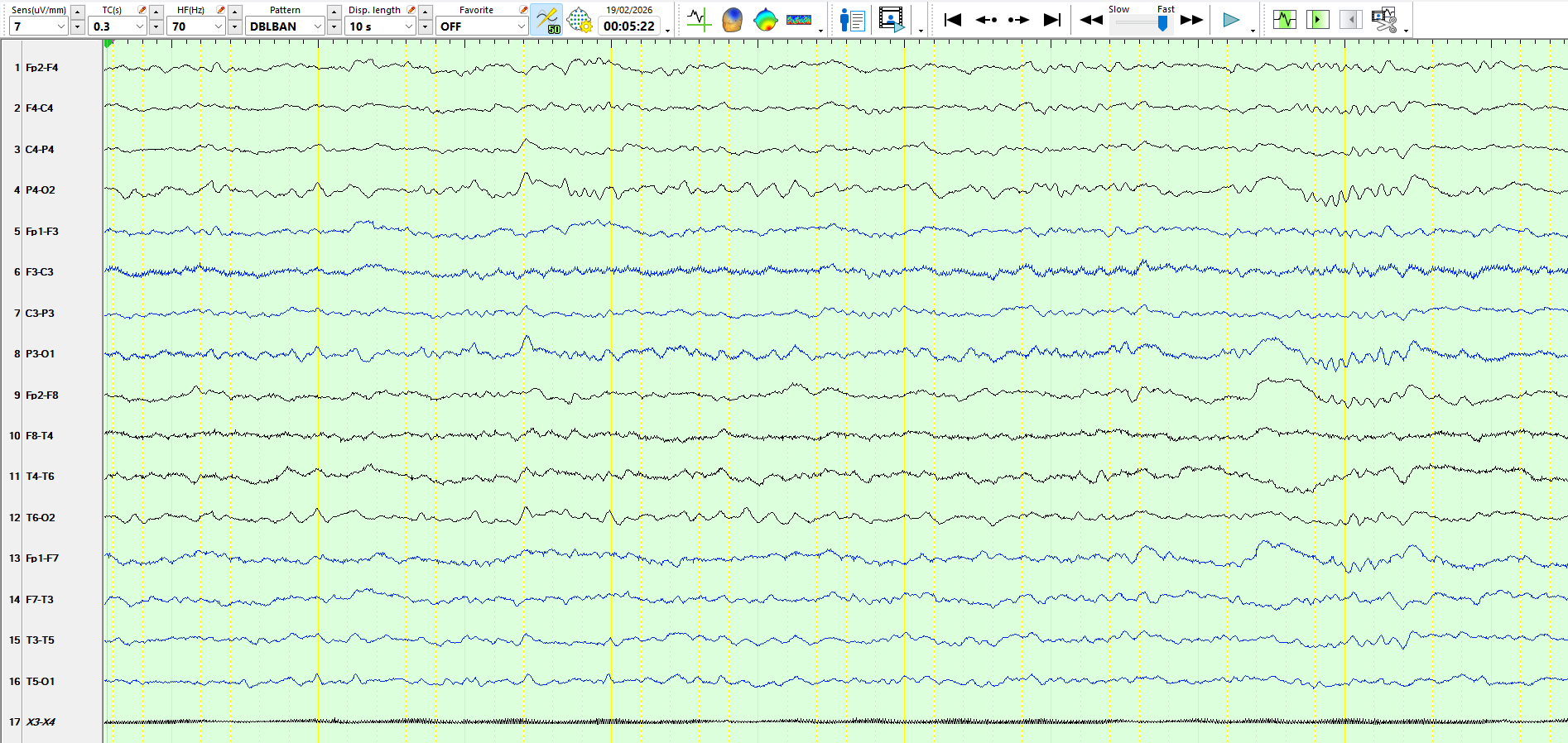
figure 6
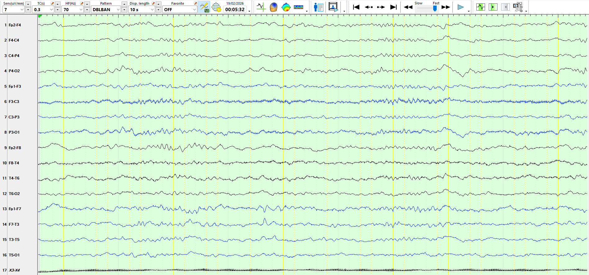
figure 7
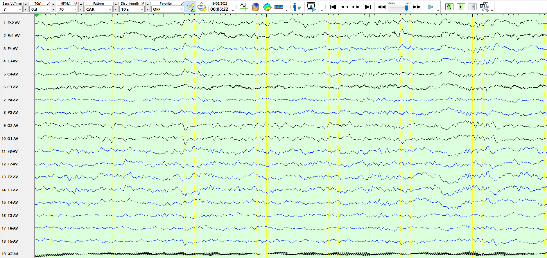
The following represents the same three pages as above, on the referential montage
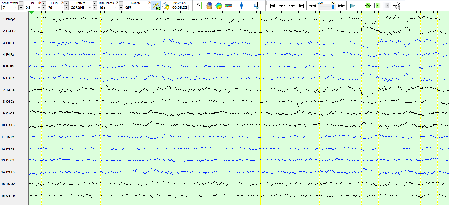
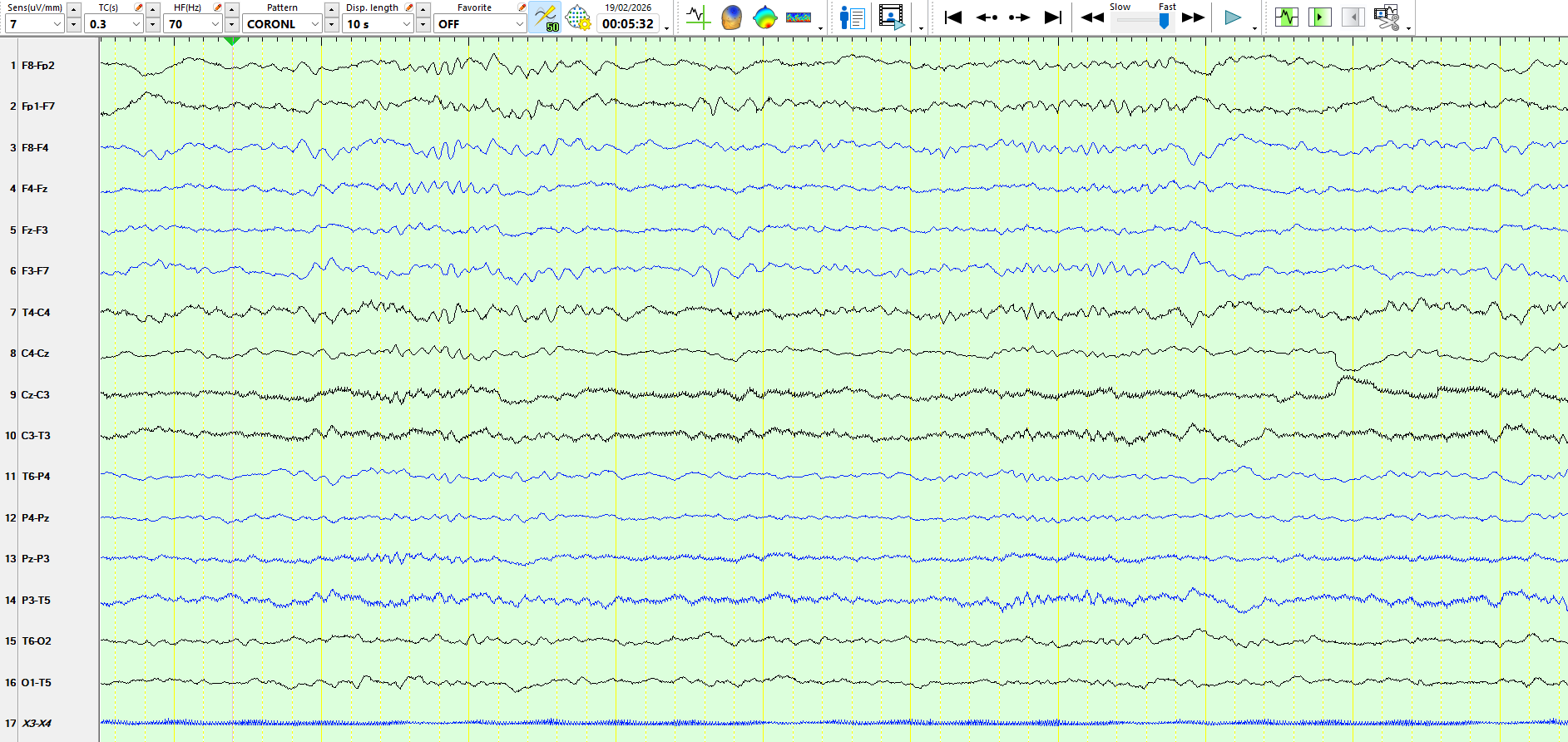
The following three pages represent the above waves on a coronal montage:
The following is a different page, taken from sleep
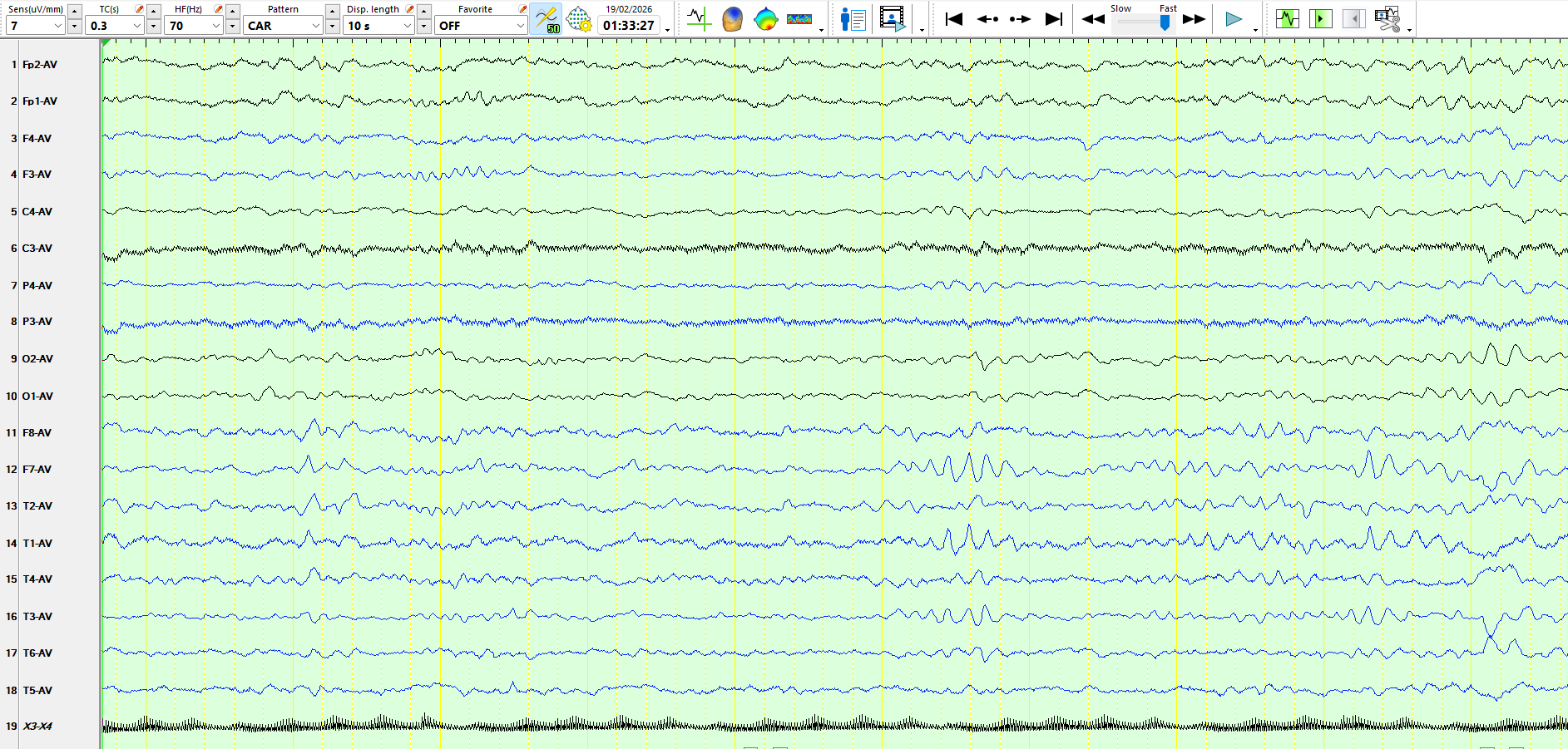
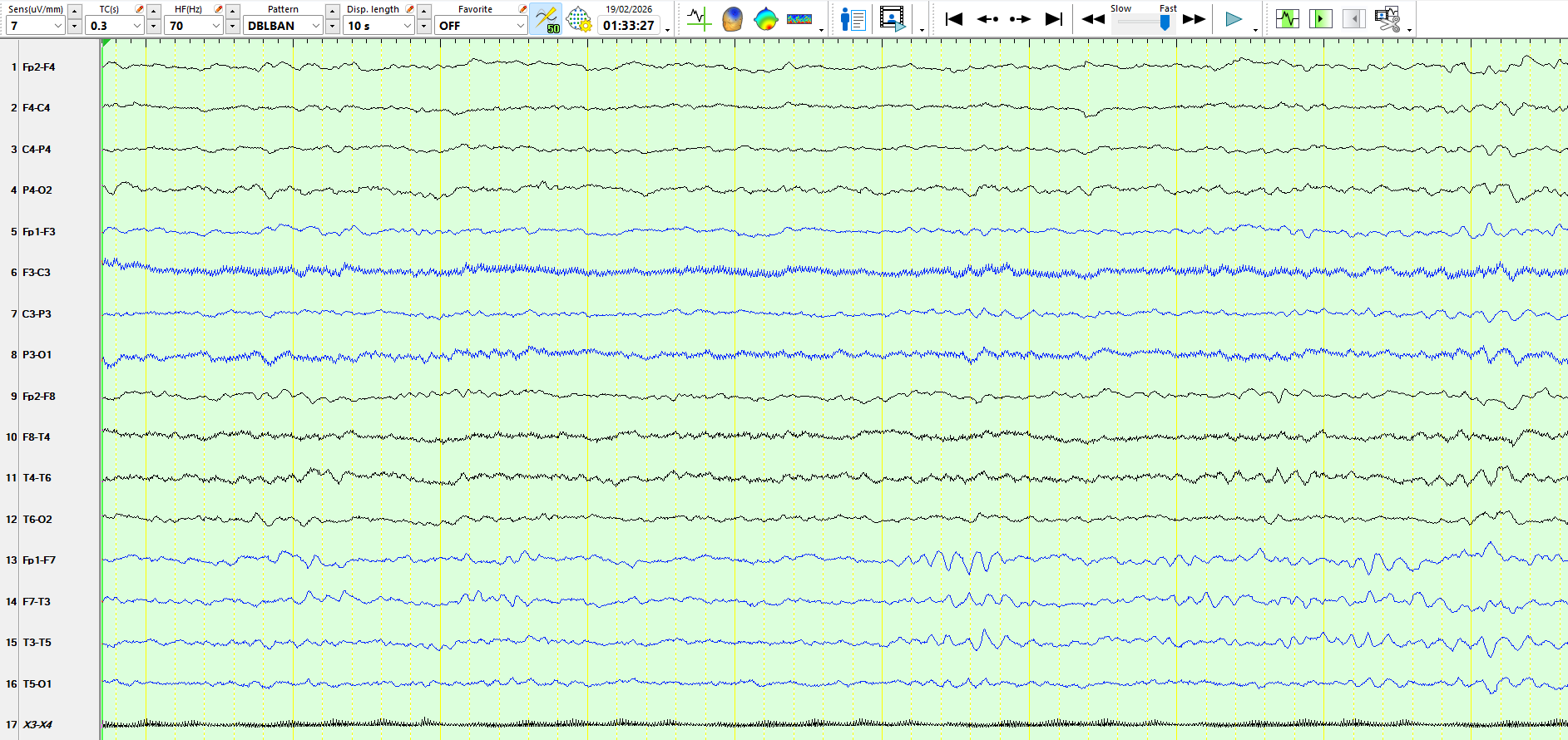
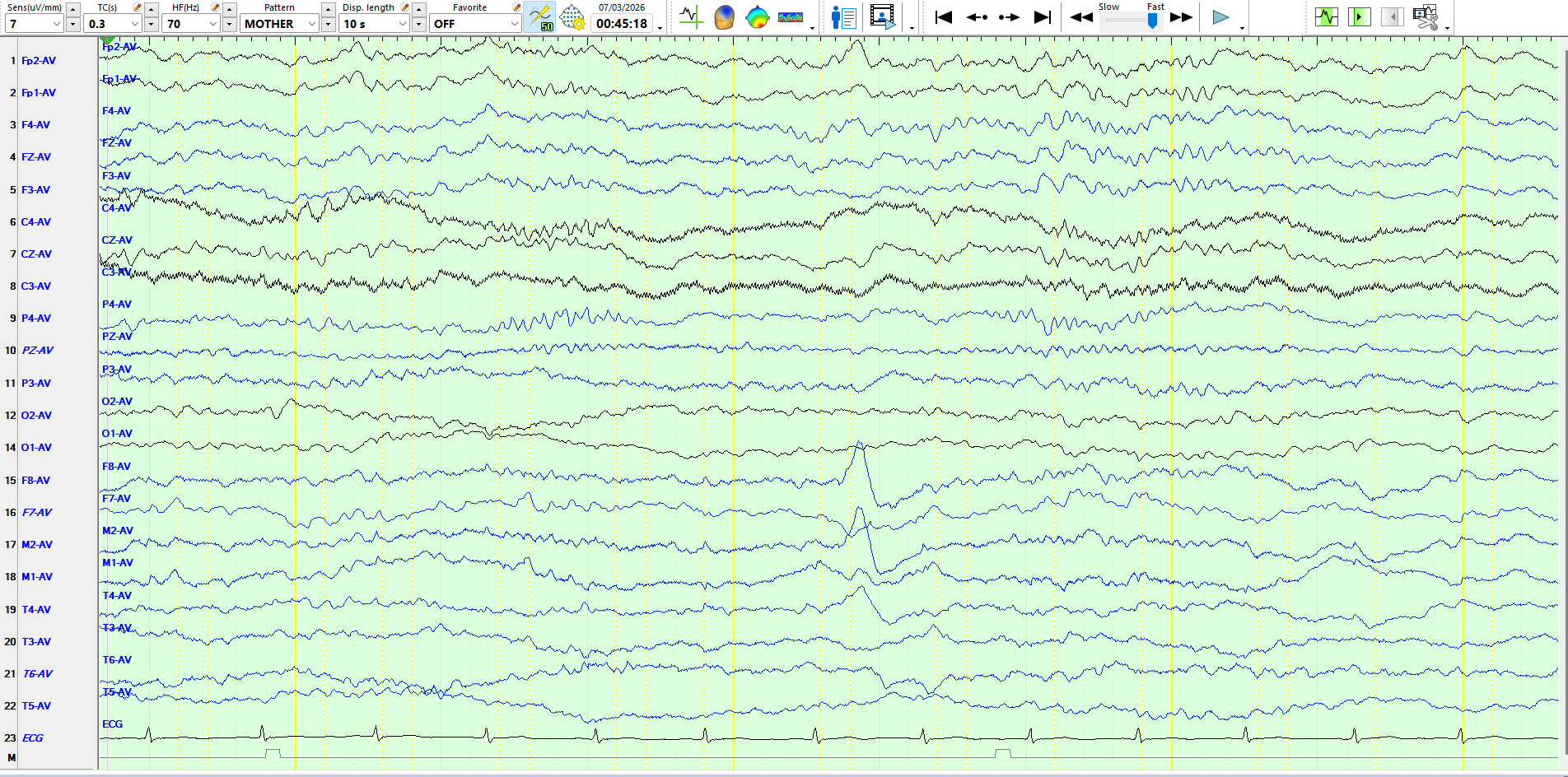
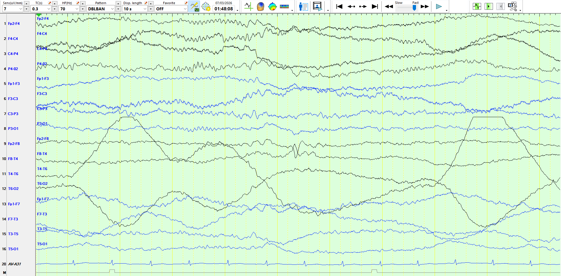
Figure 2 demonstrates rhythmic theta waves at M1-F7, somewhat sharply contoured. The clue to interpreting this correctly is context; as these waves appear, slower waves are seen in the other derivations at the same time (the entire recording slows subtly). The EEG shows no eye blinks, indicating the patient is relaxed and therefore prone to drowsiness, especially at this time of night. Although theta waves are not obvious at F8, they certainly are present. The presence of some artefact at eight and T4 further misleads the brain. Hence, these theta waves likely represent an asymmetric form of drowsiness. It is noteworthy that theta waves like this are virtually absent from the rest of the recording.
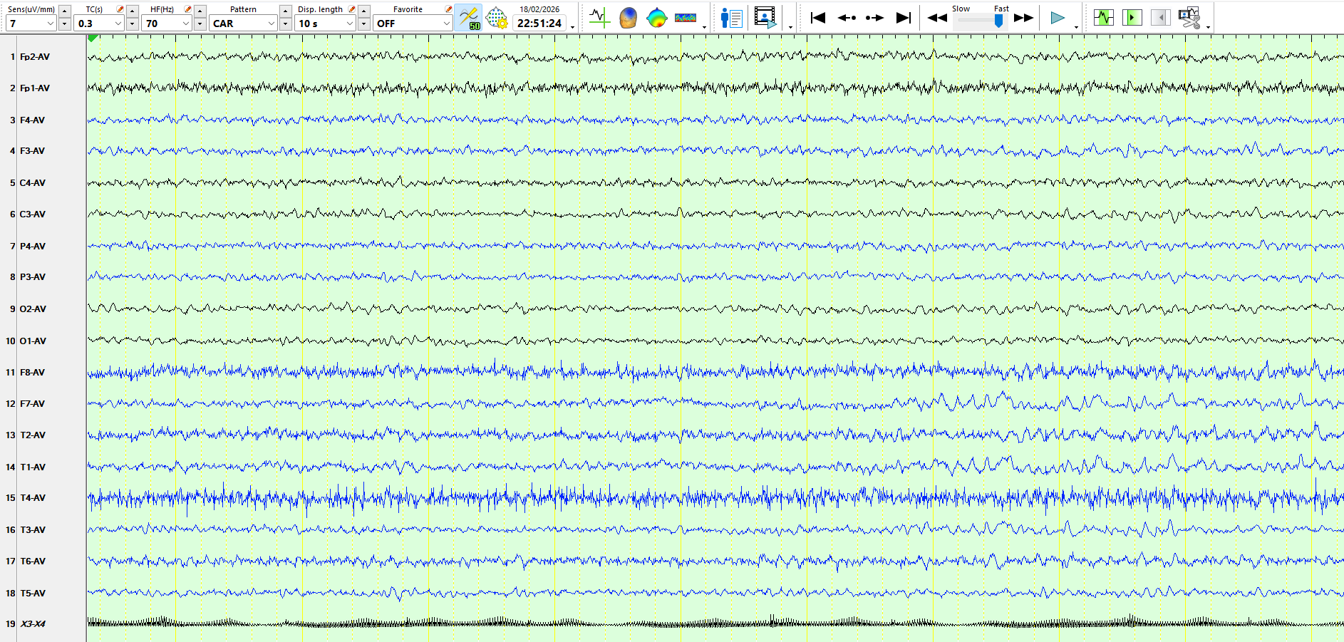
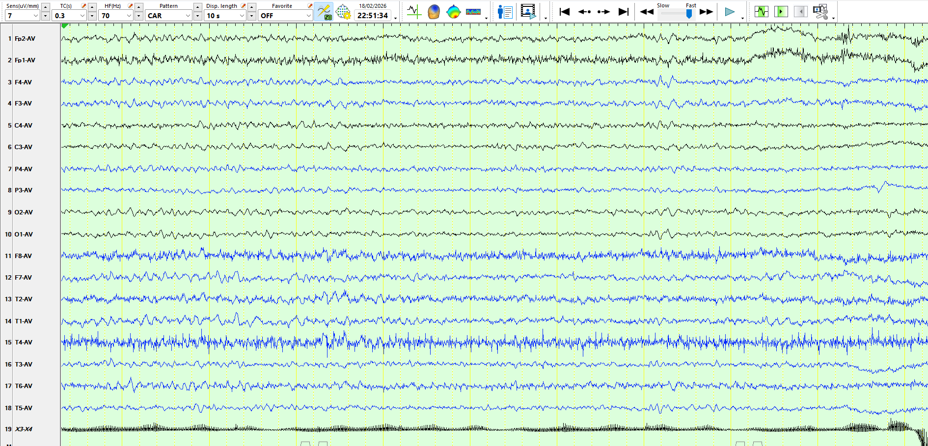
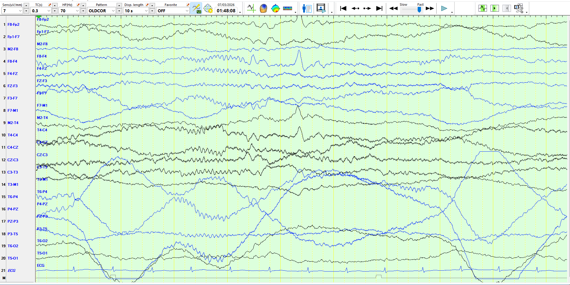
Although you might be tempted to call the sharply contoured waves at F7-T3 on page 6 a sharp wave/inter-ictal epileptiform discharges, you should avoid doing so, as
1. The wave is symmetric
2. The wave has no succeeding slow wave
3. The wave does not disrupt the background
4. The wave is not remarkable in respect of its amplitude when compared to the background rhythms
5. The final 2 pages represent sequences of waves, nearly identical to the above isolated wave. In fact, if you look at the referential and coronal montages representing the wave seen in Figure 6, you will notice that it is part of a sequence of waves.
6. Wicket spikes are notorious for appearing in drowsiness and sleep. You should be prepared for them while looking at the awake recording!
Hence, this is no more than an isolated wicket spike. Such waves are repeatedly overinterpreted in many recordings and accompanying reports I receive for review. Wicket spikes are benign temporal sharp transients that mimic epileptiform discharges, but lack a slow after-wave and do not disrupt the background.
This patient's EEG is normal.
The following EEG was performed a man in his 60s who has had seizures since his teenage years:
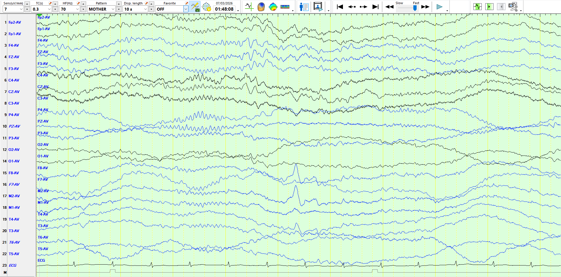
This wave is not a wicket spike because of its high amplitude, when compared to the background. It is also asymmetric and, importantly, there is no physiological wave in sleep that resembles it. Although there is no following slow wave, given the aforementioned features, this is an example of a sharp wave in sleep. These pages also illustrate why I prefer the coronal to the double banana as my preferred montage for reviewing EEGs.
Here is another page from sleep, from the same patient