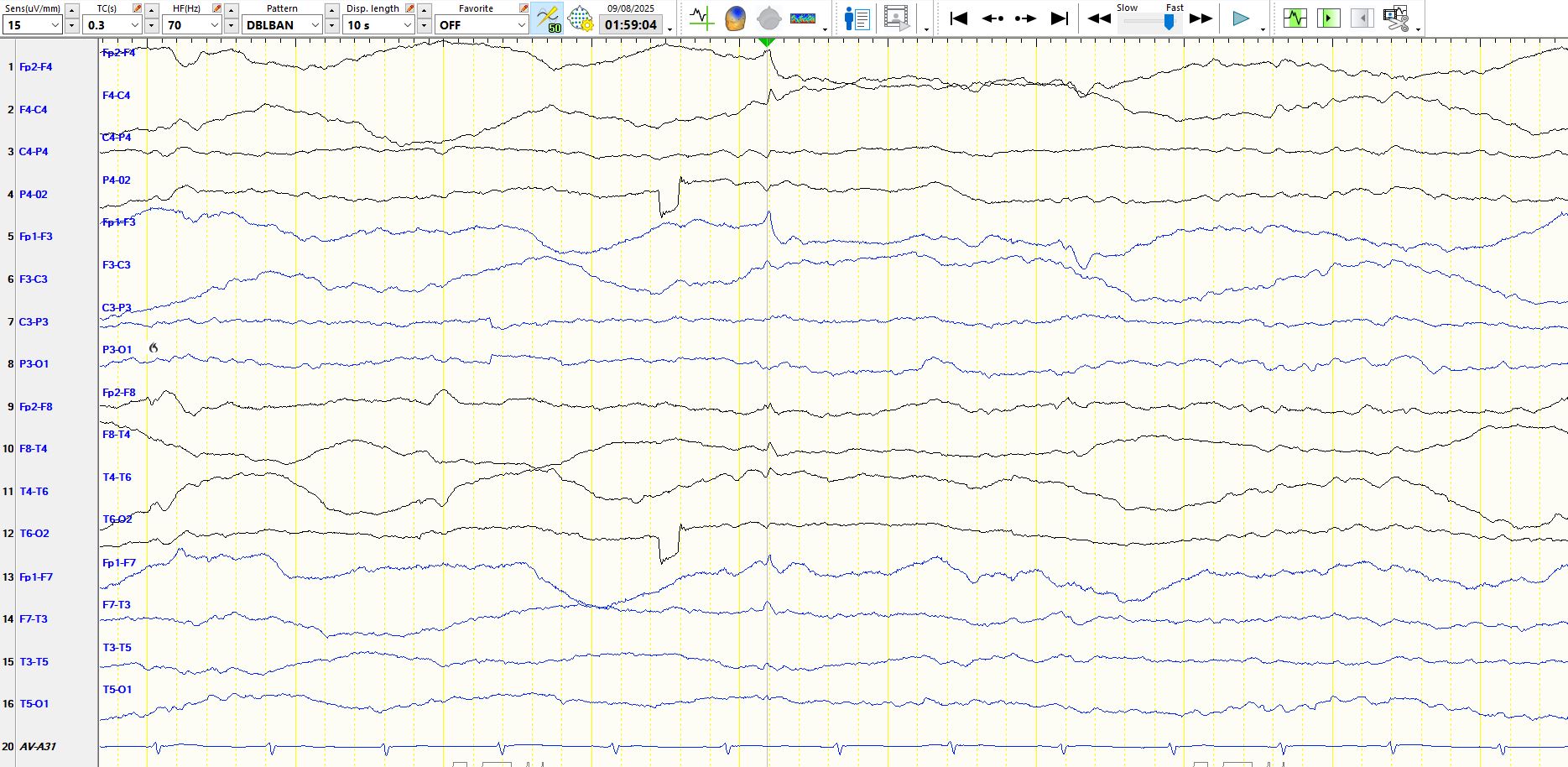
F-wave vs V-wave vs generalized spike vs left frontal spike vs other?
Aug 11, 2025The patient is 30+ years old and asleep.
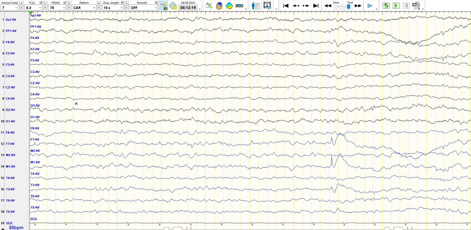
What do you make of the following wave that occurs between the 7th and 8th ECG beats?

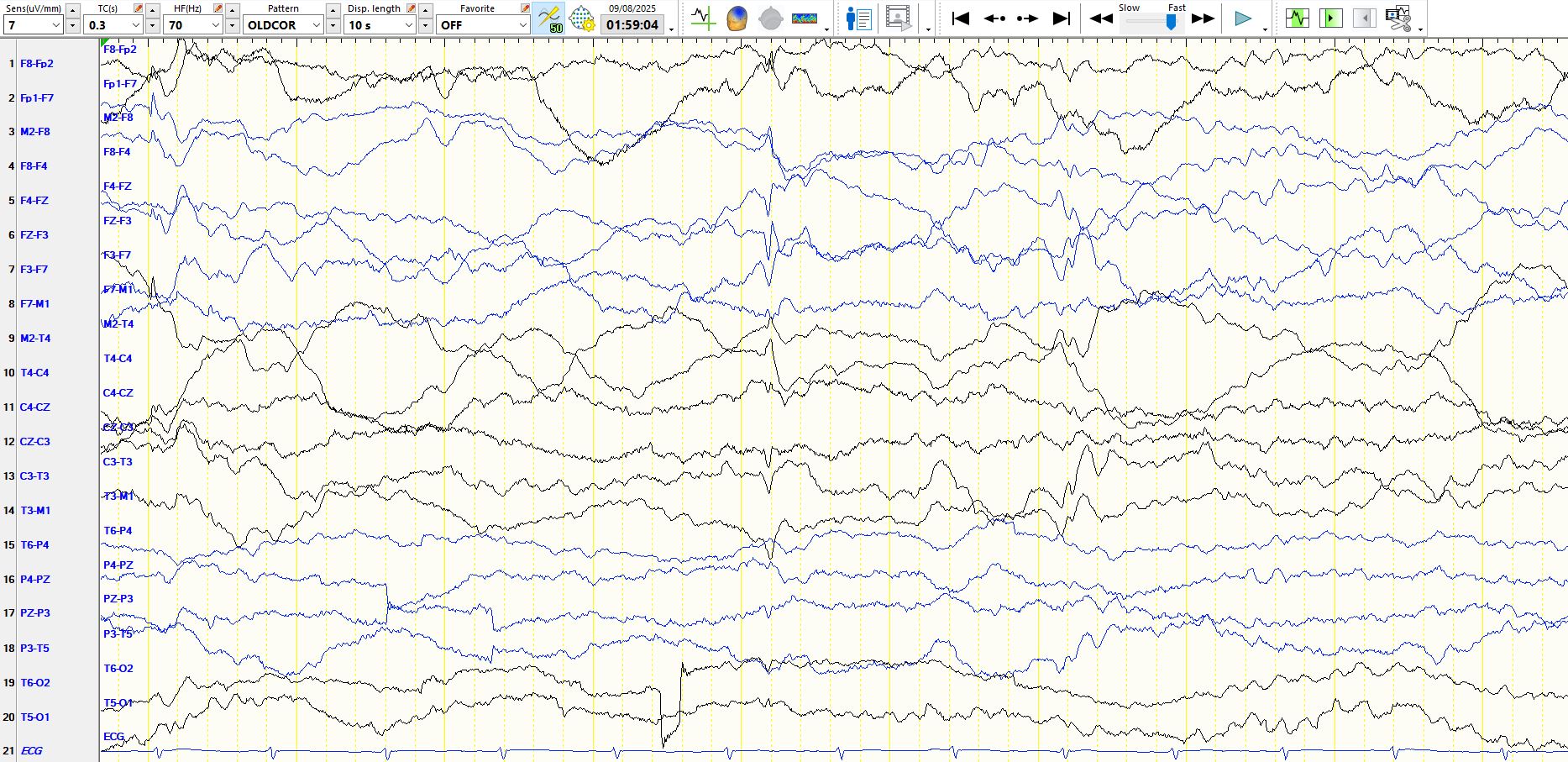
The following represents the same epoch on the coronal montage:
Based on the above, you might consider an F-wave/V-wave (they may be very sharply contoured) or a generalized spike as the most likely.
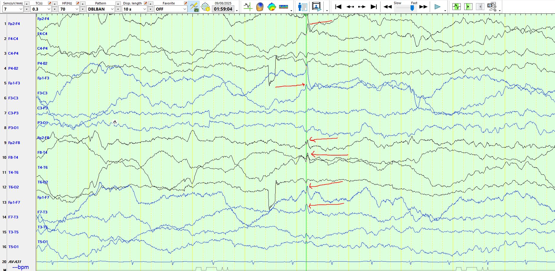
However, if you looked carefully at the top page, you might notice that the apiculate wave is not entirely synchronous in all channels. The peak of the wave corresponding to the 2nd red arrow (F3-C3) precedes that of the channel immediately above it (FP1-F3). This is not typical of a vertex wave. You might also notice that the peak of the wave corresponding to the 3rd arrow and fourth arrow is also not synchronous with that corresponding to the 2nd arrow. These findings are also not typical of a generalized spike.

I have tried to simplify things on the common average montage by removing the sagittal derivations (see below).

The opposite polarity of the left and right frontal, central, parietal and mid-temporal electrodes should be apparent in the page above and below.

On the same page above, you will notice that the discharges in the temporal regions are not coincident. There is an electro-negative wave adjacent to the 5th and 6th arrows at F7 and M1, but also at FP1. A short while later (within milliseconds) there is a similar wave at M2, F8 and FP2. Hence, these discharges are temporally associated, but probably do not represent the same discharge (discharges may sometimes be time-locked between the hemispheres). The first of these discharges is almost equi-potential at F7 and FP1 and M1 and with opposite polarity over the right frontal-central-temporal region. Hence, this is a left fronto-temporal electronegative spike, most likely originating in the left lateral frontal region.
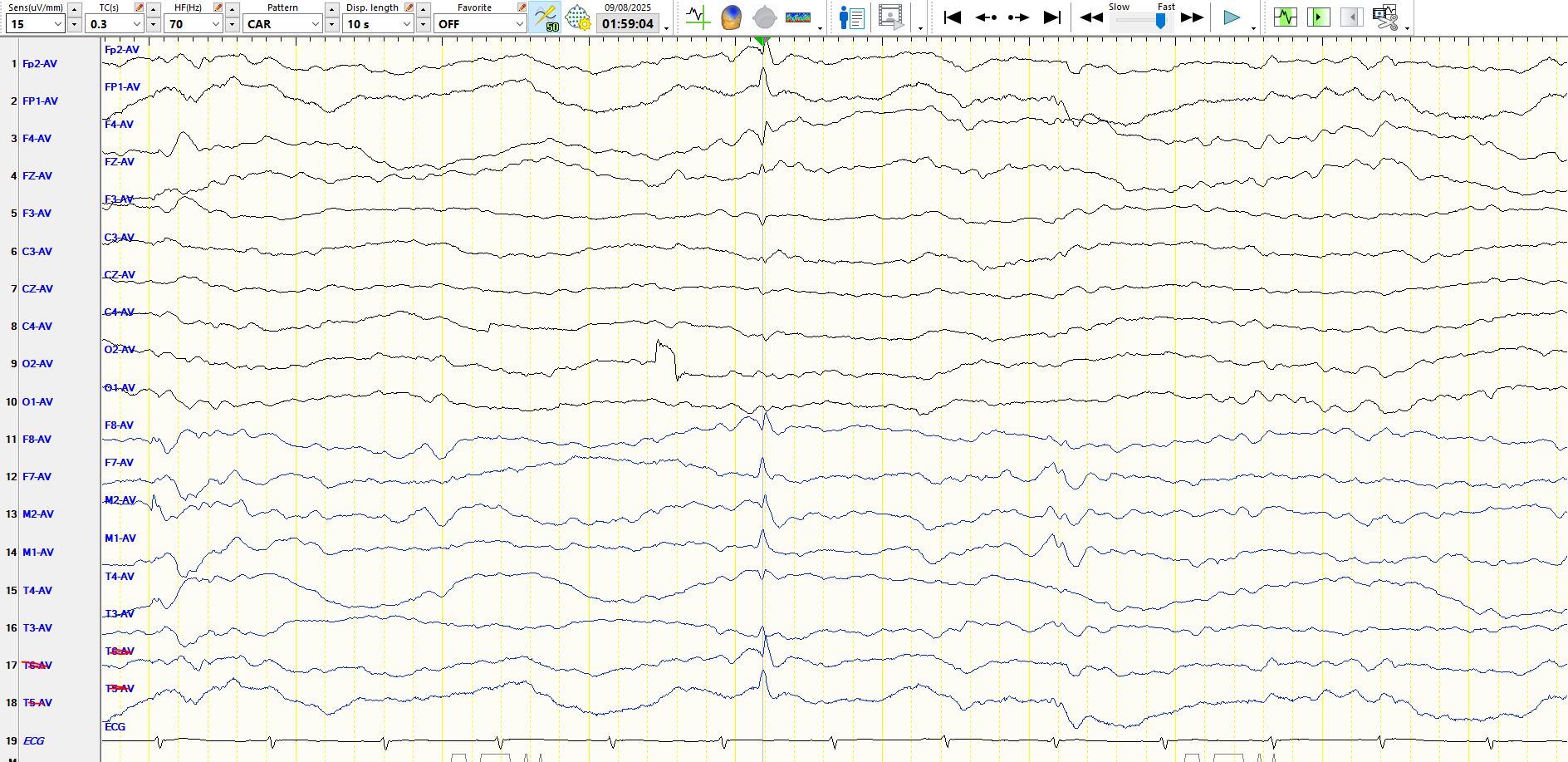
To try and distinguish the amplitude differences between the prefrontal and temporal electrodes, I have moved the prefrontal electrodes to the back of the head in place of T6 and T5 on the following image and the sensitivity has been changed to 15 µV per millimetre. This illustrates the point that the 2nd wave is a right prefrontal spike, primarily electro-negative at FP2 (FP2-F8-M2).
The following is the bipolar AP montage, with the sensitivity set at 15 uV per millimetre to permit another way of visualizing cancellation:
The following is the common average montage, with the waves displayed at 10 µV per millimetre.
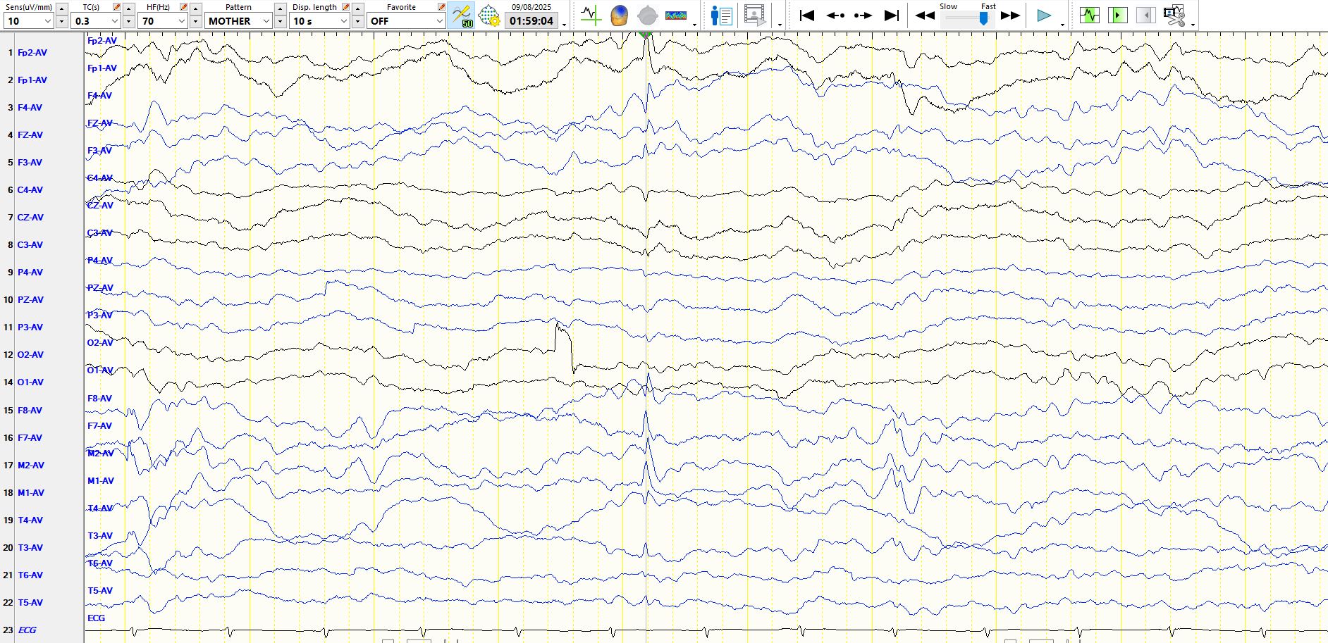
In conclusion, the above likely represents a left lateral frontal spike, immediately followed by a right prefrontal spike. You might not have guessed that when looking at the top image.
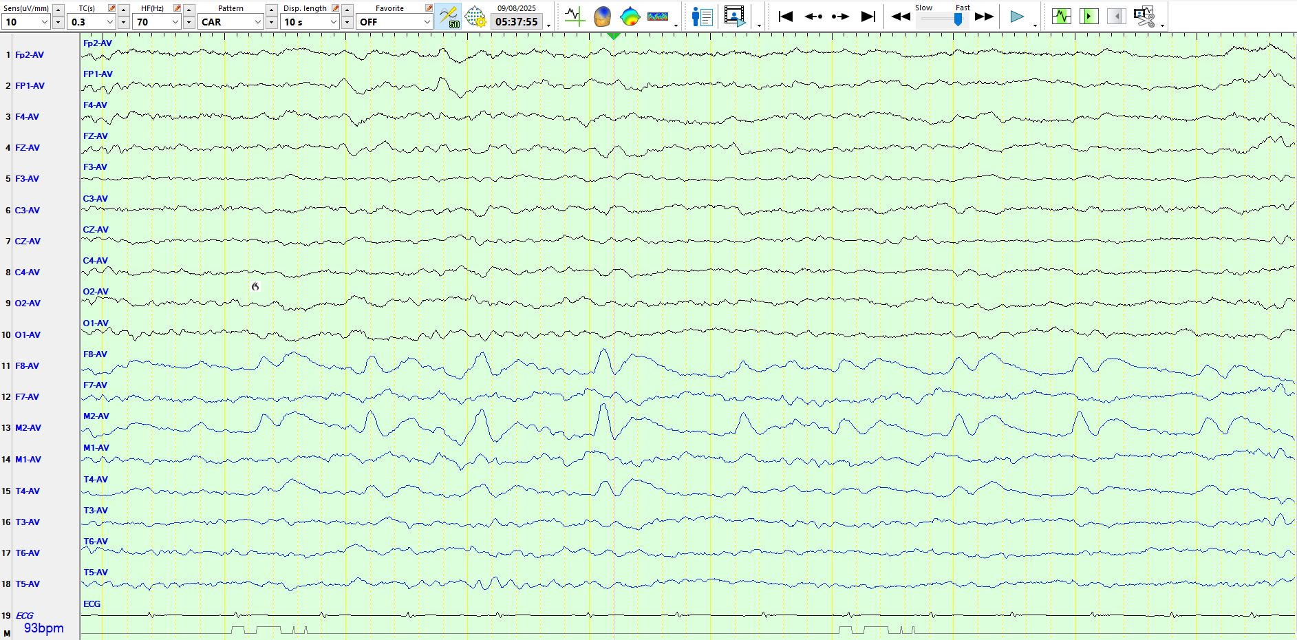
And the following?

The following is a spike at FP1-F3, followed by spike-and-wave at M1-F7, while there is a spike-and-wave in the 3rd second at F8-M2. The amplitude is higher than you think, as the sensitivity has been reduced to 10 µV per millimetre.

The following is a train of sharp and slow waves at M2-F8. The maximum duration of these sharp waves is 180 ms. This still conforms to the current definition of a sharp wave (70-200 ms).
Just for good measure, here is a left temporal spike-and-slow-wave. The spike below is a lot sharper than the above "sharp waves". Spikes are defined by a duration of less than 70 ms.