
More little bumps
Aug 01, 2025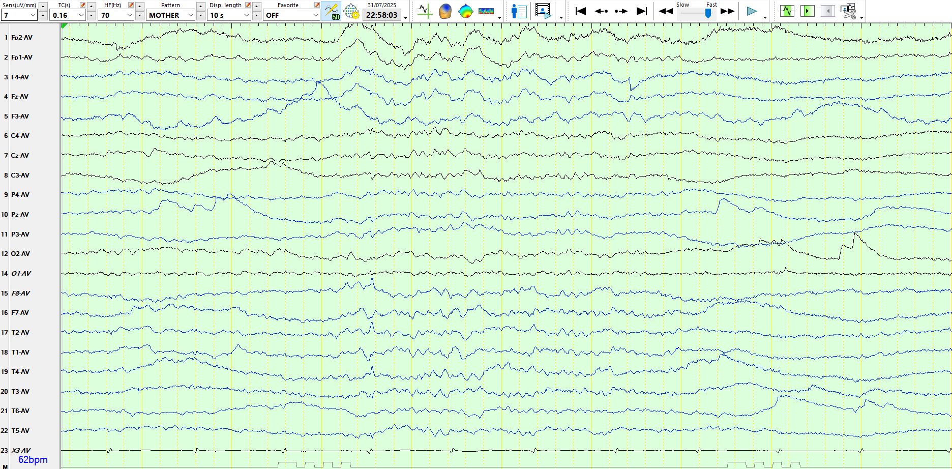
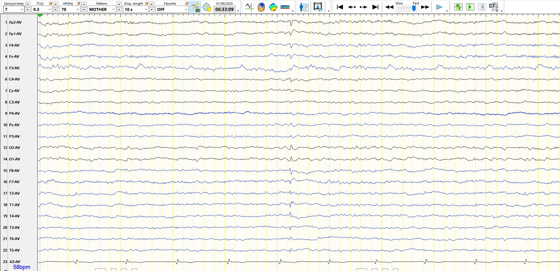
The above is a summation of waves that includes the 4th ECG beat. However, it has a large field and hence is more than just the ECG beat. There are suspicions of a small sharp spike that is coincident with this wave.
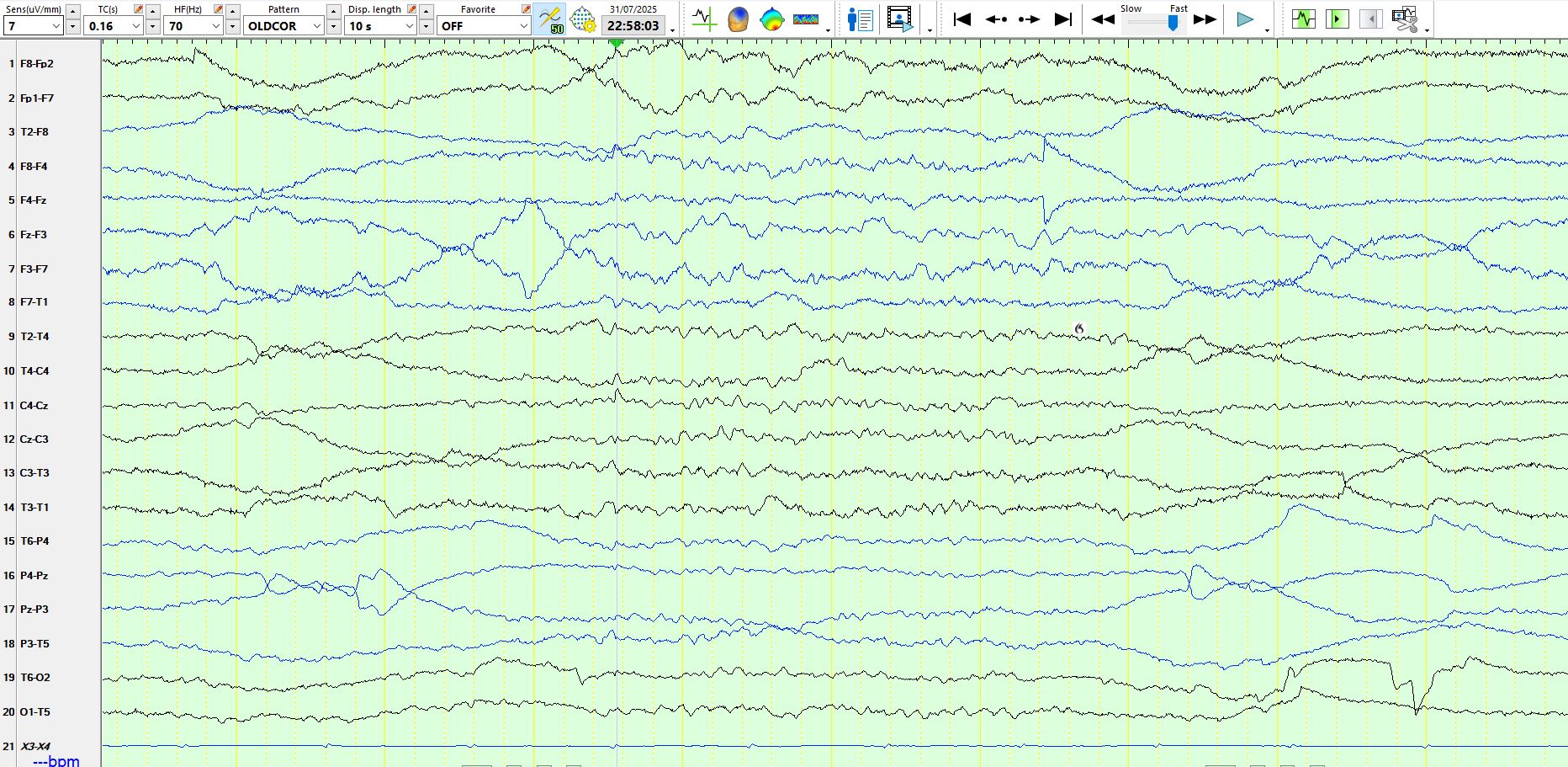
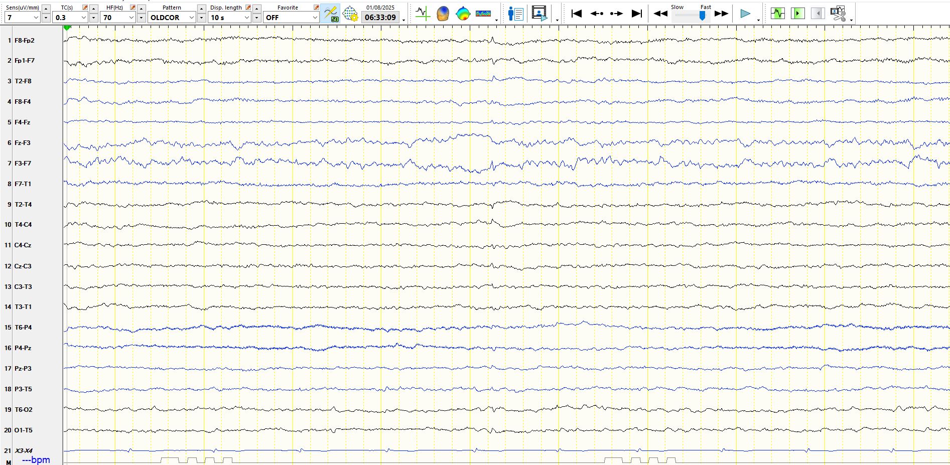
The coronal montage, above, substantiates this hypothesis. The wave is barely visible and has a large field, typical of a small sharp spike.
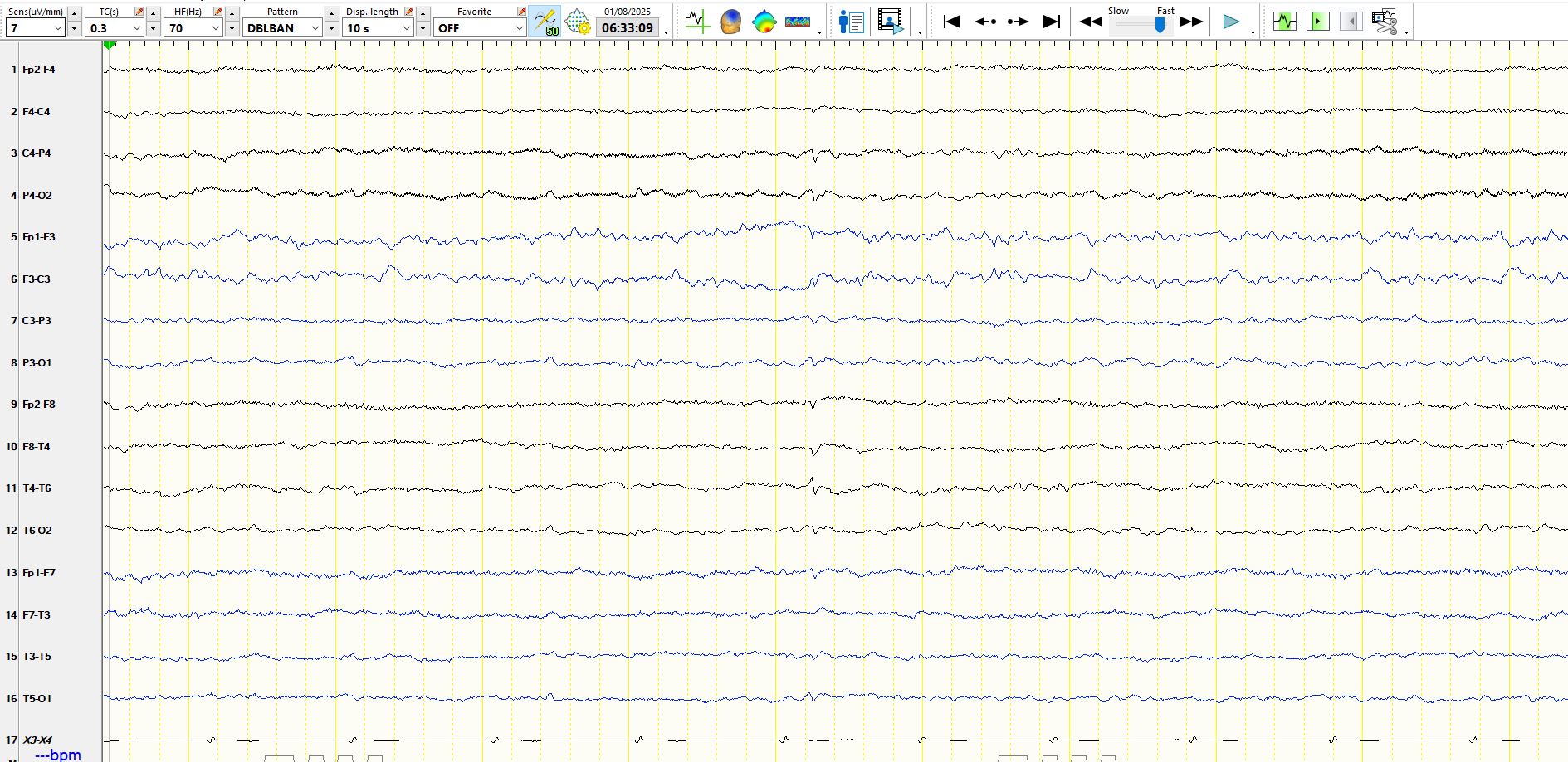
The patient has many small sharp spikes. The following 3 pages represent the same page, viewed in different ways; the third page is a representation of the same waves on the coronal montage.
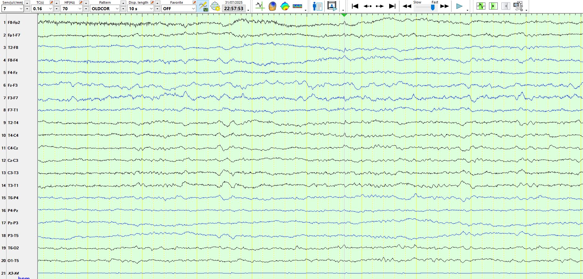
In the above examples, notice the proximity of this wave to the ECG beat and large field on the coronal montage, which provides assurance that this does not represent an ECG beat and provides corroborative evidence for a small sharp spike.
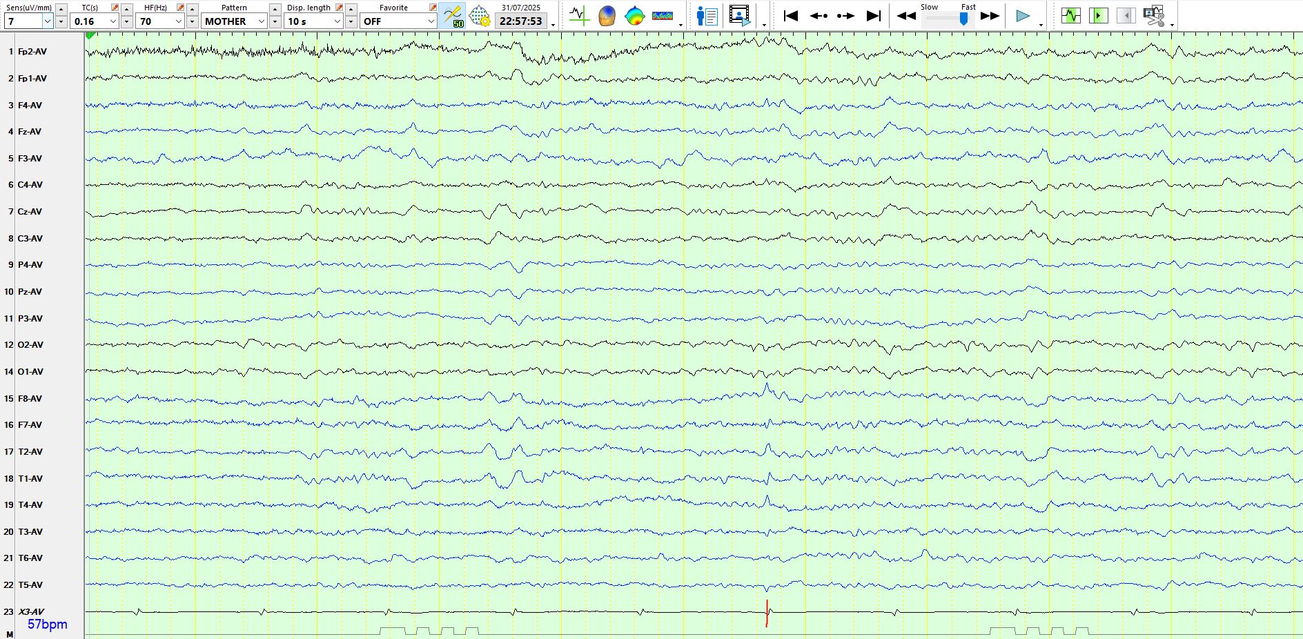
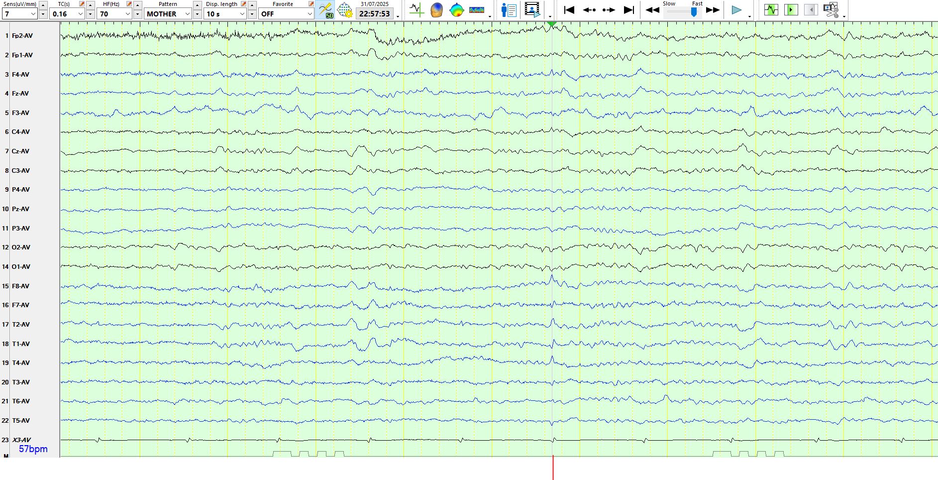
The following is taken from a different patient:
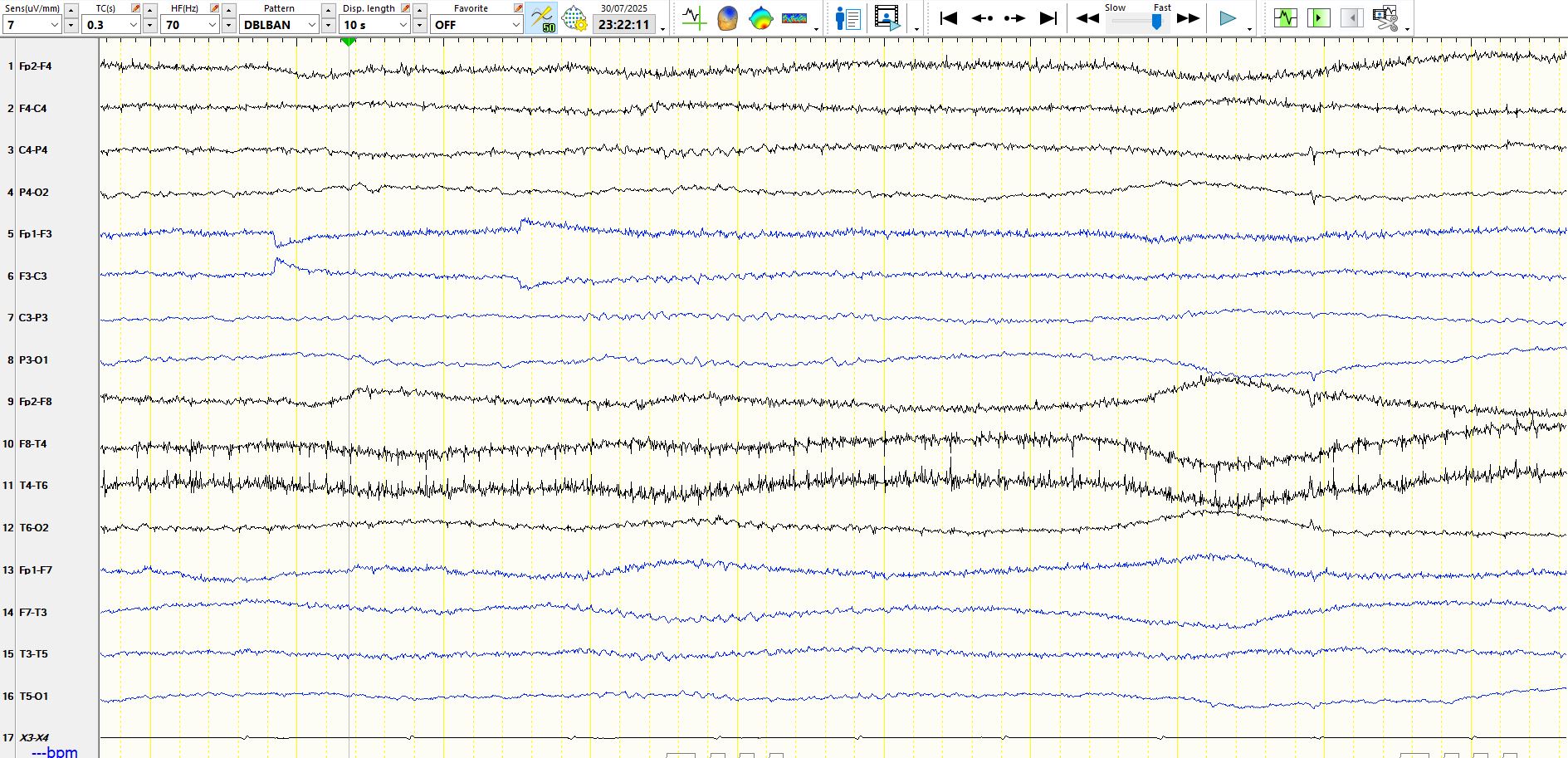
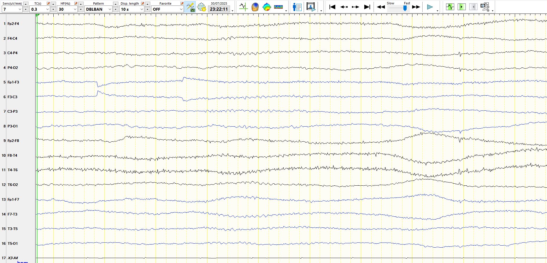
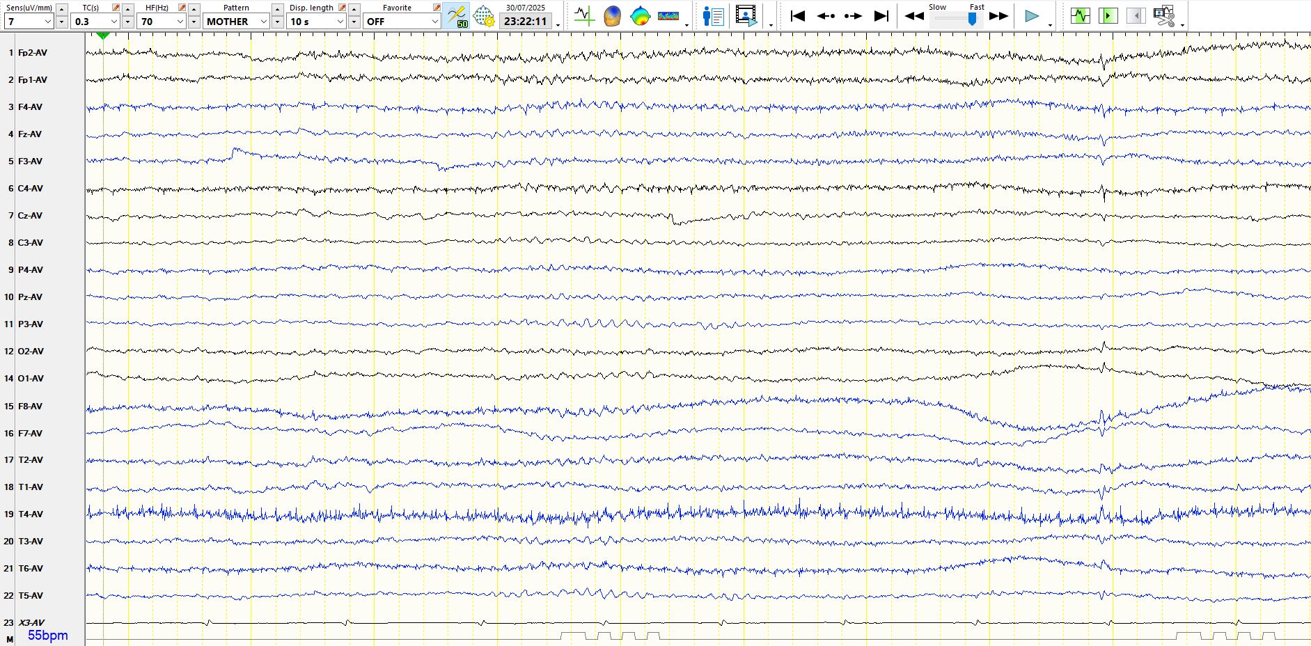
The above pages represent the same 10 seconds. The high frequency filter has been changed on the 2nd page. The 3rd page demonstrates the appearance of the discharge in the 8th second on a common average montage. You will notice that the discharge has an unusually sharp morphology when the high frequency filter is left at 70 Hz. When the high frequency filter is reduced to 30 Hz, the wave starts to resemble that of a cortical origin and is not dissimilar to a small sharp spike; this wave likely is an electrostatic artefact.
Here is another example of a small sharp spike. The same spike is shown on 3 different montages:

The key elements are:
1. its low amplitude
2. its characteristic biphasic morphology
3. the absence of a slow wave
4. its large field.