
Bursts of theta (v1)
Aug 24, 2025The following is a common phenomenon. The million-dollar question is, what is its significance? Attempt to arrive at your own conclusion, before reading my thoughts at the bottom. To reiterate, mine are not necessarily correct, so please feel free to comment at the bottom of the page.
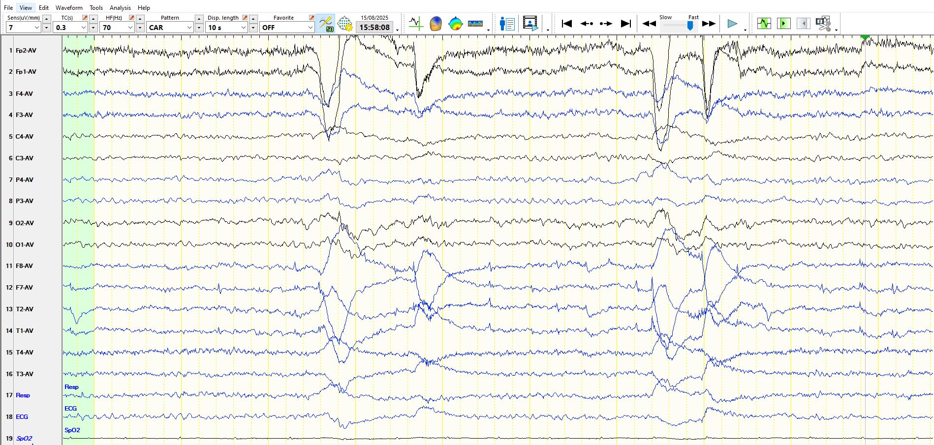
Figure 1:
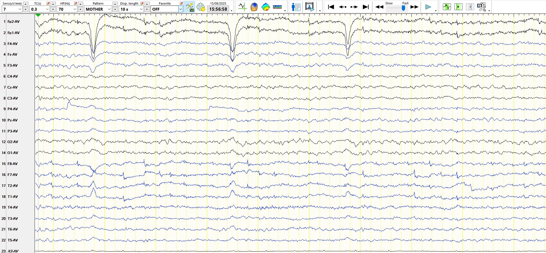
The page above demonstrates that the patient is awake, as alpha rhythms can be seen, there are eyeblinks and there are lateral rectus spikes. There is the subtle appearance of theta at PZ during the last two seconds of the same page, but "lateral rectus spikes" continue until the end of the page In F8, M2, F7 and M1.
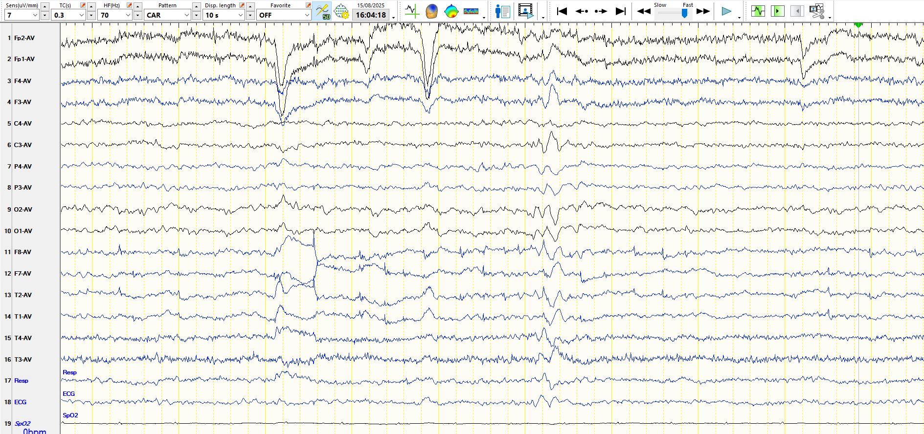
Figure 2:
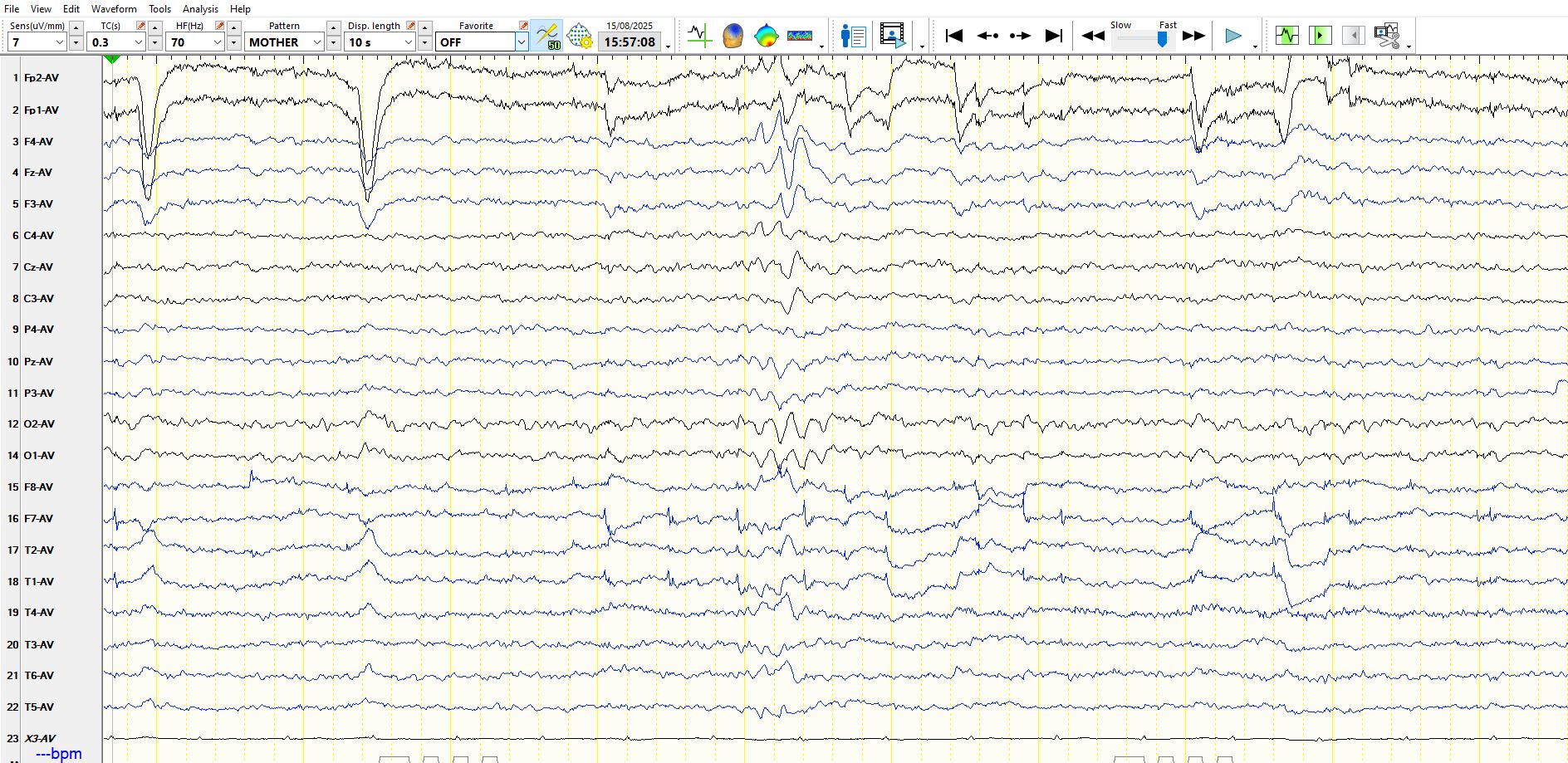
On the page above there are eyeblinks, lateral rectus spikes and preservation of EMG artifact; there is also a burst of frontally predominant theta waves in the middle of the page. The above montage is not ideal for considering these waves and hence the bipolar montages are required. The above the waves represented on the coronal montage can be seen below.
Figure 3:
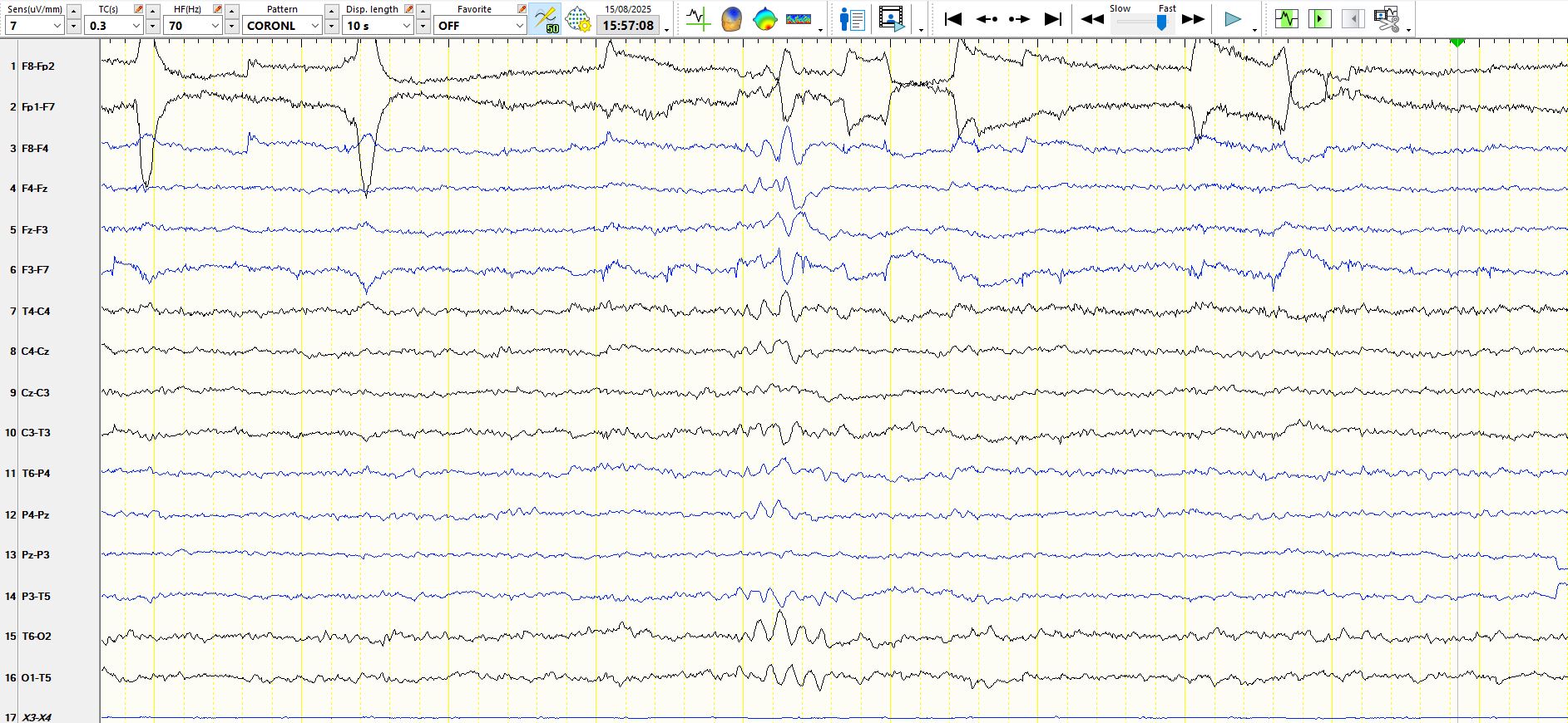
On the coronal montage, above, the theta waves clearly appear over the frontal, central and posterior head regions, providing assurance that these do not represent spatially restricted frontal discharges.
Figure 4:
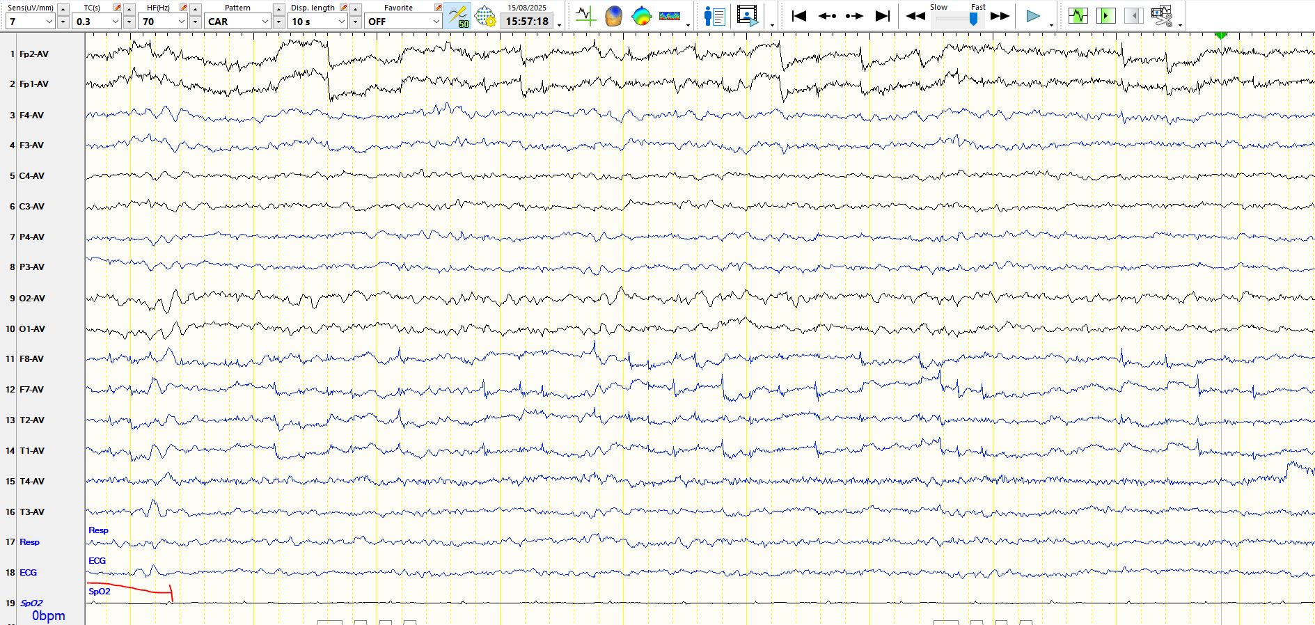
Figure 5:
The bursts of theta waves were seen on multiple occasions. There is another example below, on this occasion slightly higher in amplitude over the left frontal-central region.
Figure 6:
You might want to know the age of the patient; a 20 odd year-old student. The preponderance and frequency of "lateral rectus spikes" is typical of someone who is reading. The first clue about the nature of these can be found in figure 1; in the final two seconds the alpha rhythms attenuate and the early appearance of theta frequencies over the parietal regions indicates minimal drowsiness.
In figures 2 and 3 the bursts of theta waves emerge from the background, progressively increasing in amplitude and appearing widely over the brain, including posteriorly.
Figure 4 is intended to juxtapose nine seconds of the patient reading with the fleeting appearance of drowsiness during the first second of the same page. On this occasion the theta is of low amplitude, but the point is that this page provides corroborative evidence that the patient is drowsy around the time of the bursts of higher amplitude theta waves. Of note, such bursts did not appear the next morning when the patient was awake and reading.
Figure 5 demonstrates the appearance of this patient's normal rhythms during full arousal.
The recording is obtained in the middle of the afternoon, when the genetically determined circadian rhythms modestly reduce the threshold for drowsiness and sleep. Hence, it is unsurprising that mild drowsiness might occur while the patient is reading at this time.
I suspect that bursts of medium amplitude frontally predominant theta waves are the most over-interpreted physiological waves. They are normal. As everyone knows, you can be drowsy when you read or study!
For what it's worth, this patient has functional seizures with a myriad of associated other functional symptoms.
More to follow on this subject.