
Discharges with a variable appearance, SSS vs multifocal spikes or both?
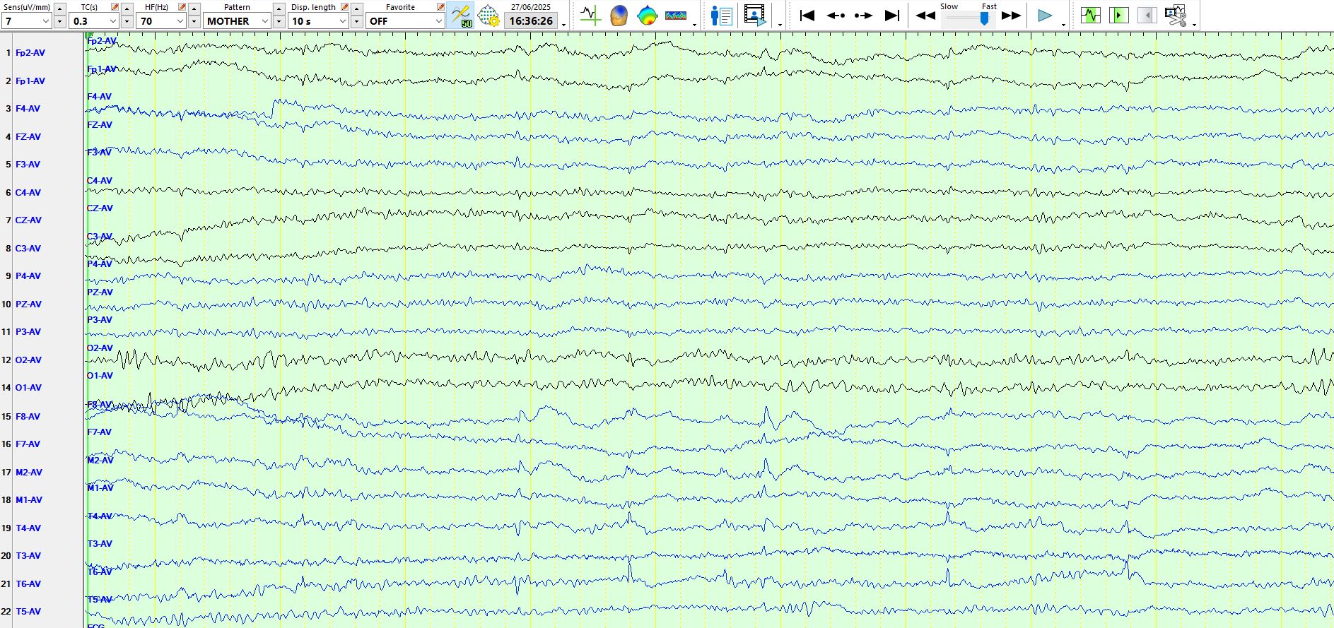
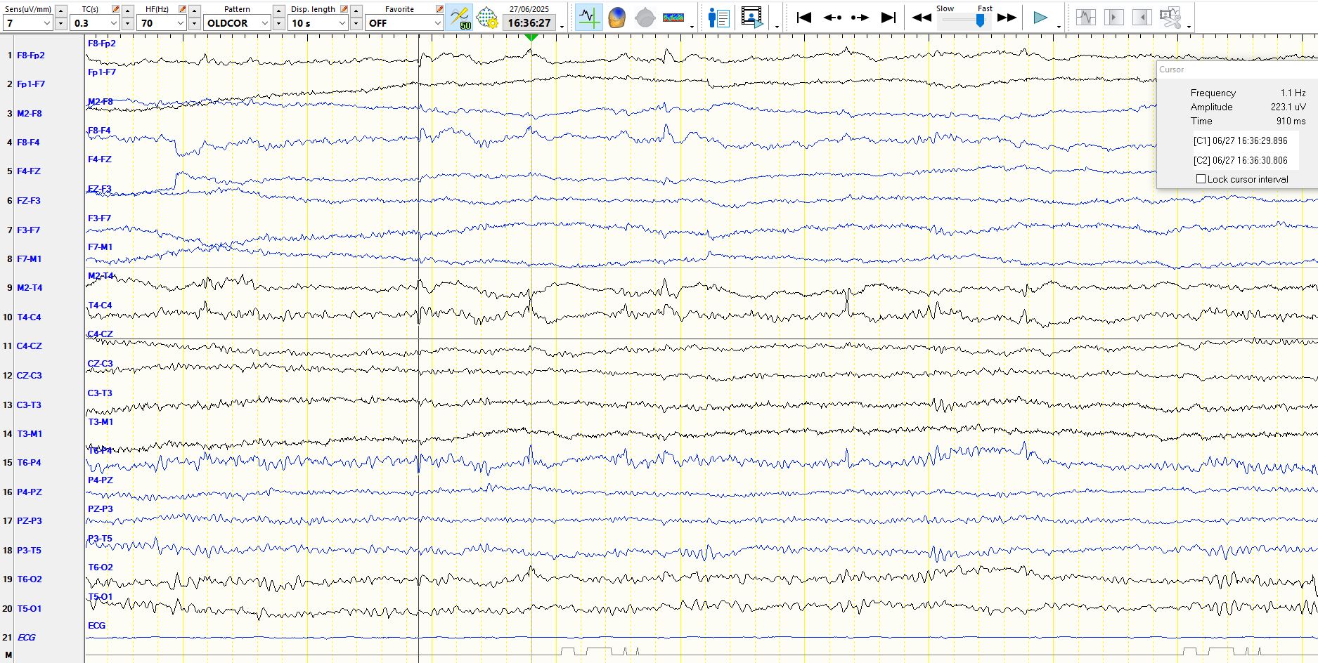
Jul 05, 2025Consider the following page of a young adult. Try and identify the nature of the discharges that correspond to the red lines at the bottom of the page. The patient is asleep.

In the image above, what do you make of the first and fourth discharges corresponding to the red lines? What do you make of the discharge corresponding to the fifth vertical red line?
Here are more discharges from the same patient on the following page:
The above discharges highlight some of the difficulties of distinguishing small sharp spikes, a normal physiological variant, from inter-ictal epileptiform discharges. In the top image, the discharge corresponding to the second vertical red line can be confidently called a spike-and-wave at M2-F8 but notice that the discharge is also seen at T6-T4, if anything larger in amplitude at T6 than at T4. the last vertical red line on the top image demonstrates a very similar morphology to the discharge corresponding to the second vertical red line and is clearly primarily electro-negative at T6. If you then consider the first and fourth vertical red lines in the top image, these are largely of opposite polarity and are therefore electropositive at T6-T4, while electro-negative at F8-M2. These therefore represent the two ends of a dipole. This steep dipole from the back to the front of the temporal lobe is unlike a small sharp spike, which typically has a large field. However, without the distinct slow-wave at times, one might be left wondering and uncertain about these waves; the best course of action is then to note them in the report, but not as part of the clinical interpretation and certainly do not call them inter-ictal epileptiform discharges.
So, what does one make of the variable morphology and variable apparent distribution? These likely represent the same dipole and this dipole only needs to shift a little, either at the front or the back, medially or laterally, superiorly or inferiorly and the appearance will change. At times you see the electropositive discharge only at T6 and on other occasions only at F8 and on other occasions at both.
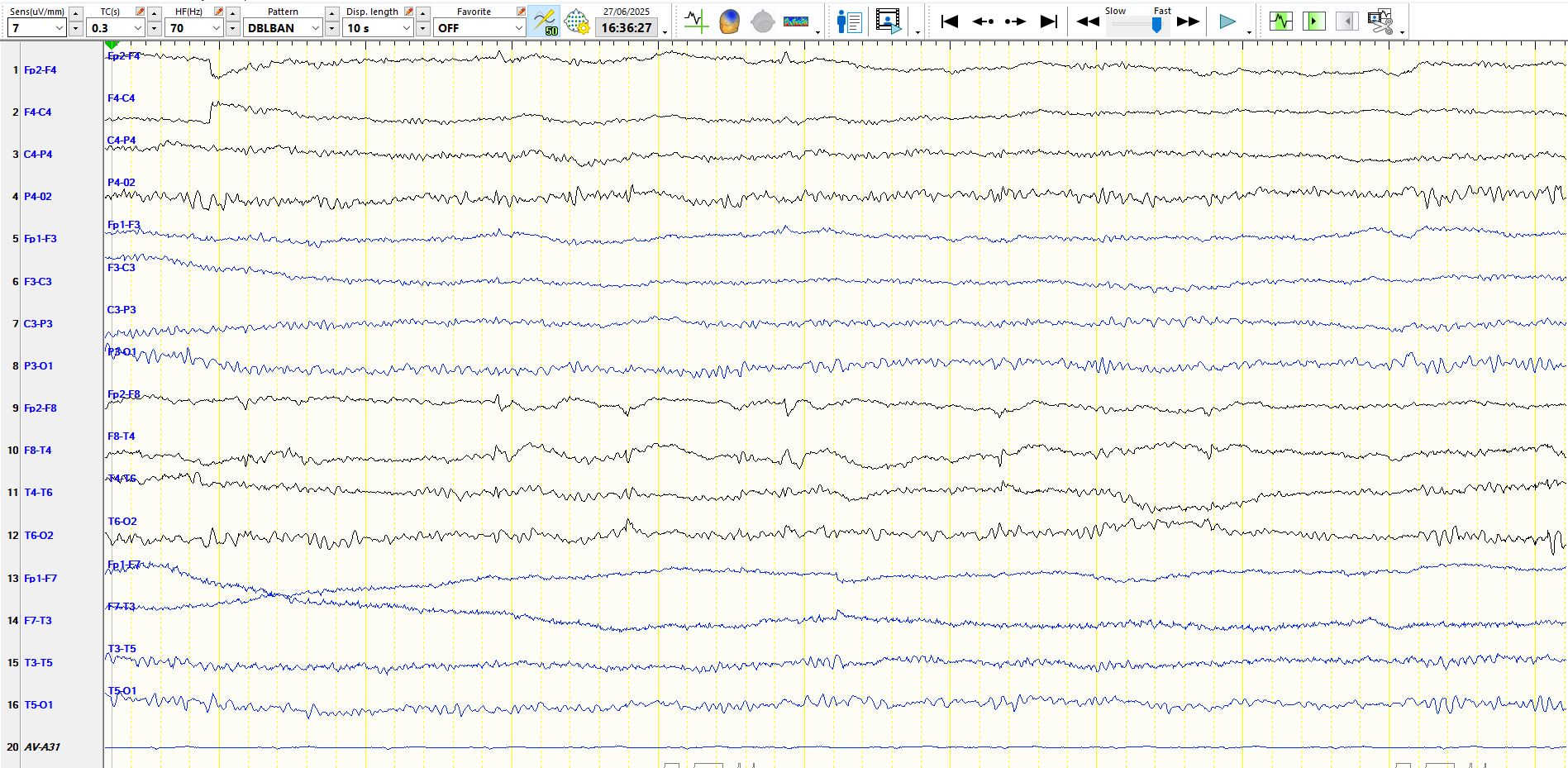
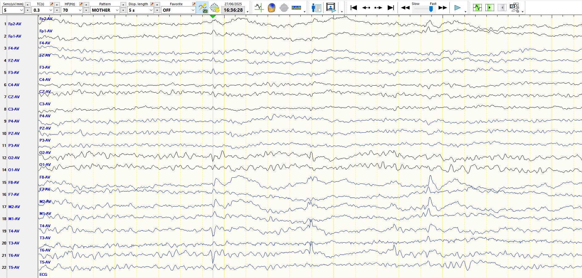
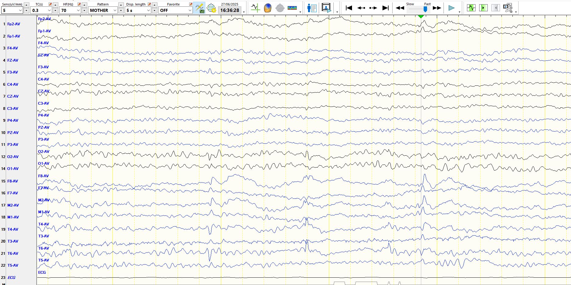
The phenomenon is further illustrated by a page represented on referential, anterior-posterior bipolar and coronal bipolar montages below. Notice that on bipolar montages the dipole is remarkably monomorphic when compared to the referential montage, supporting the hypothesis above. It likely is the same thing, it just does not look like this on the referential montage! And the restricted field means that these are not small sharp spikes. The presence of the slow waves settles the matter. These are particularly well seen on the coronal montage below.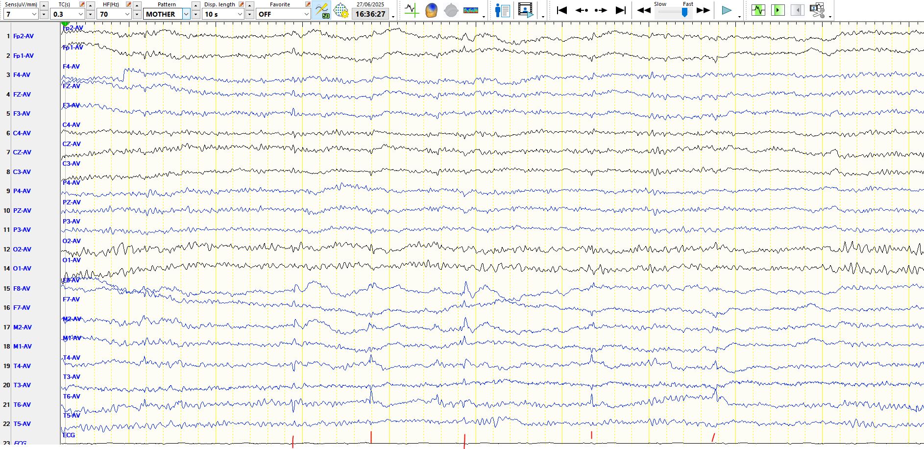
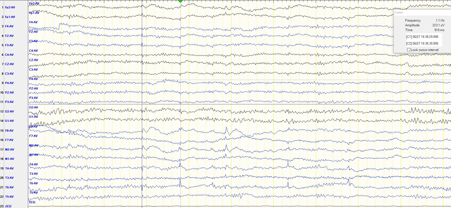
If you are having difficulty seeing the dipole, have a look at the following page at five seconds per page, rather than the 10 seconds per page above. Notice the opposite polarity at FP1-F3-C3-FZ. The electropositive at discharge at T6-T4 can also be seen at O2, providing strong reassurance about its nature and location, while the temporal association of the slow waves, which appears at F8-M2, adds further weight to the argument that this is a single dipole
Just for good measure, this is how the patient's seizures started
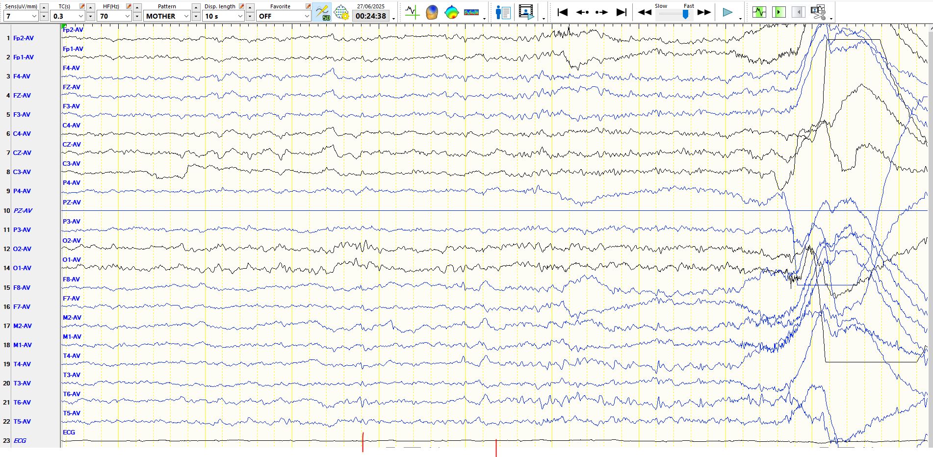
In the image above, the first red vertical line denotes an electric positive spike at T6-T4, while the second red vertical line denotes the onset of the seizure, primarily at T6-T4.
