
Suppression of EEG rhythms
Aug 28, 2025This discussion related to an EEG performed on someone in ICU refers ICU, "Not waking up, seizures? Brain dead?".
To reiterate, judgements about suppression can be quite tricky, doubly so in the ICU setting, and, if there is any doubt, a repeat EEG, preferably at least 24 hours later, should be obtained. After the above EEG the physician informed me that the patient was "clinically brain-dead".
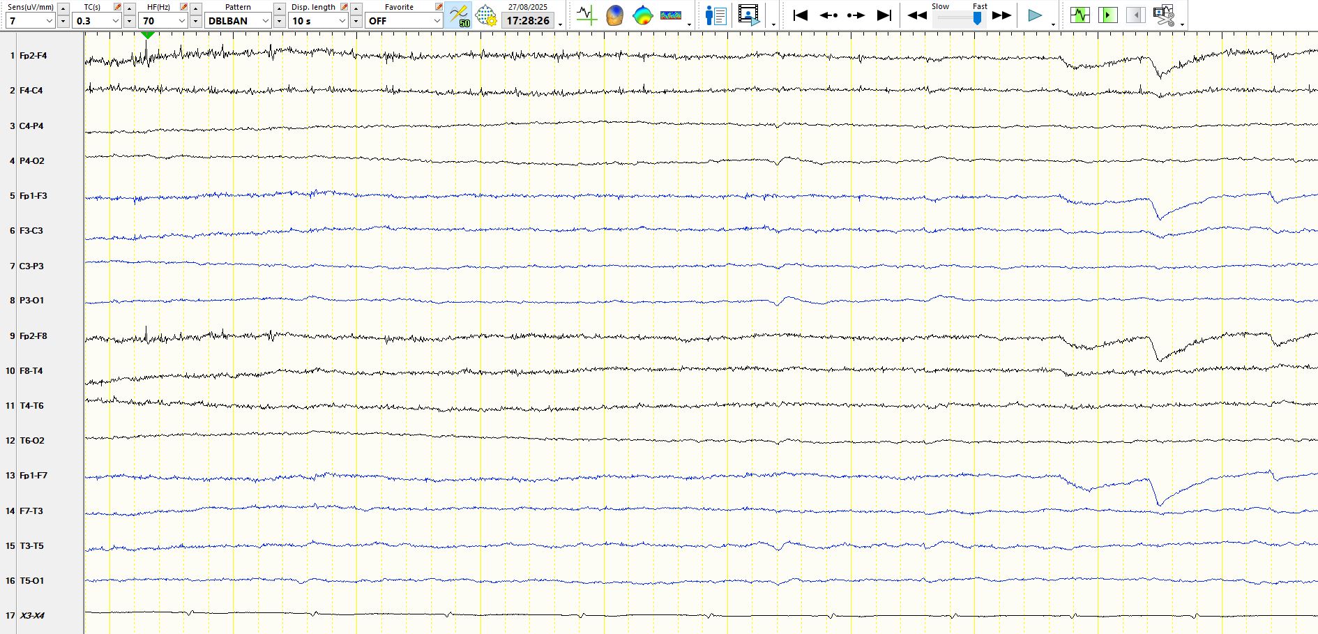
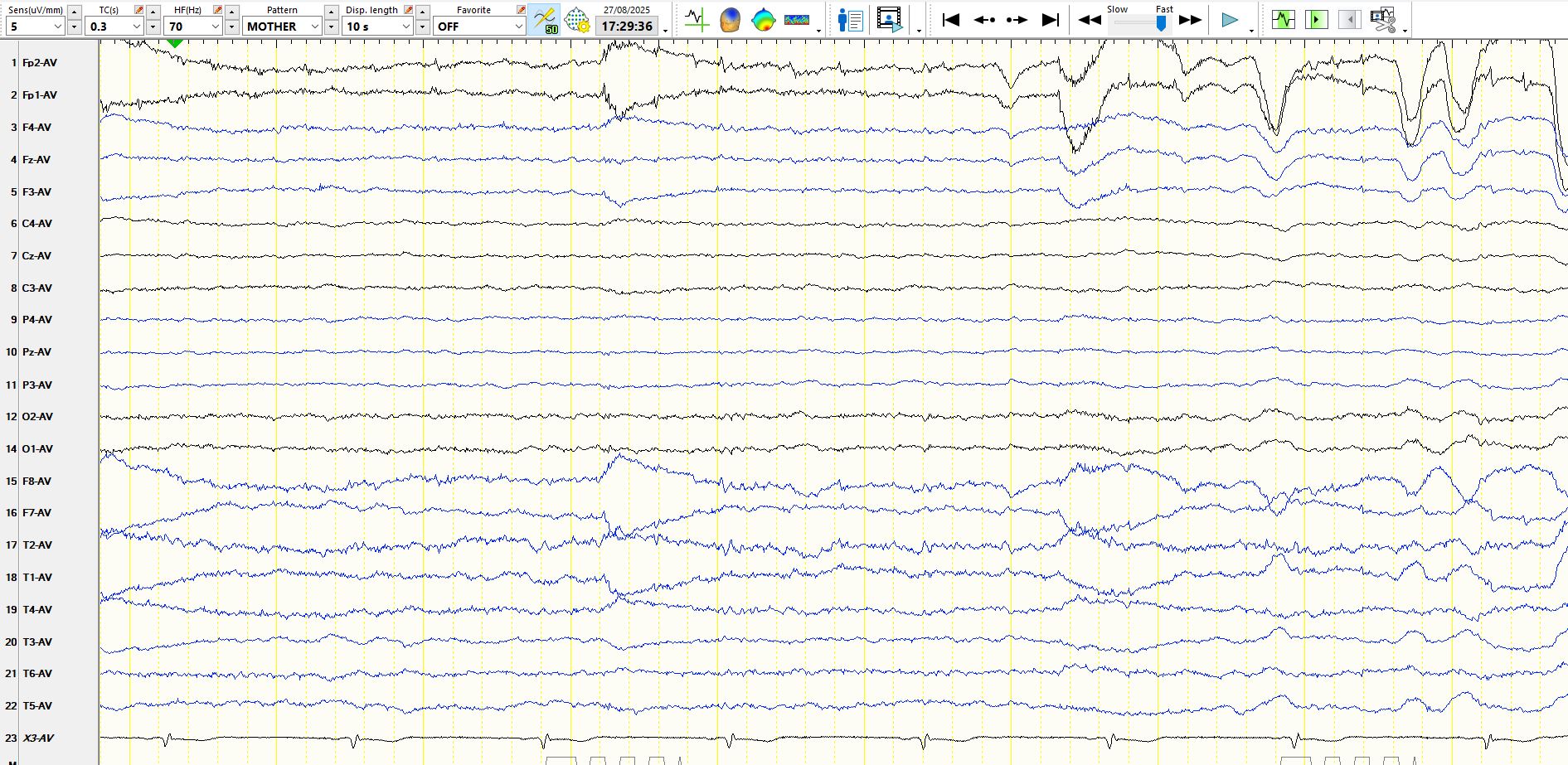
To illustrate the problem further, consider the following EEG from a young adult patient who is wide awake, interactive and known with left hemispheric Sturge-Weber Syndrome.
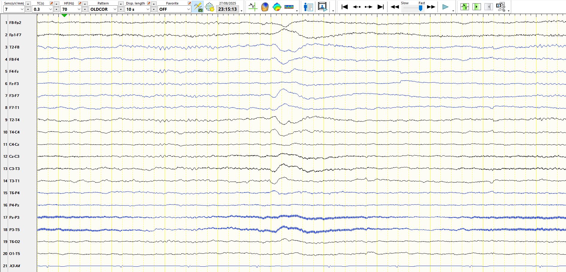
Bipolar anterior-posterior montage, 7 µV per millimetre:
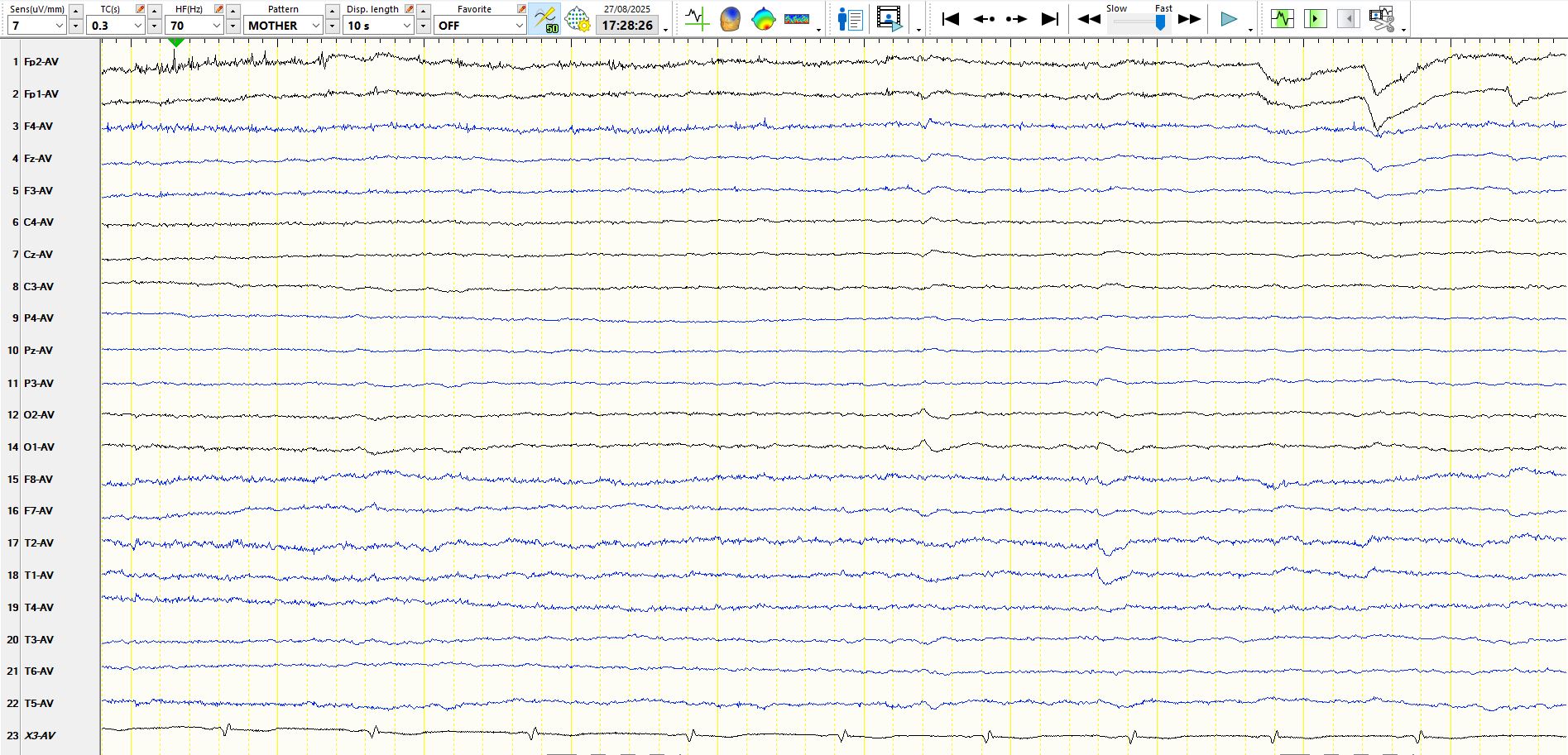
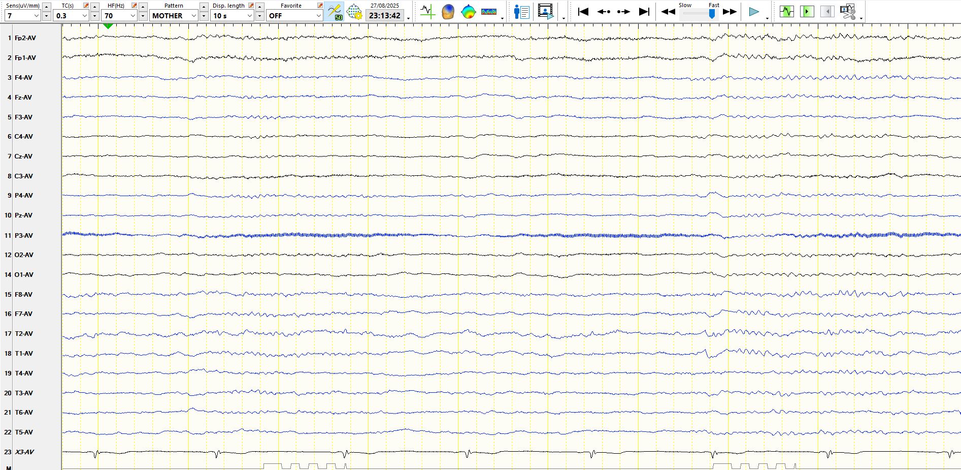
Common average montage, 7 µV per millimetre:
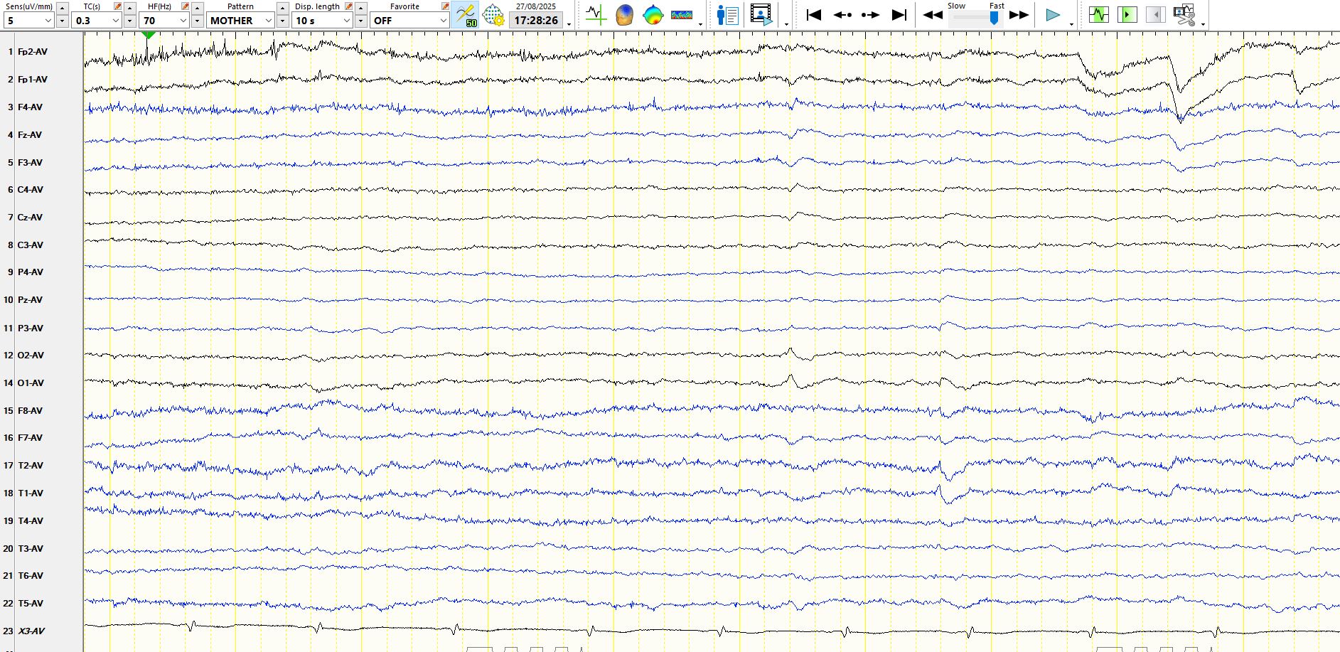
Common average montage, 5 µV per millimetre:
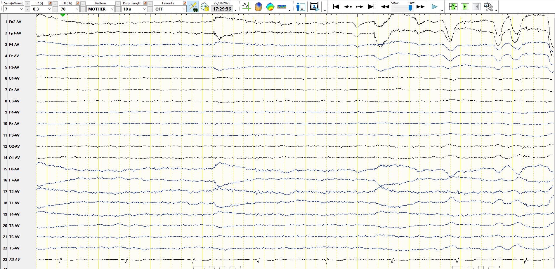
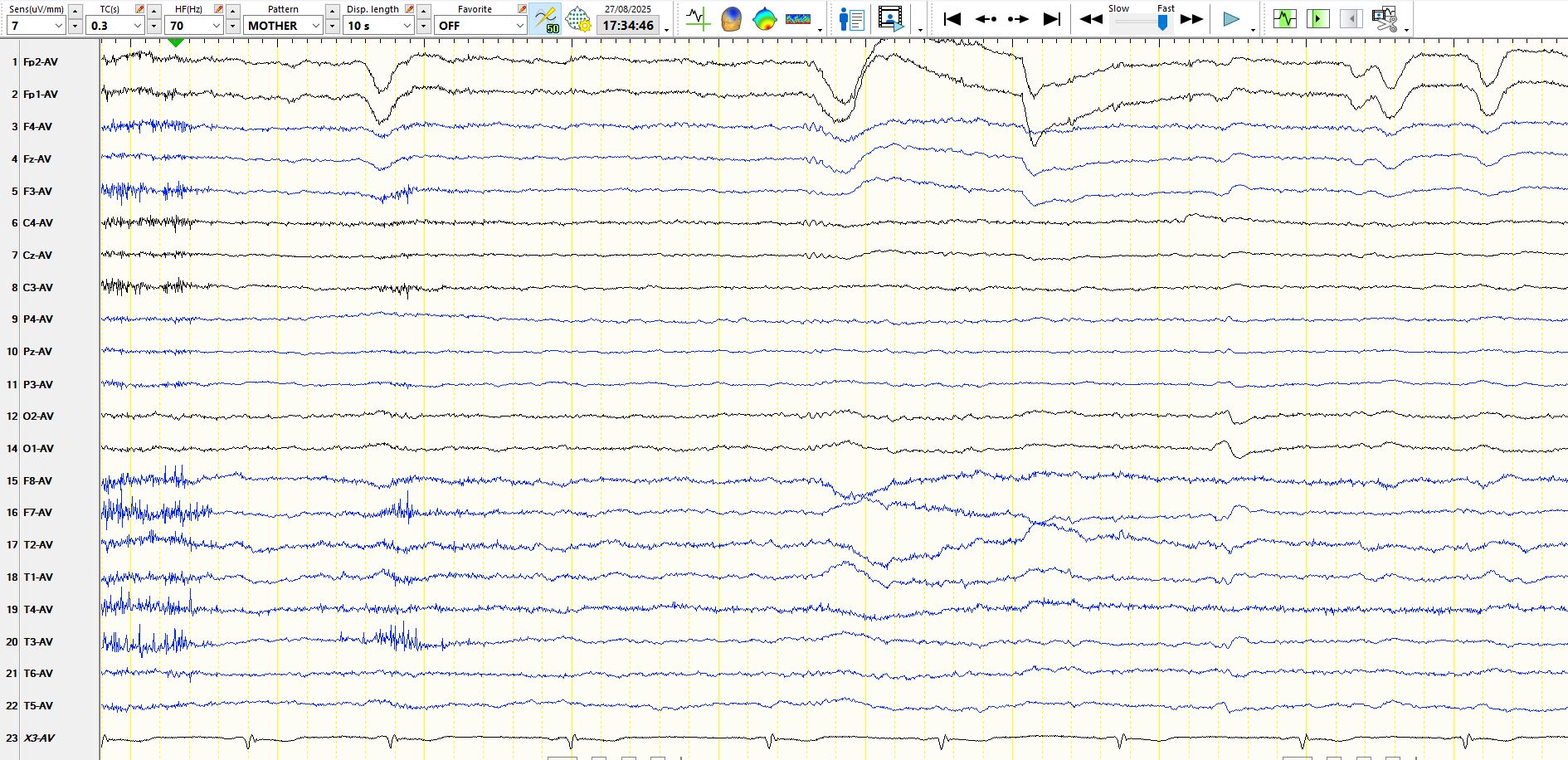
The next 10 seconds:
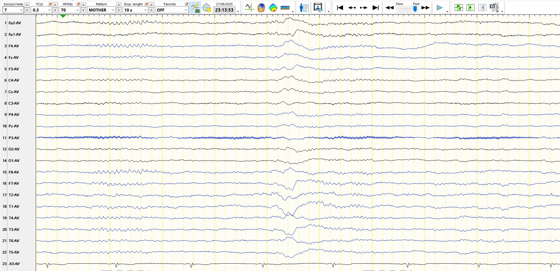
The above page, but at 5 µV per millimetre:
The next 10 seconds at 7 µmol per millimetre:
The following pages illustrate the EEG during sleep.
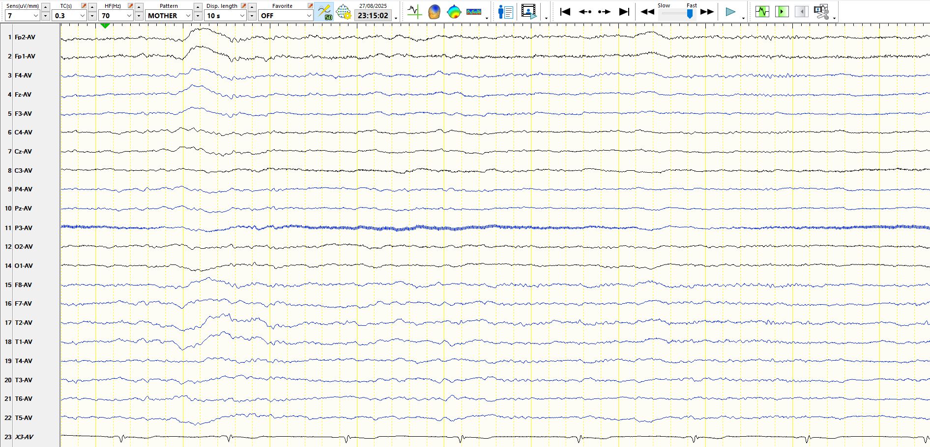
During wakefulness the recording is low in amplitude, but there are definite beta frequencies that wax and wane and these represent the dominant background rhythm. You can see these most clearly when the recording is largely devoid of artefact, as in the last page above while awake. There are low amplitude delta waves at 01-T5-02. If there is any doubt in your mind about the veracity of these waves, especially the beta frequencies, notice that the patient has beta frequencies, K-complexes and spindles during sleep; the recording during sleep is normal. Unsurprisingly, the amplitudes of the background frequencies during sleep are relatively low.
This example illustrates a few points, namely that:
1. the background rhythms may be remarkably low in amplitude in some individuals (one should always consider the possibility of hypothyroidism or extensive bilateral subdural haematomas as a rare cause of suppression of normal rhythms)
2. if there is doubt, one should pay careful attention to spontaneous fluctuations over time and the appearance of the EEG following stimulation by the technologist
3. every attempt should be made to remove environmental artefact, especially in the ICU setting, as passing judgement about the presence or absence of low amplitude beta frequencies may sometimes be impossible
4. in the normal state, background EEG rhythms are often, but not invariably, more easily seen during sleep
Incidentally, suppression is defined in Kanes' 2017 glossary as follows: "Entirety of an EEG record showing activity below 10 mV (reference derivation)."