
Arising from the background
Sep 01, 2025Do the following represent alpha rhythms that are propagating anteriorly? Or is this drowsiness? Could these waves be spikes? Could this be a benign phenomenon?
figure 1:
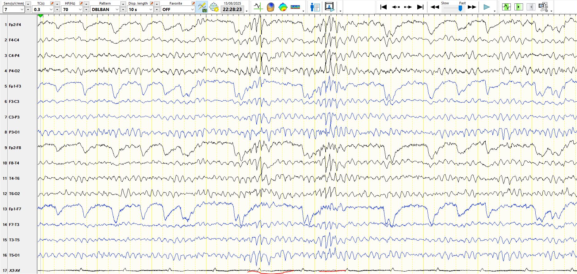
Figure 2:
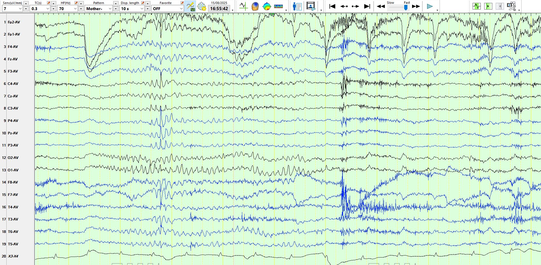
Figure 3:
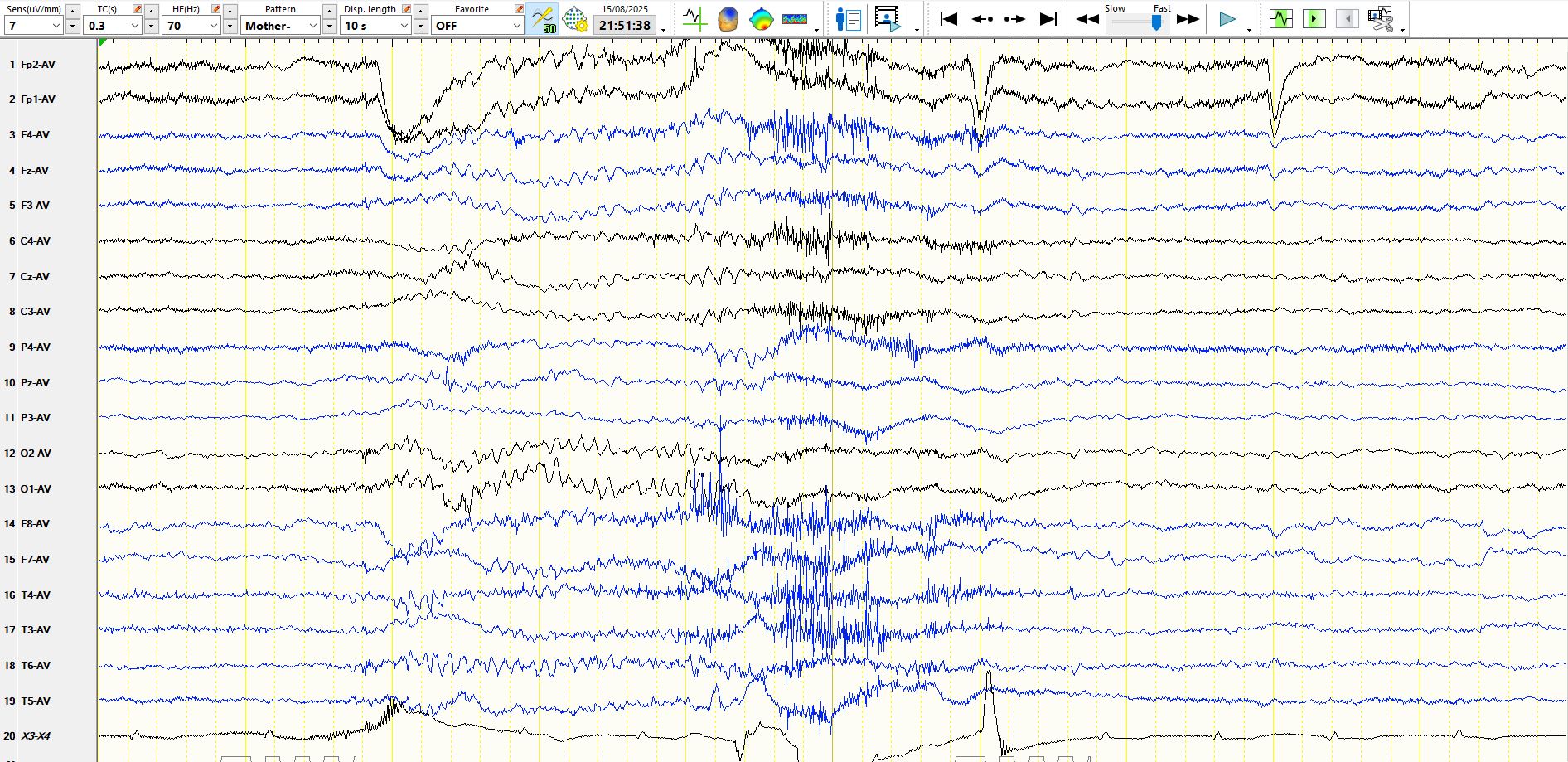
Figure 4:
And then, what do you make of these waves in figure 5?
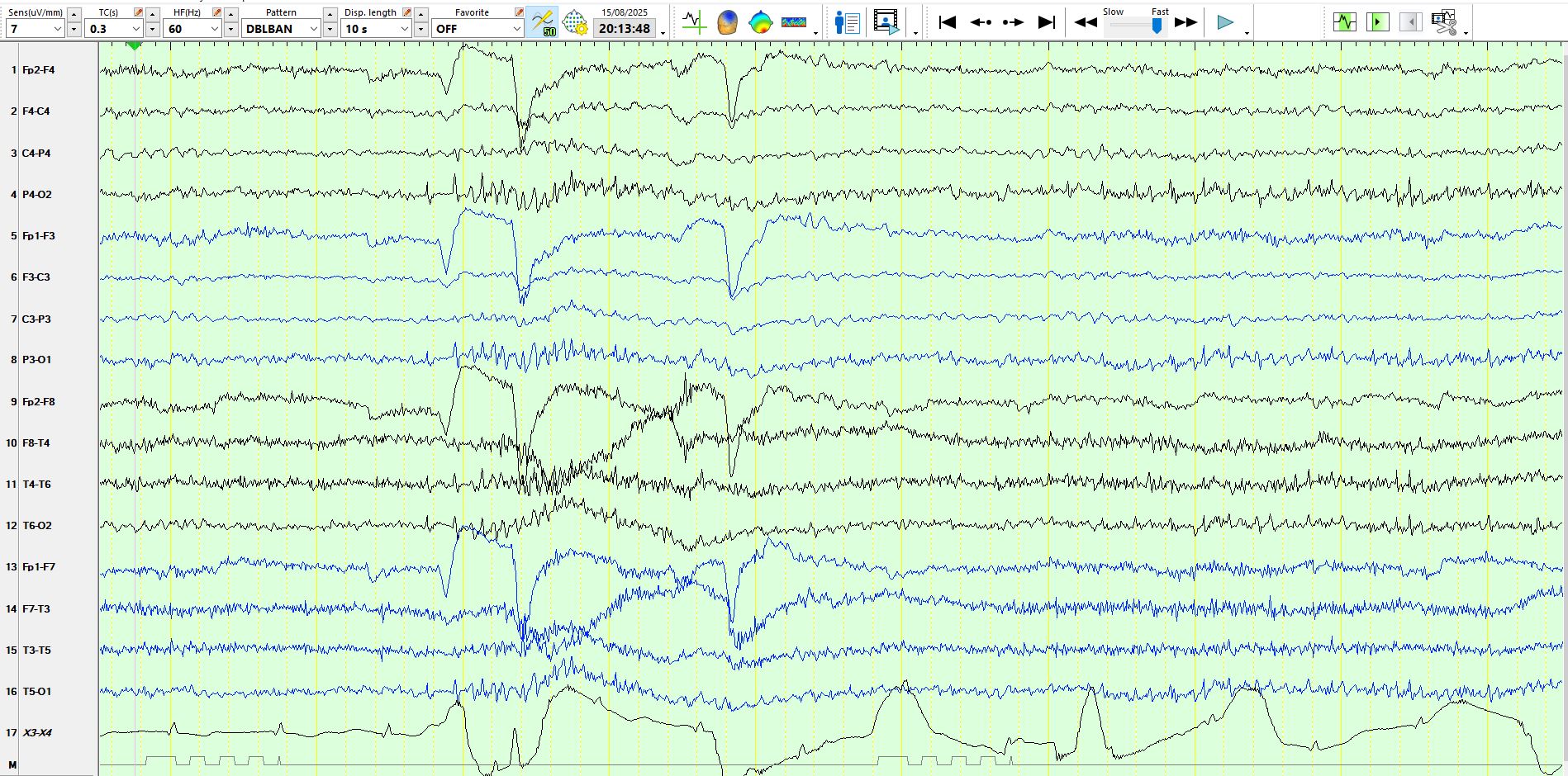
Figure 6 - Coronal montage:
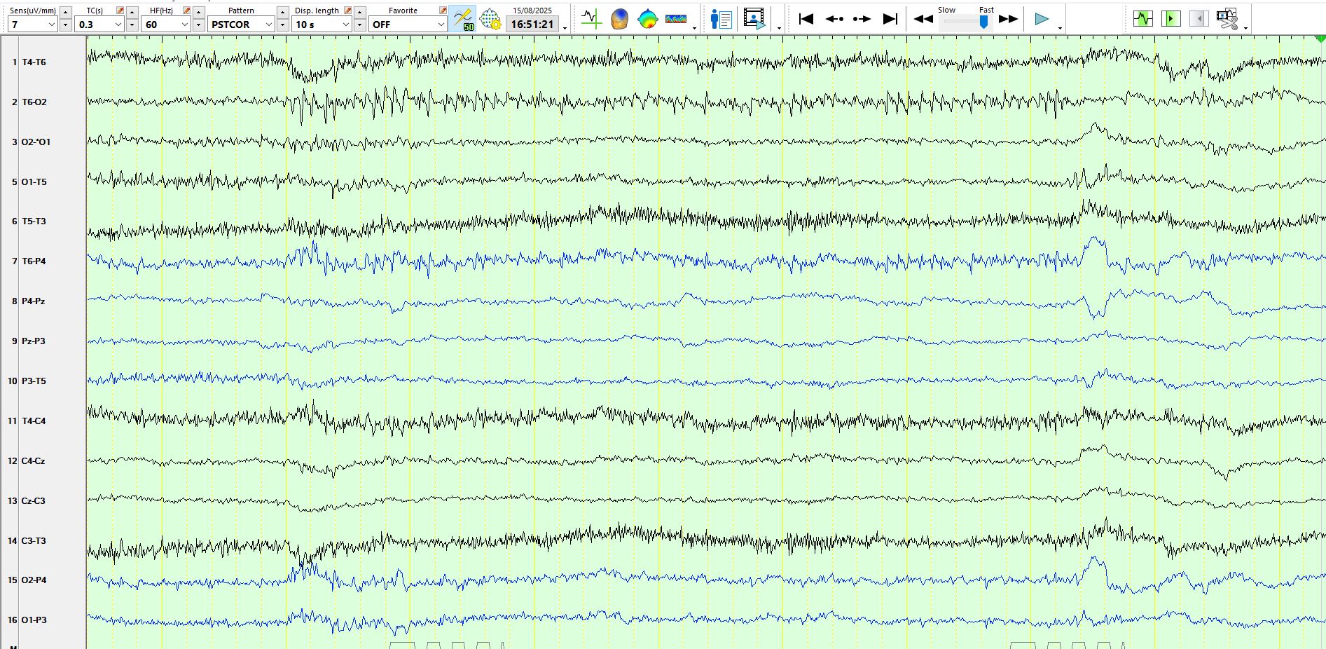
Figure 7:

Figure 8:
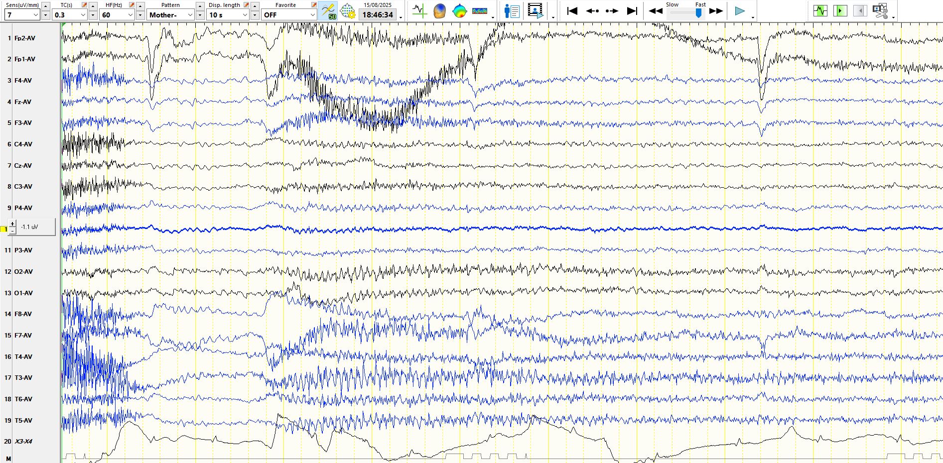
Figure 9:
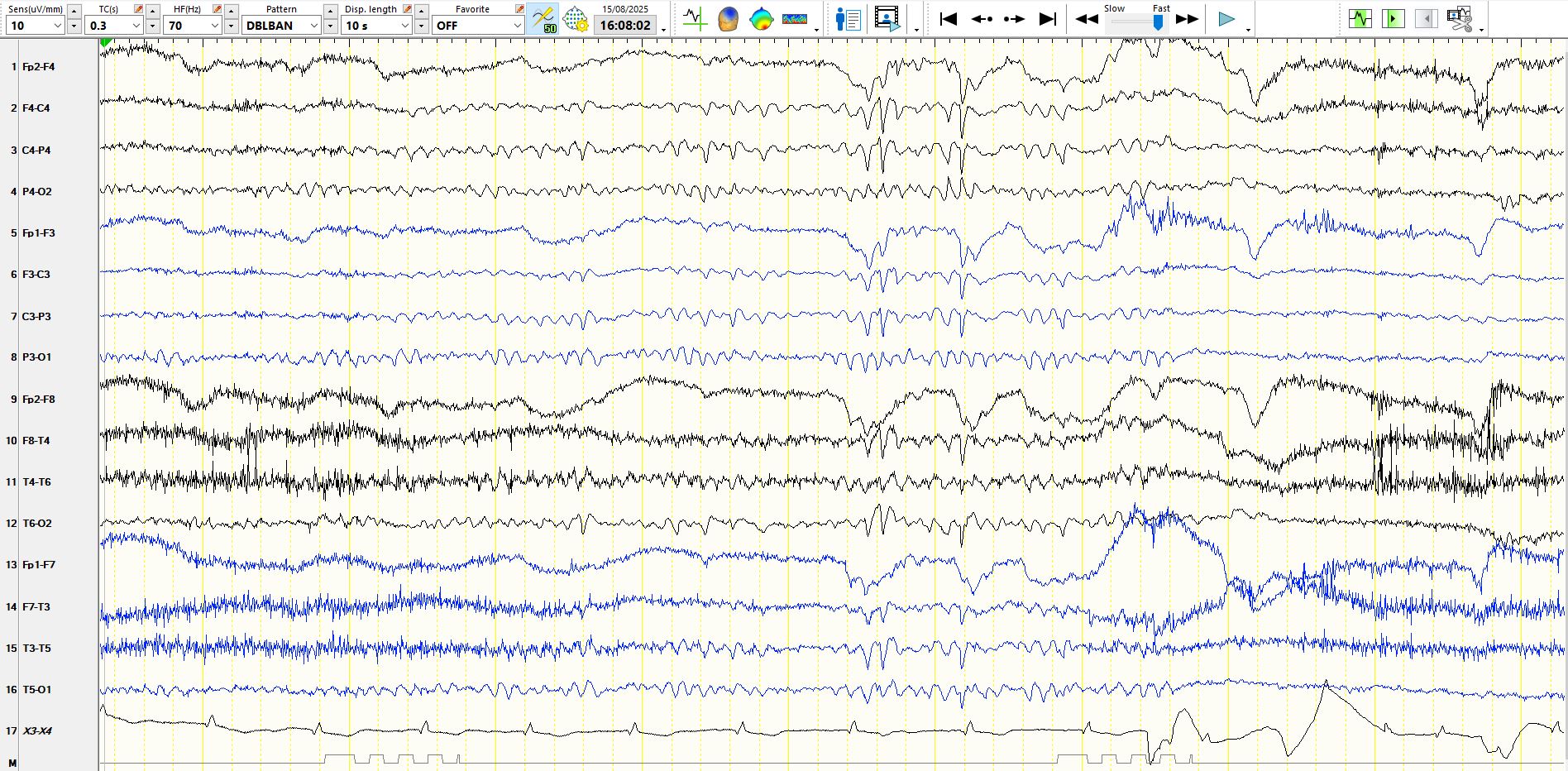
Figure 10:
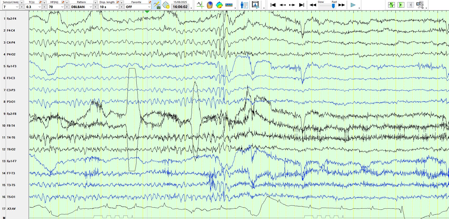
The following two pages demonstrate the same 10 seconds on two different montages, namely the bipolar and the referential (figure 11):
This is the referential representation of the above page. figure 12:
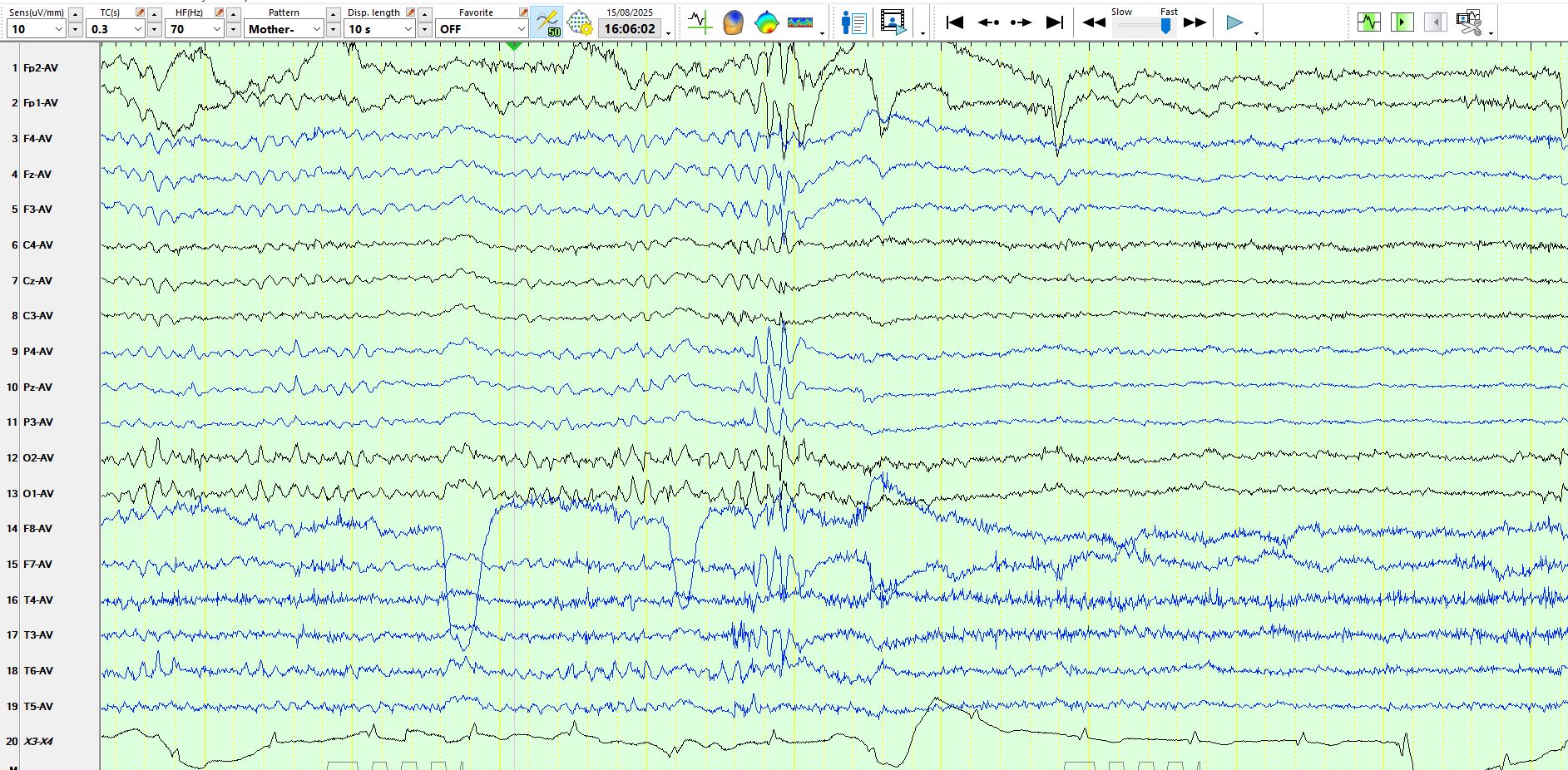
Fig 13:
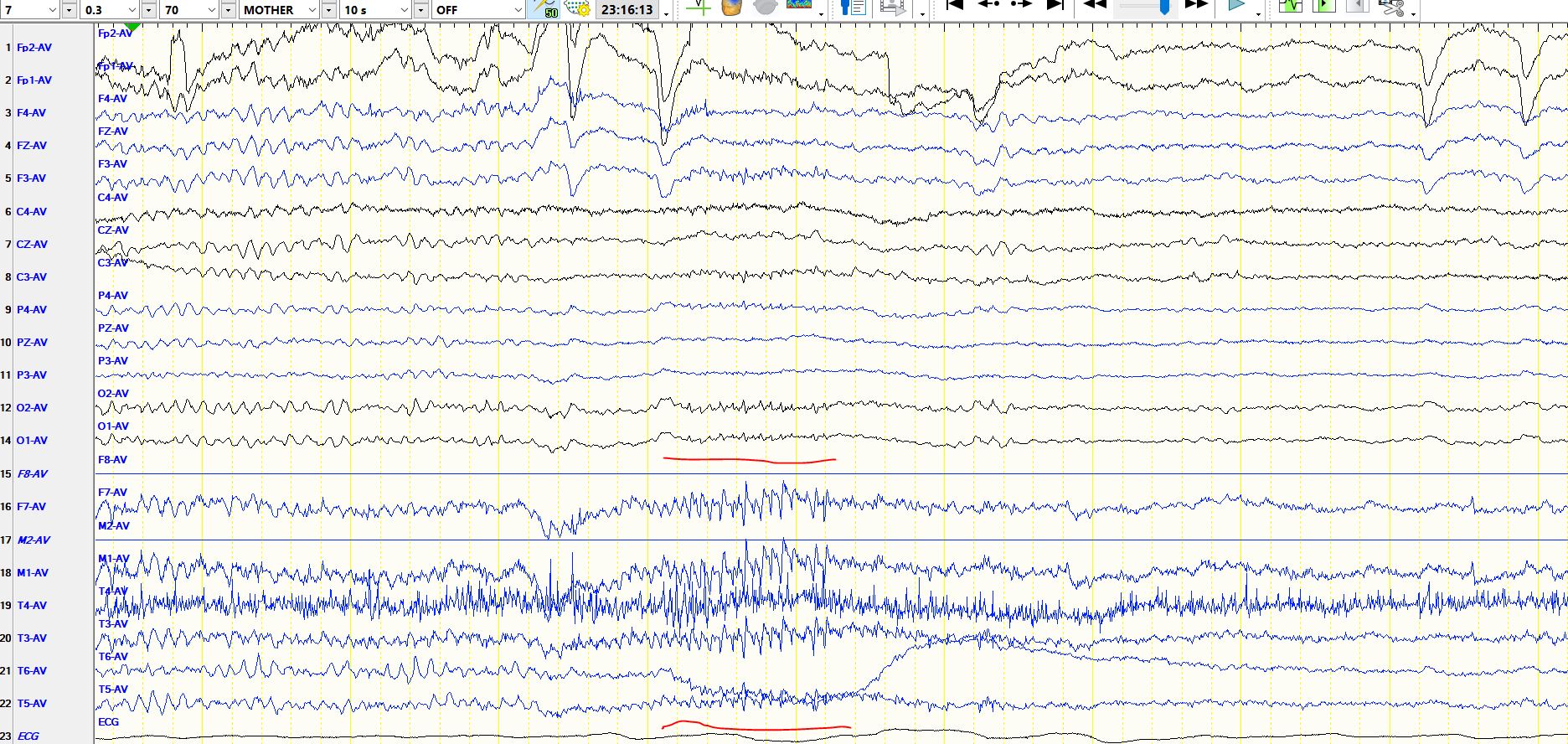
Fig 14:
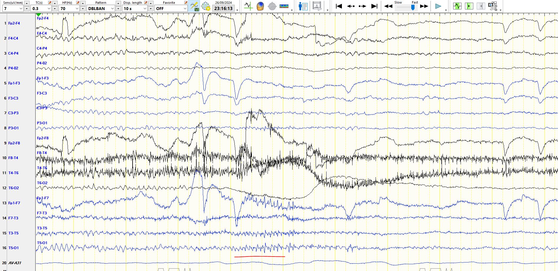
Fig 15:
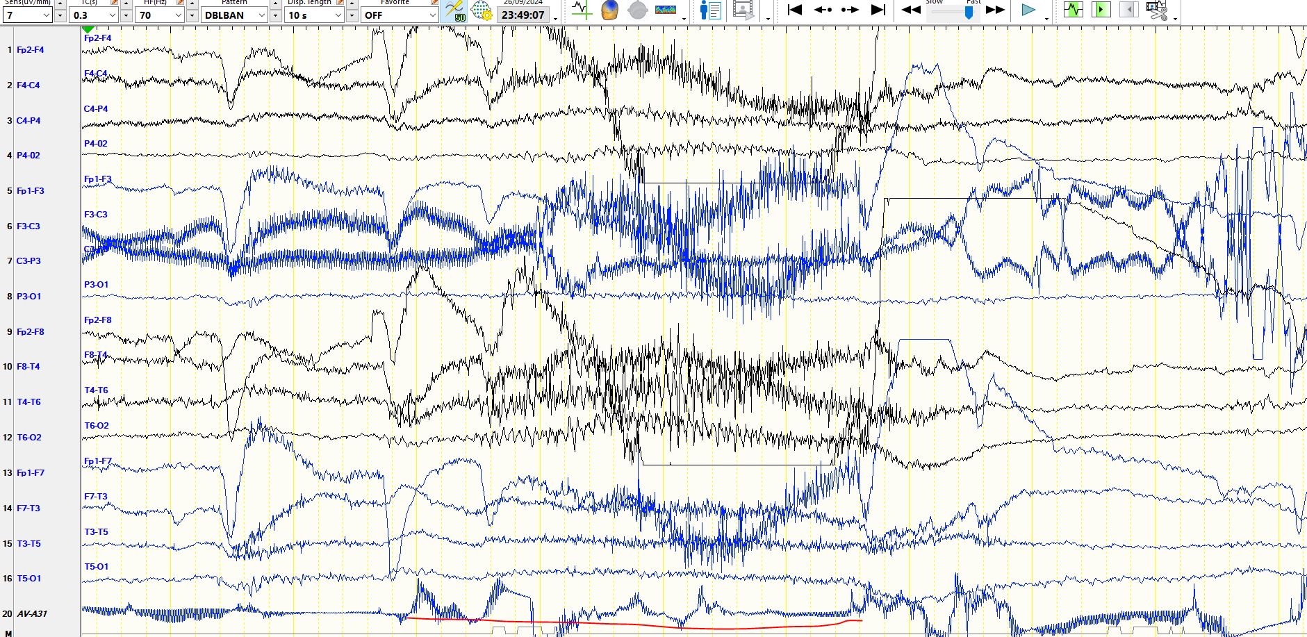
Fig 16:
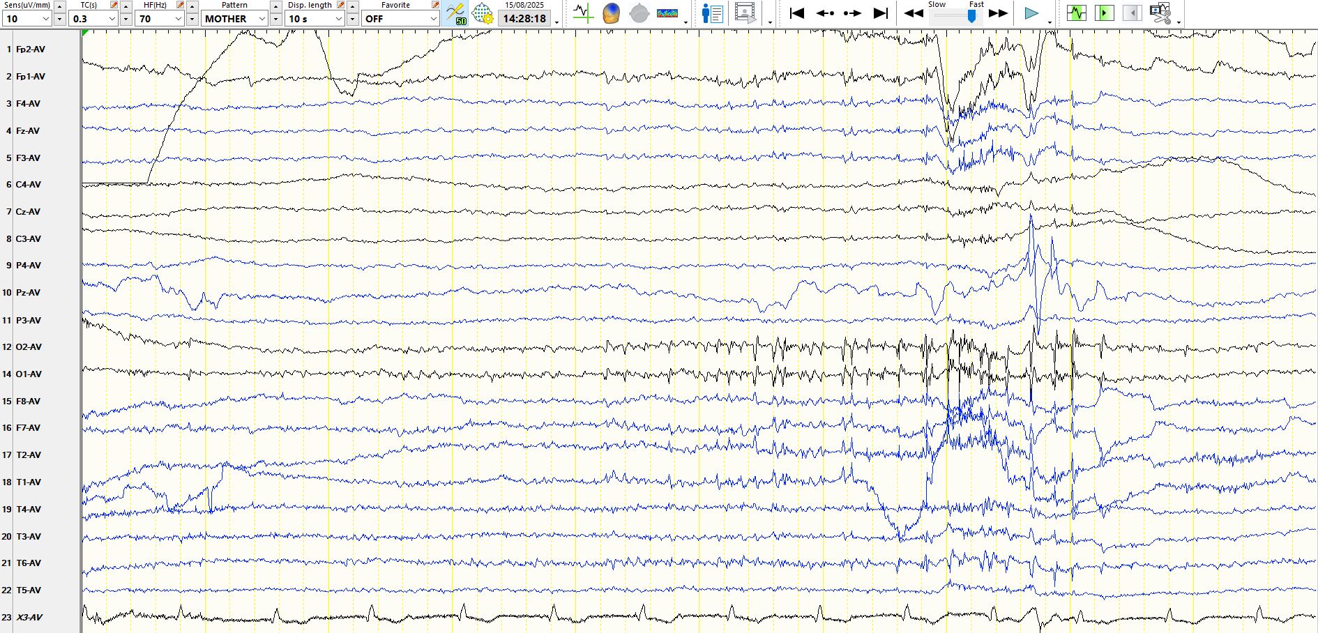
Fig 17:
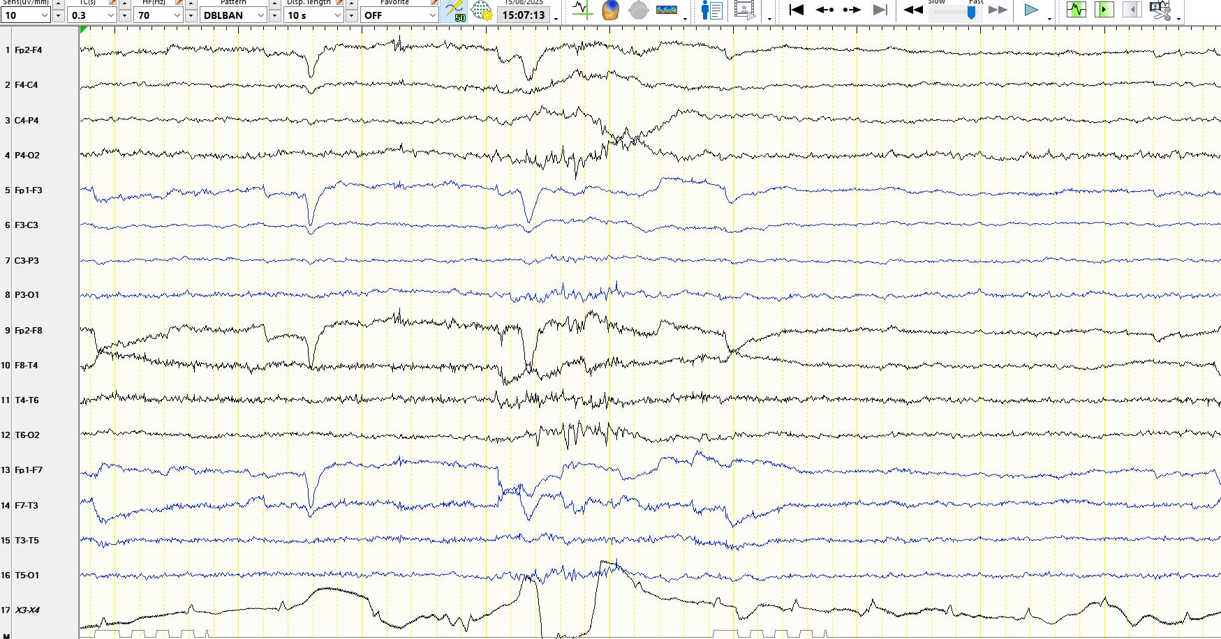
Fig 18:
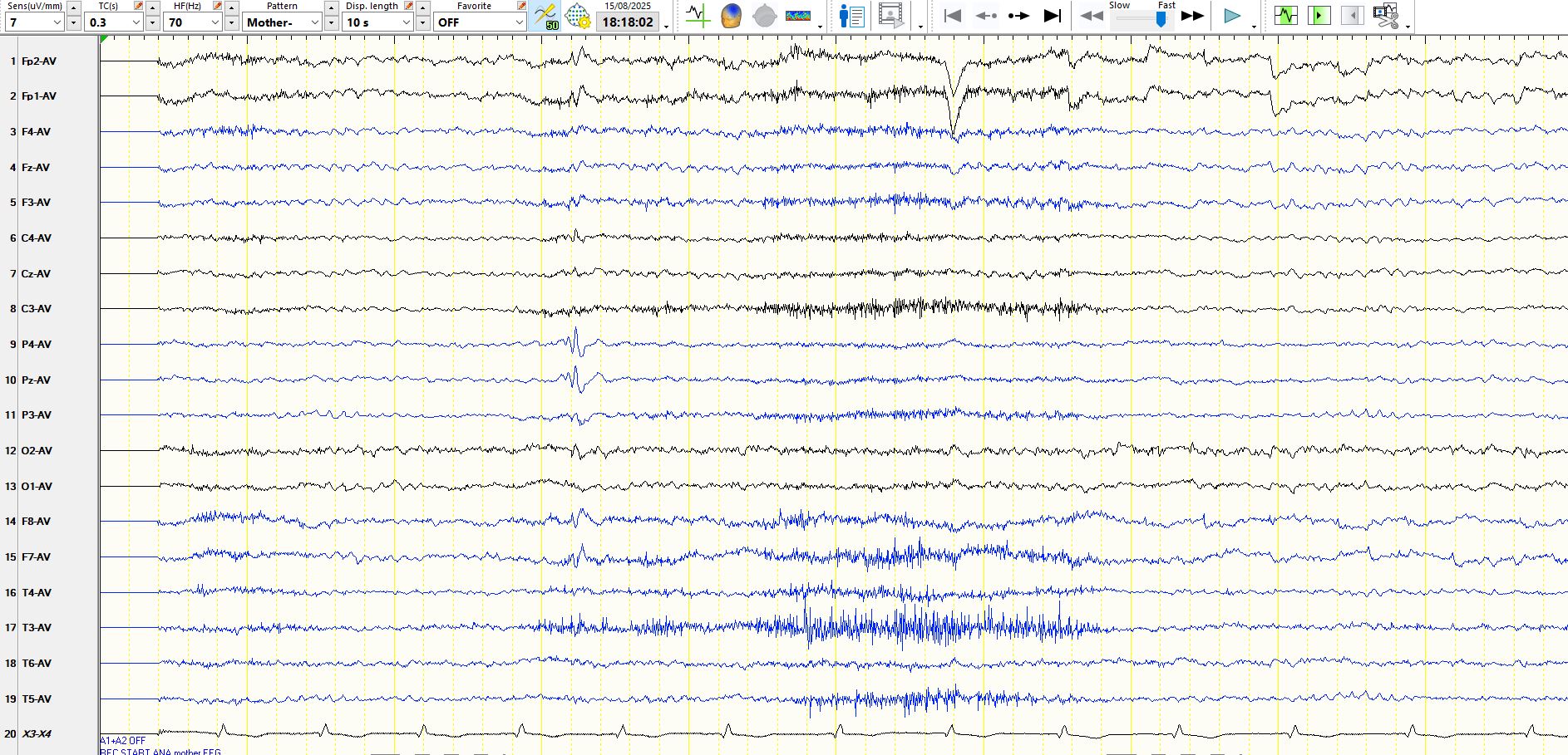
Fig 19:
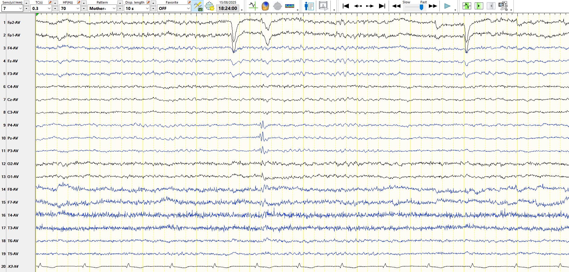
Fig 20:
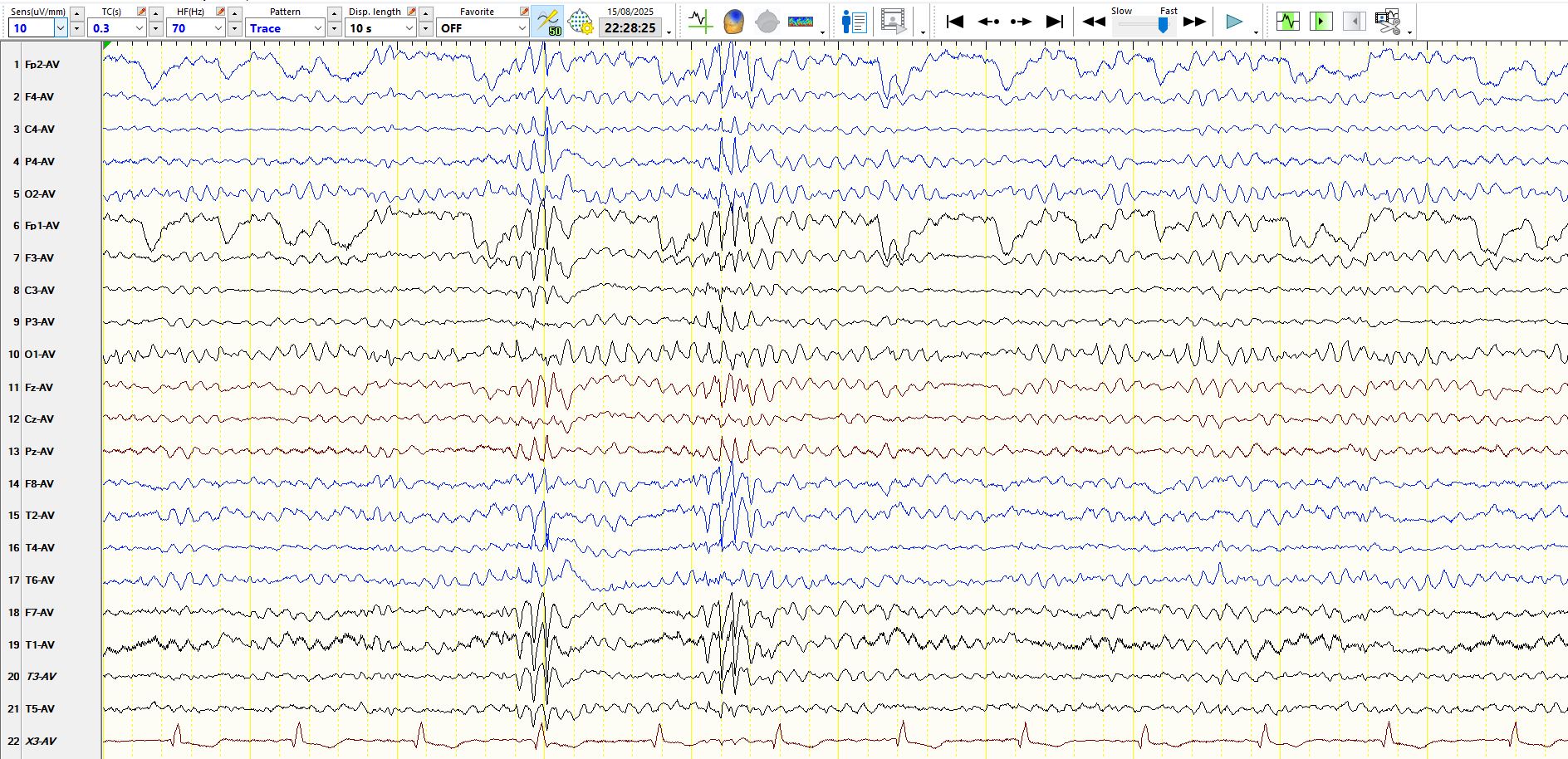
Fig 21, Sleep:
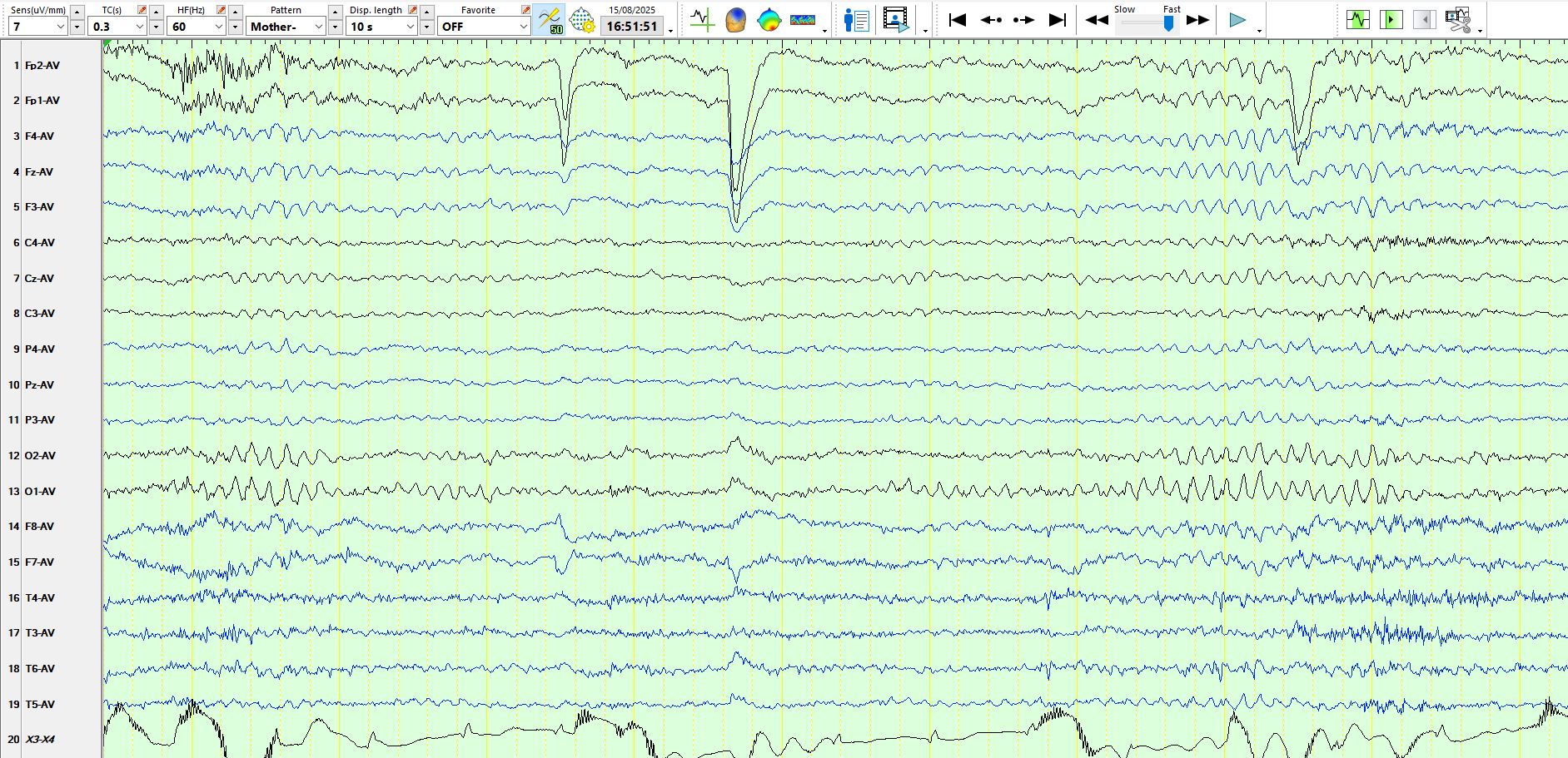
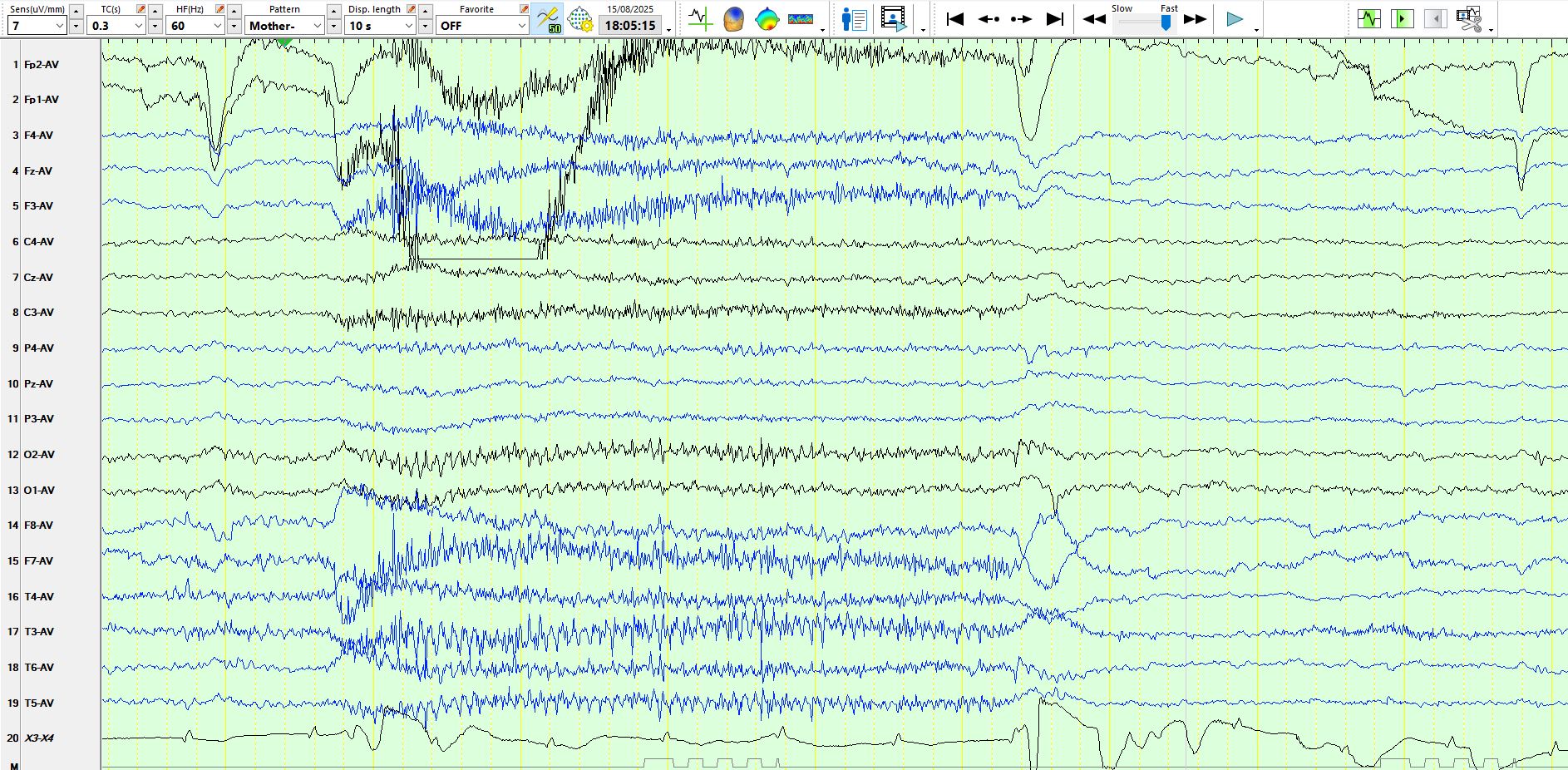
Figures 1, 2, 9, 11 and 12 are of interest as the obvious waves exhibit a progressive increase in amplitude and appear to arise from the background rhythms. Moreover, the frequencies of these sequential waves resemble the background rhythms and do not disrupt the background rhythms. Initially my instinct was to be wary of over-interpreting these. However, these rhythms i) become sharply contoured and ii) exceed in amplitude anything in the background by some margin. Generally speaking, these criteria alone would be insufficient to call a sequence of waves spikes, but these waves simply do not look normal! I repeatedly asked myself at what point in the sequence of waves does one say that "this is a spike" and therefore be obliged call them spikes; I'm not sure! This is a good example of interpretation of waves that can cause one a lot of discomfort, and discomfort should be a reason to avoid calling them spikes. In this instance any argument is settled by the presence of other waves, such as those in drowsiness (figure 20) and during sleep (figure 21). Hence, with the benefit of seeing the latter waves, there is reassurance about the interpretation waves that arise from the background; one certainly learns from EEGs like this.
Note the "focal" spikes in figure 18 and 19, not an uncommon finding in people with "generalised" epilepsy. Figure 18 is intermediate between the focal spikes seen in figure 19 and the generalized spikes seen elsewhere.
In Figure 2, there is "alpha squeak" immediately following eye-closure, especially at 02, 01 and T6, followed by a slight reduction in the frequency of the alpha rhythm.
The EEG also of interest because it is diagnostic of Epilepsy with Eyelid Myoclonia (Jaevon syndrome). If you start with figure 5, you will notice that the eye-closure precipitates a brief run of spikes over the occipital regions. This relationship can be seen in many of the subsequent epochs, with spiking of a variable duration and location. Some are largely seen over either temporal region, others largely over the occipital regions and others have a larger field. It is this distinctive association between the eye-closure and spiking that settles the diagnosis.
The other point of interest is the morphology of the repetitive spiking in response to eye-closure (figures 5-10, 13-1, 17); this resembles EMG artifact. One could easily mistake these discharges for EMG artifact and therefore miss important, at times prolonged discharges, some of which arguably represent electrographic seizures, such as in figures 5-8, 10, 16. I was sitting with the patient at the time of figure 5 and asked him whether he had felt anything; he reported with an emphatic "yes, like little seizures, I feel a bit dissociated". This makes the point that the distinction between a clinical and an electrographic seizure is often utterly arbitrary and context dependent.
The EEG also makes the point that inter-ictal epileptiform discharges may vary markedly in their morphology.
There is no eyelid myoclonus on the EEG in this instance; it is present in about half of patients, according to this review Epilepsy with eyelid myoclonia: A systematic review and meta-analysis - PubMed. About 78% of patients have a photoparoxysmal response on EEG. Perhaps unsurprisingly, focal spikes are also seen in about 33% of patients.
He is a mid 20-year-old adult who developed absence seizures about 15 years ago, and has had occasional tonic-clonic seizures since; he recently had his first tonic-clonic seizure in a few years. He seeks out the sun whenever he can and his parents feel that he is photosensitive. Nevertheless, repeat photic stimulation has never provoked any abnormalities on EEG during the five EEGs performed over the past 15 years, with photic stimulation done during the eyes-open and closed state.
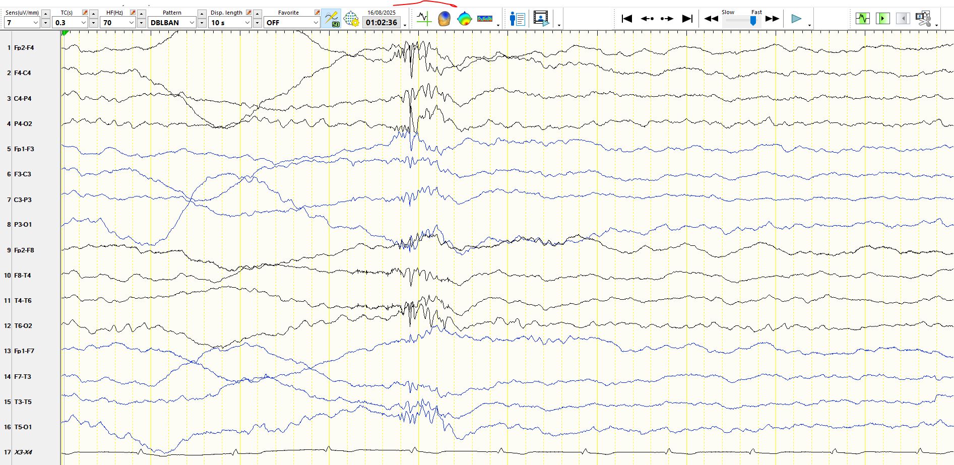
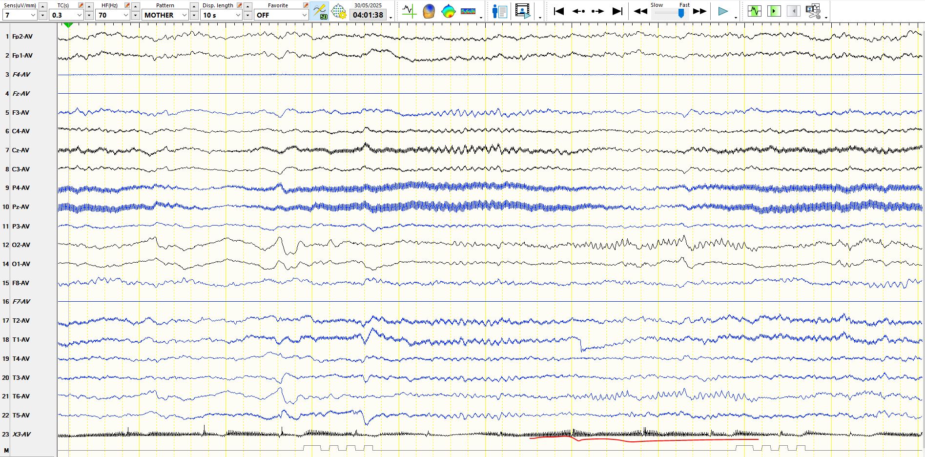
The final point is that bursts of generalised alpha frequencies may sometimes represent inter-ictal epileptiform discharges. A reminder, focal alpha frequencies may sometimes also represent epileptic seizures, as is the case in the image below from this case 17, born prem, given O2, blind, MRI reported as normal
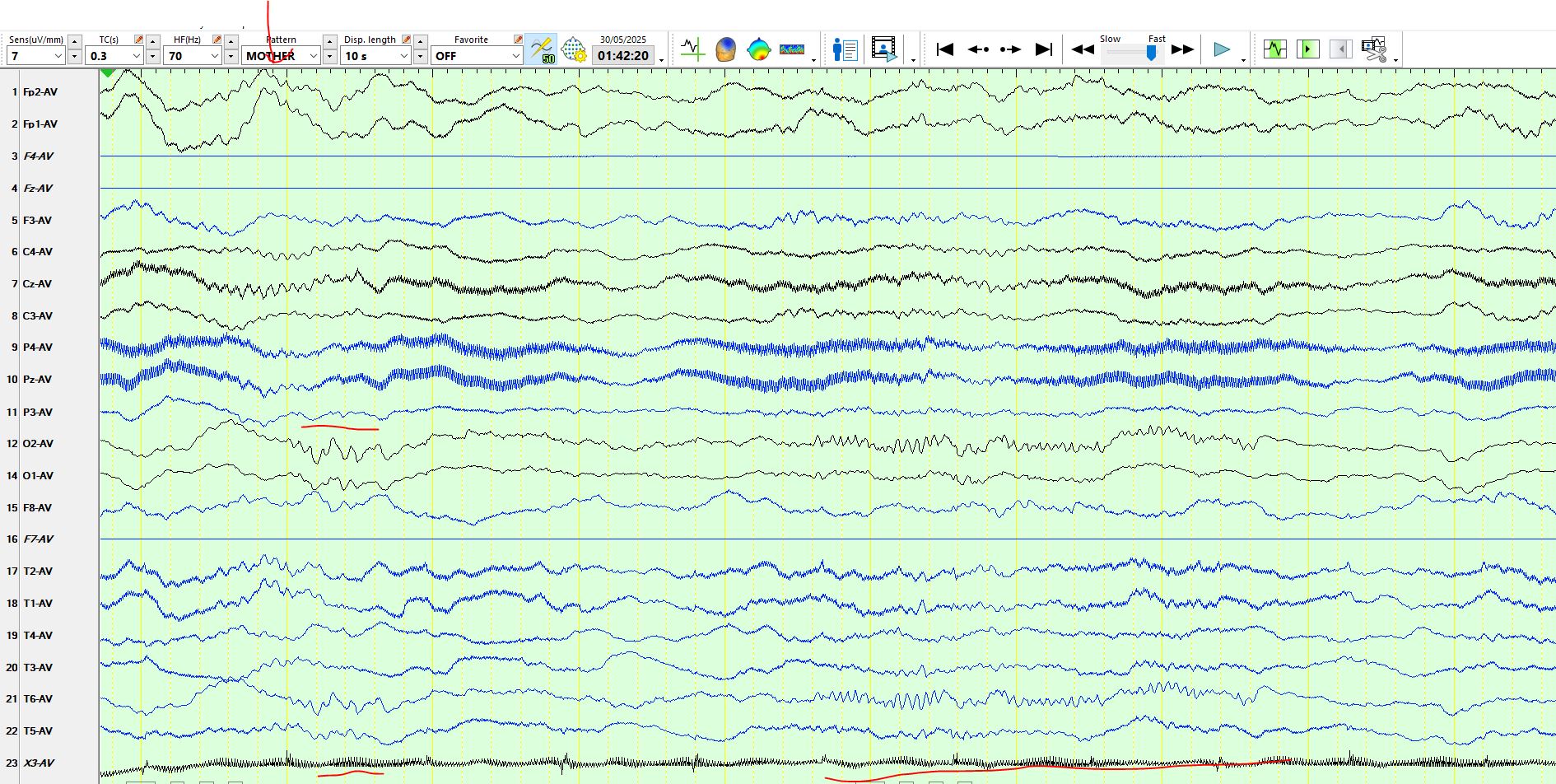
The vertical red arrow above delineates sleep spindles, the first horizontal red line delineates sequential spikes at 02-T6 and the long horizontal line demonstrates a prolonged alpha frequency discharge at 02-T6, arguably representing an electrographic seizure lasting approximately three seconds.
Here is a similar discharge:
The above discharges are frequently seen in people with focal cortical dysplasias.