
27y, proven left TLE
Jun 01, 2025This EEG below is of interest and reinforces the point made in this post: 41y, TLE
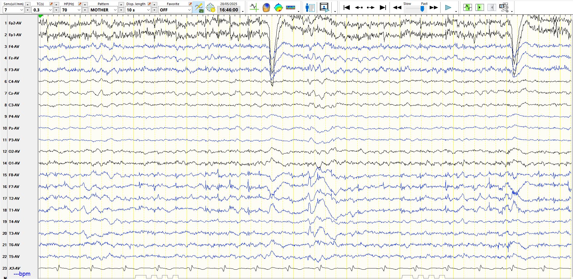
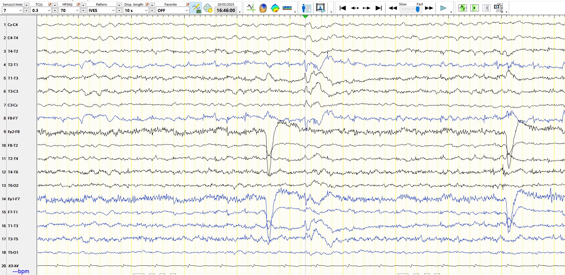
On this occasion the patient is awake. F7, T3, M1, F8, T4 and M2 are excluded from the reference. There is a spike and M1-F7-T3, but notice that the electronegative field extends to M2 and that it is positive at the vertex (see above and below)
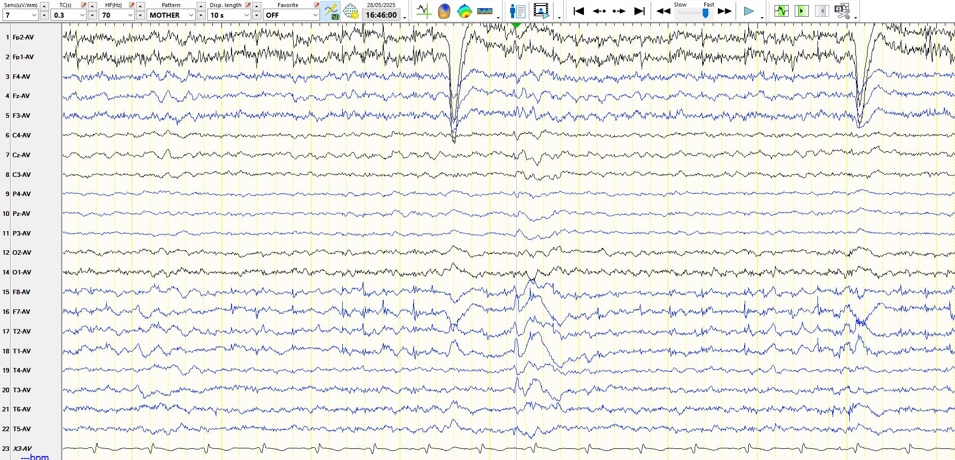
This is also apparent on a bipolar montage that is both in the coronal plane and AP plane (see below)
And below is a subdural recording of a 21-year-old. The first eight electrodes are located from medial to lateral over the right inferior temporal region anteriorly, the second eight are located posterior to these on the right, the next 16 are over the left inferior temporal region, while the remainder are over the right frontal region. Notice the field involves both basal temporal regions, almost synchronously, with the spike on the left preceding that on the right by matter of milliseconds. The spike does not involve the frontal regions, although there is widespread relative suppression following the spike (a topic for another day).. On scalp EEG this spike would appear at M2 and M1 and probably also involved F8 and F7. This is what the above spike might look like on subdural EEG. Now go back and look at the above scalp EEG recording from a different patient and you will see a similar subtle difference in the spikes over the left and right temporal regions.
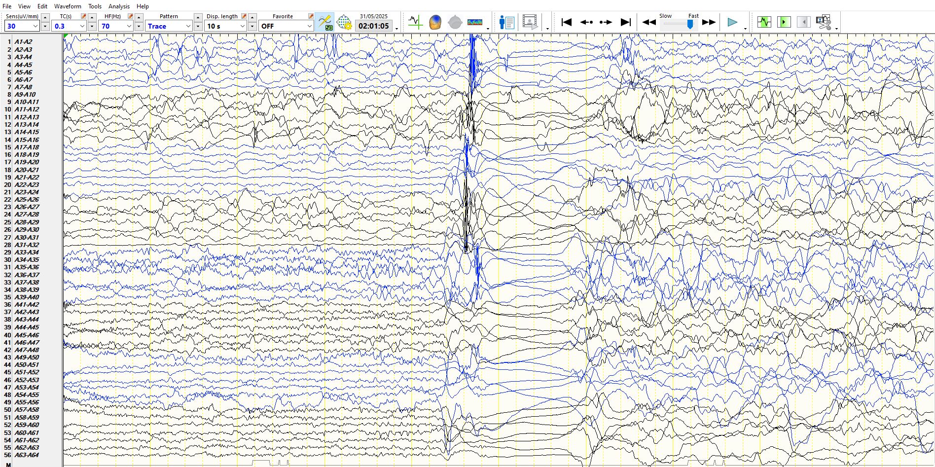
Most of this patient's sparks appear over the right inferior temporal region (often in very long trains), while a minority of the spikes appear on teft.. Occasional spikes appear bitemporally.