
Theta wave vs spike vs eye movement artefact
Jul 03, 2025Most of the time there should be little difficulty in distinguishing these, but sometimes there are waves which mimic sharp waves and spikes. Have a look at the following waves at F8 and M2
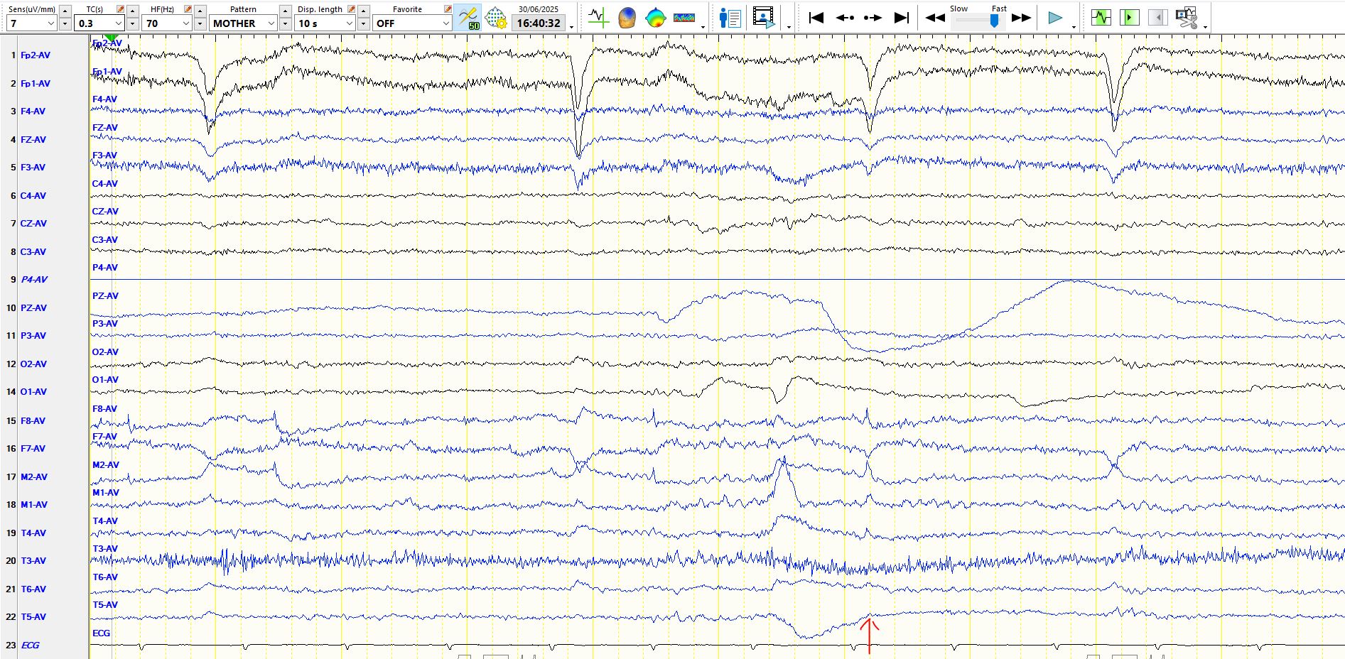
In the above image, multiple "lateral rectus spikes" can be seen, most of which have a distinct morphology and the apex is characteristically very sharp. The discharge corresponding to the arrow has a broader base. The deflection likely is a composite of changing polarity corresponding to the movement of the eyeball and the very sharply-contoured component generated by the muscle (EMG). In this instance the arrowed deflections correspond to an oblique eye movement, upward and to the left. The downward deflection at FP2 and FP1 indicates an electropositive discharge in these derivations, consequent upon upward movement of the eye which is electropositive at anteriorly. Similarly, F7 is electropositive, indicating some leftward movement of the eye, which is electropositive anteriorly. Not unexpectedly, F8 demonstrates the opposite deflection. The deflections at F8 and F7 are not the result of reference contamination of eye movements, as FP1 and FP2 are excluded from the reference and the most remote electrodes from FP2 and FP1 do not demonstrate a discharge of opposite polarity.
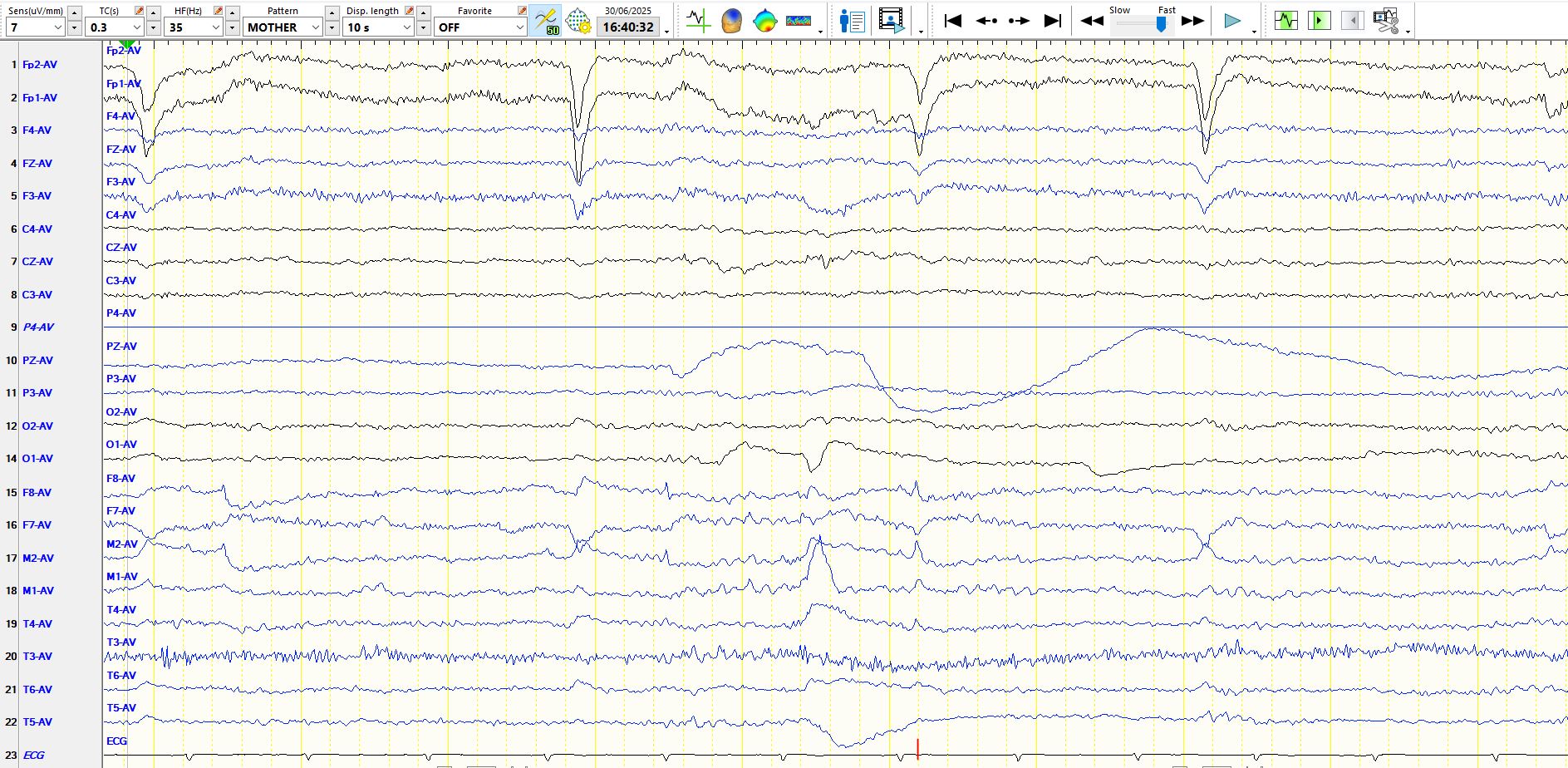
In the image above, the high frequency filter has been changed from 70 Hz to 35 Hz in order to remove the "EMG", mainly to show you the dangers of leaving the high frequency filter on. In this case the wave resembles a cortical discharge at F8 and M2. There are many physiological waves and artefacts which may be transformed into discharges identical to sharp waves and spikes by such a change in the high frequency filter.
Bottom line? Ensure that the EEG recordings are routinely performed with no filters turned on or run the risk of sending innocent men to prison!