The brain is not only AP and coronal
Sep 11, 2025The following illustrates a relatively common issue:
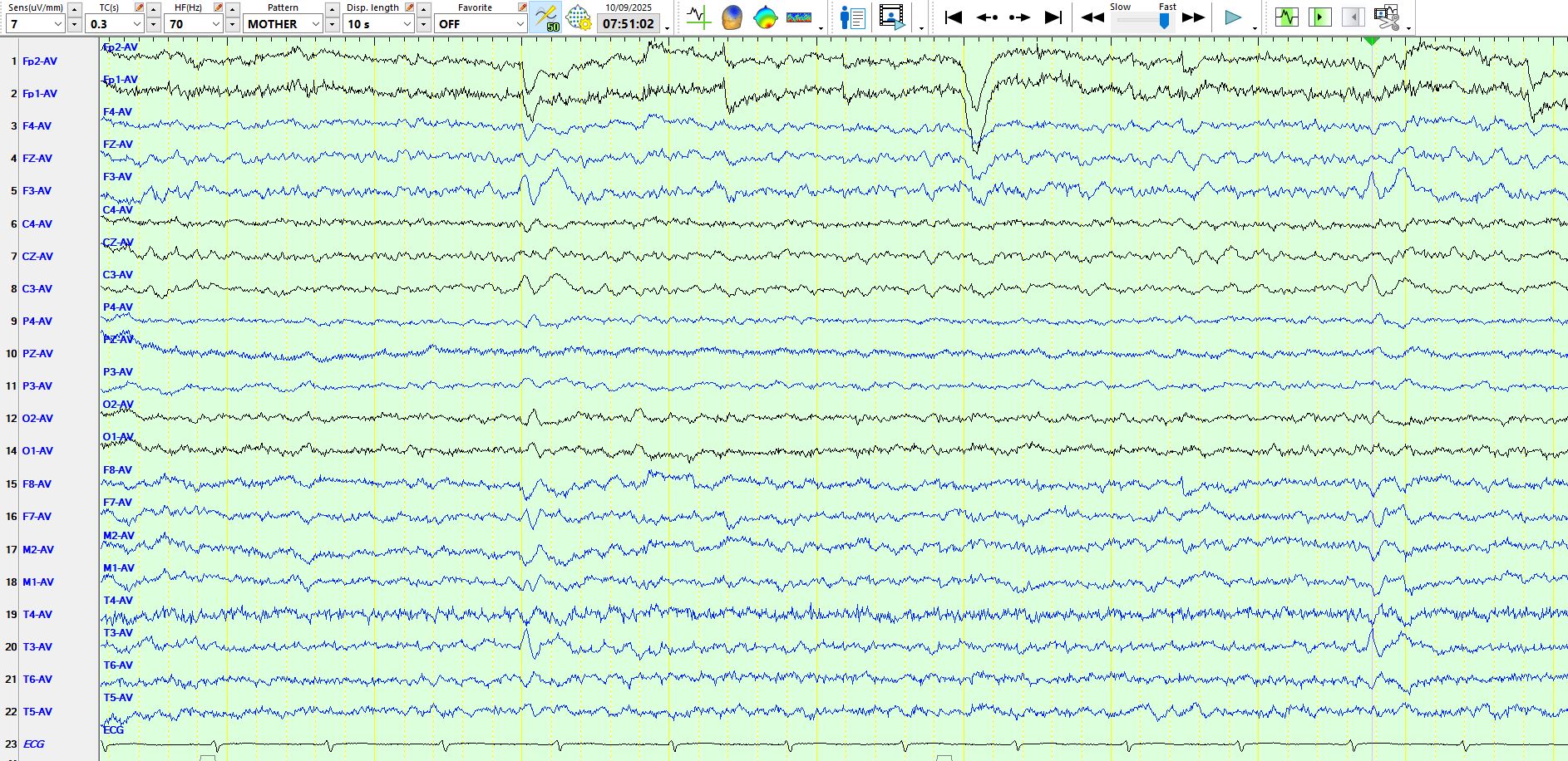
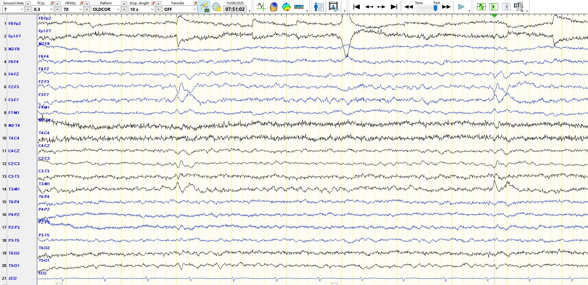
In the above image, the sharp wave and, less clearly, the slow wave, demonstrate phase reversal at F3 and T3. Because F7 is closer than T3 to F3, one might wonder whether F7 and T3 or F3 and C3 have been transposed. I checked on this and it is certainly not the case; there are ways of checking on transpositions even after the EEG is done; This will be a subject of a post sometime in the future. You can check on the above conclusions by reviewing the EEG in the coronal montage:
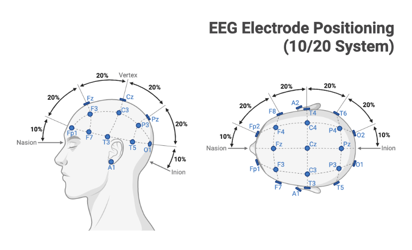
When the top page is represented in the coronal montage, the conclusions are unchanged.
The next question is whether the discharge is primarily electro-negative over the left frontal or peri-Sylvian temporal region. To answer this question, I have added an extra channel at the bottom, comparing with F3 with T3; see my red annotation.
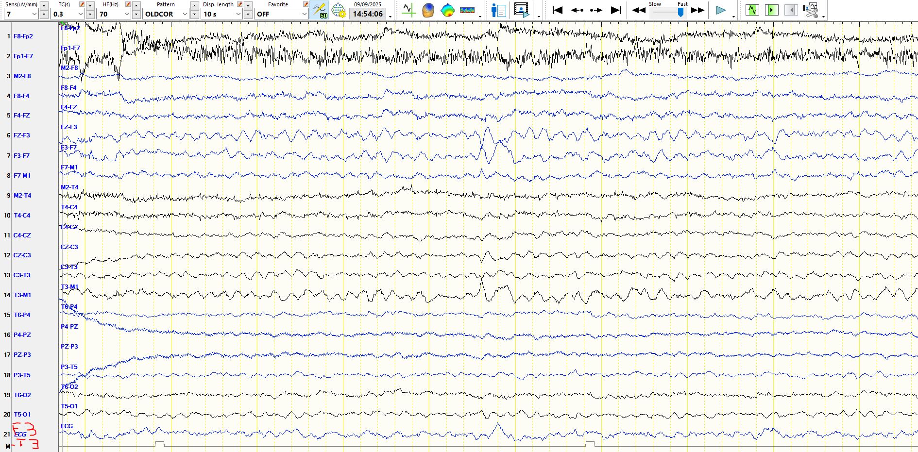
The spike is equi-potential at F3 and T3 (there is no deflection in Channel 21), indicating that the spike is probably primarily generated somewhere between these two electrodes, perhaps halfway. As you can see, the aftercoming slow wave is more electronegative at F3 than at F7, but this does not materially change the conclusions. Hence, the discharge likely originates somewhere in the region of left anterior peri-Sylvian cortex, likely above the left Sylvian Fissure.
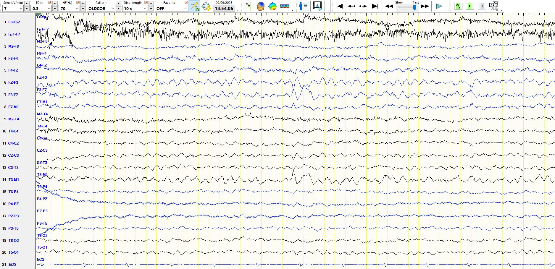
The above page represented in the referential montage: