
T1, T2 and M1, M2; the basics
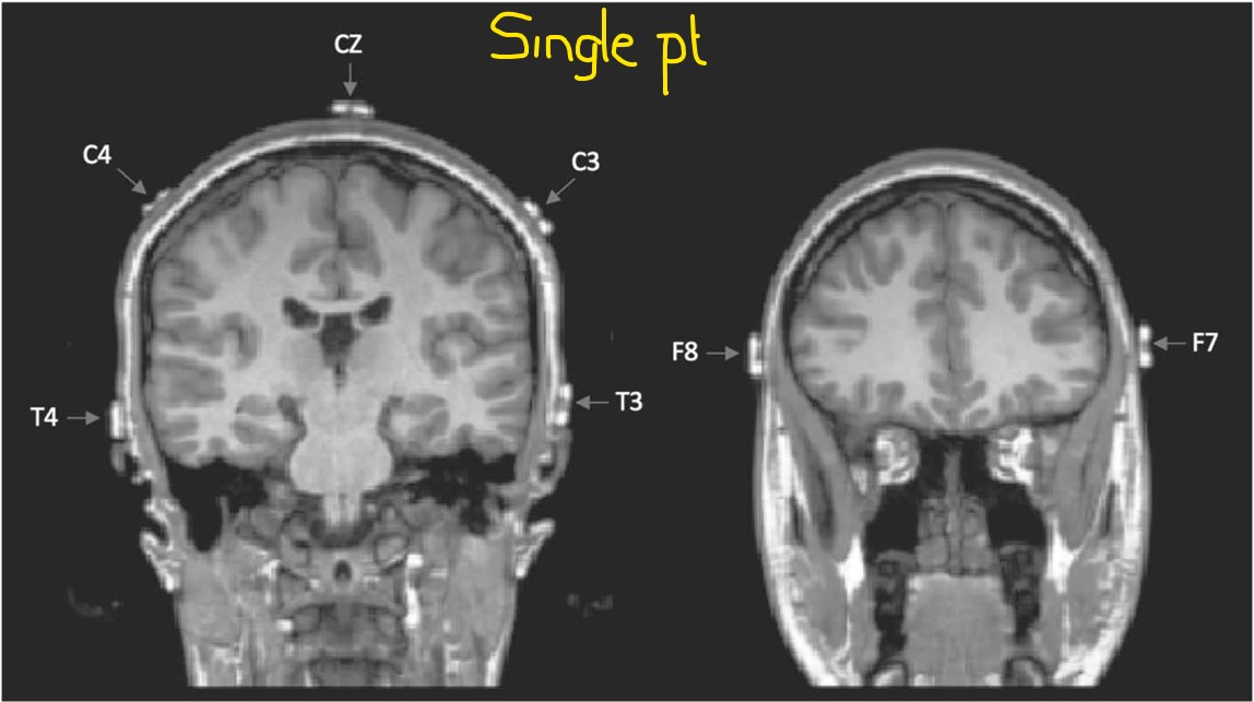
Sep 14, 2025What is the point of these electrodes? Have a look at the following images from this study, Relationship Between EEG Electrode and Functional Cortex in the International 10 to 20 System - PubMed, and note the relationship between the F8 and F7 electrodes and the temporal pole. Note also the location of T3 and T4 in relation to the inferior temporal gyrus; you may also appreciate the variation in the placement of electrodes on the left and right-hand sides.
The average placement for the group is shown below. Note the variable location of F8 and F7, designated by the yellow arrows.
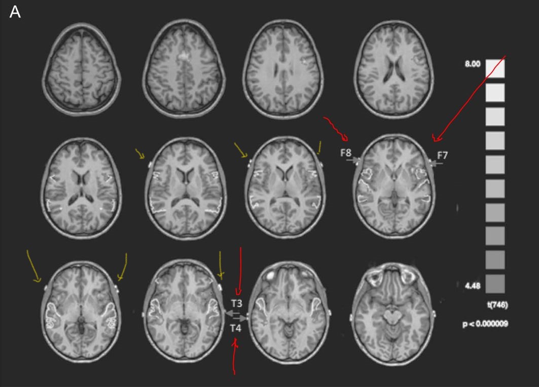
As you know, interictal epileptiform discharges and seizures most commonly originate within the medial temporal region. Unfortunately, F8/F7 and T4/T3 electrodes are not located over the left inferior temporal region! The average location of F8 and F7 is further forward and higher up than you might imagine; they provide more electrophysiological data from the left lateral frontal region than from the left temporal pole, which in turn is at some distance from the medial temporal lobe.
The above images demonstrate that T3 and T4 overlie the mid-temporal region in the anterior-posterior plane and most commonly lie over the middle temporal gyrus, and sometimes even over the left superior temporal gyrus. These electrodes do not face the inferior temporal region.
To overcome the limitations of the above electrodes, inferior temporal electrodes (A1, A2, T1, T2, M1, and M2) have been studied and shown to provide significantly greater sensitivity for inferior temporal spikes. Various studies have examined T1/T2, M1/M2, and T9/T10, demonstrating that these have the highest sensitivity for detecting such spikes, but do not differ in their yield. Multiple electrodes for detecting spikes in partial complex seizures - PubMed. A1 and A2 are located over the mid-temporal region and, therefore, are not ideal for detecting spikes over the inferior, anterior temporal region.
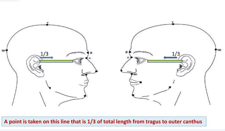
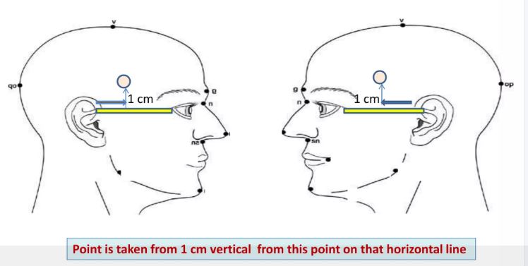
This is how T1 and T2 should be placed (images courtesy of Google):
The following picture from this publication provides a good idea of how low "inferior temporal electrodes" need to be compared to F7 and T3 (the latter is labelled as F9 below). You may notice that T9 is lower than T1 would be if it were to be placed on the individual below. I prefer M1/M2, which are virtually identically located to T9/T10, to T2/T1, but there is no difference in yield. It is probably quicker and easier for a technologist to place M1/M2. The mandibular notch electrode is placed in front of the ear, just below the zygomatic arch, within the mandibular notch. This is also the surface marking for the insertion of sphenoidal electrodes, which are directed towards the foramen ovale. These are little used.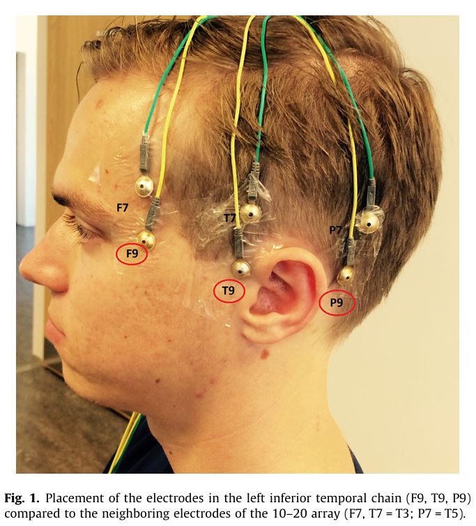
The bottom line?
1. If you want to record electrophysiological information from the inferior temporal convexity, you should place electrodes at M1 and M2 or at T1 and T2.
2. If you don't, you are overlooking the part of the human cortex where you are most likely to see spikes or seizures on the scalp EEG.