Low amplitude spike and wave don't mean it ain't an IED
Jul 19, 2025Part of the vaccine against the (false positive) spike protein on the EEG is the use of amplitude as a criterion. However, no criterion is absolute, but in disregarding it you need to be very, very, very sure. Get it? If in doubt, disregard it. Here is an exception:

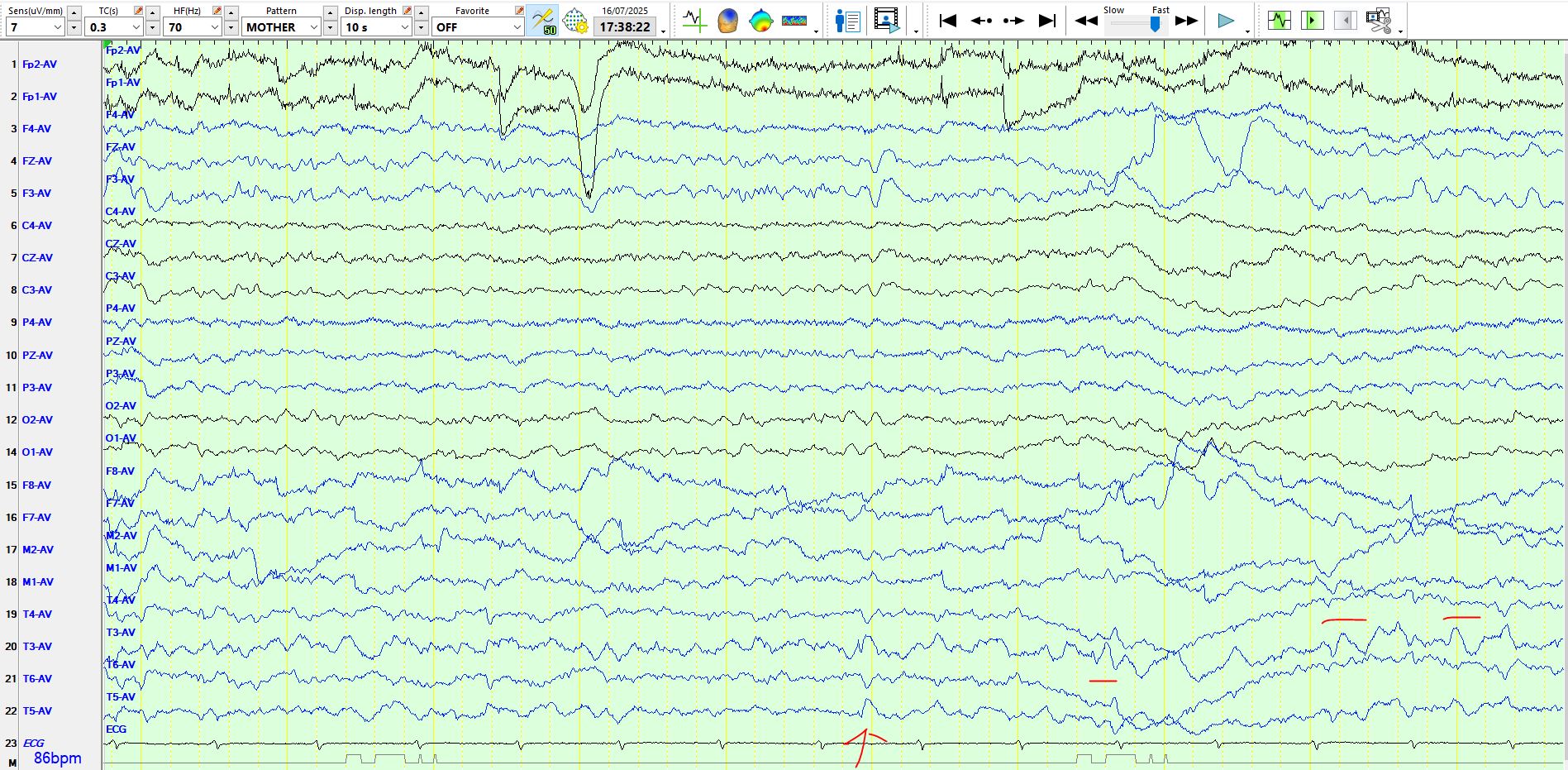
The second red vertical line demonstrates a low amplitude spike-and-wave at T3-T5. This on its own should mandate some caution, just in case the low amplitude spike is an artefact that coincidently occurs in the same channels as the theta wave and just in case its timing is terrible. This does not negate the presence of a sharply contoured theta wave at T3-T5 (first vertical; it is not an IED), theta waves (horizontal red lines) and delta waves (wavy horizontal line) at T3-T5. Incidentally, do not miss the theta wave at F3 immediately after this spike-and-wave. It is suggested that you hold your horses and await reproducibility of the "spike and wave discharge", before calling it an inter-ictal epileptiform discharge.

In the above bipolar representation of the same page, you can see the same low amplitude wave at T3-T5 and there is even a suggestion of a similar wave at F7 (? EMG).
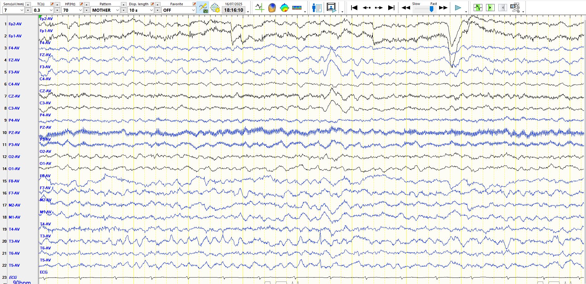
The above page demonstrates a waveform that is almost a carbon copy of the spike-and-wave seen previously (vertical red arrow). The horizontal red lines denote waves at T3 that individually would not be considered to represent spikes; however, if you compare their morphology to the spike and wave, you might be persuaded to believe that they are of the same family. This raises another important point; spikes in a particular area may vary considerably in their morphology. The reasons for this are complex but suffice it to say that intracranial recordings often demonstrate a large array of waves of varying morphologies and amplitudes and subtly different distributions that summate in complex ways to produce variable waveforms on scalp EEG.
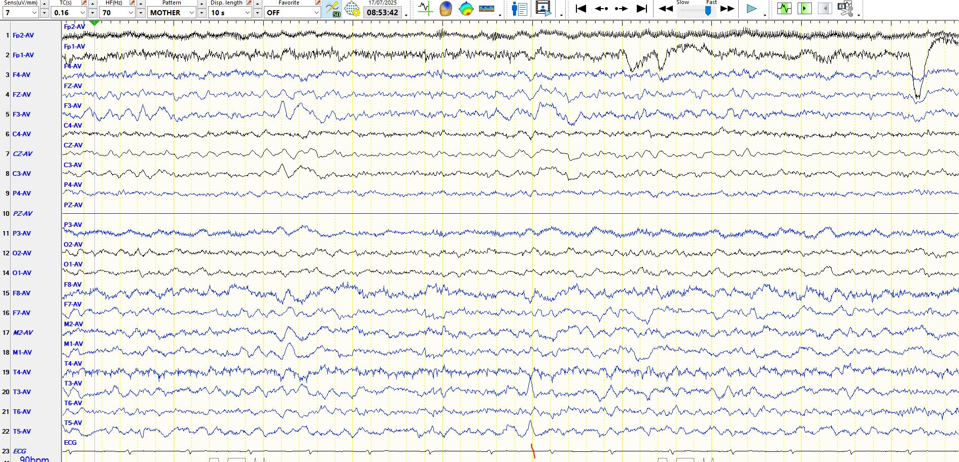
There are abundant sequential theta waves at T3-T5, some of which are sharply contoured, yet do not represent IED's, as they are the background! In the middle of the page is a remarkable needle-like wave at T3-T5; it is noteworthy because it has a distinct morphology from the spike-and-wave discharges highlighted in images further above (to reiterate, spikes in the same place do not always have the same morphology). This makes the point that spikes on EEG may be remarkably sharp. The latter results in them resembling environmental, electrode and EMG artifacts. This is where one has to be especially careful of the high frequency filter.