Arousal, resembles a seizure
Jul 18, 2025The asymmetry of the following rhythms during transient arousal resembles a seizure at T4-T6. The following are three consecutive pages.


In contradistinction to a seizure, the above rhythms do not evolve. The parallels for the asymmetry are to be found in the recording during wakefulness.
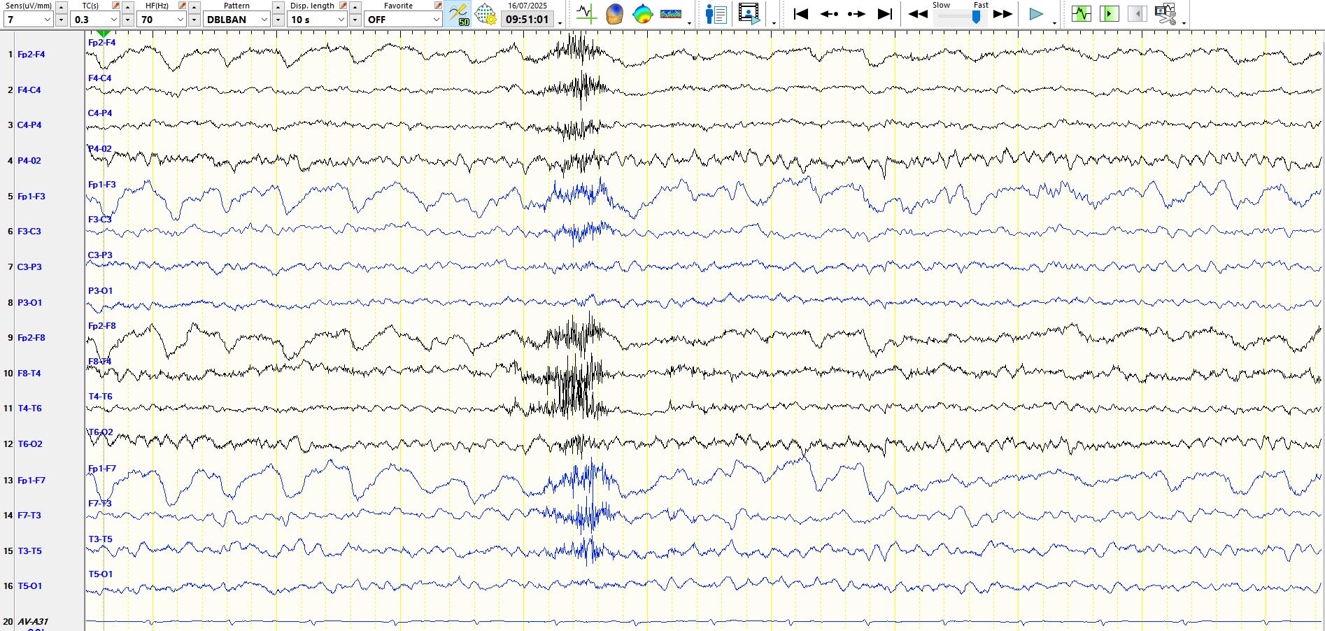
There are delta waves at F3-C3 and left hemispheric theta waves, disproportionately well-developed at T3-T5, in the above epochs. Some of the waveforms in sleep resemble asymmetric F-waves and V-waves, which they are not. The asymmetry of theta frequencies is more easily seen when the alpha generators are turned off, as in the following page:
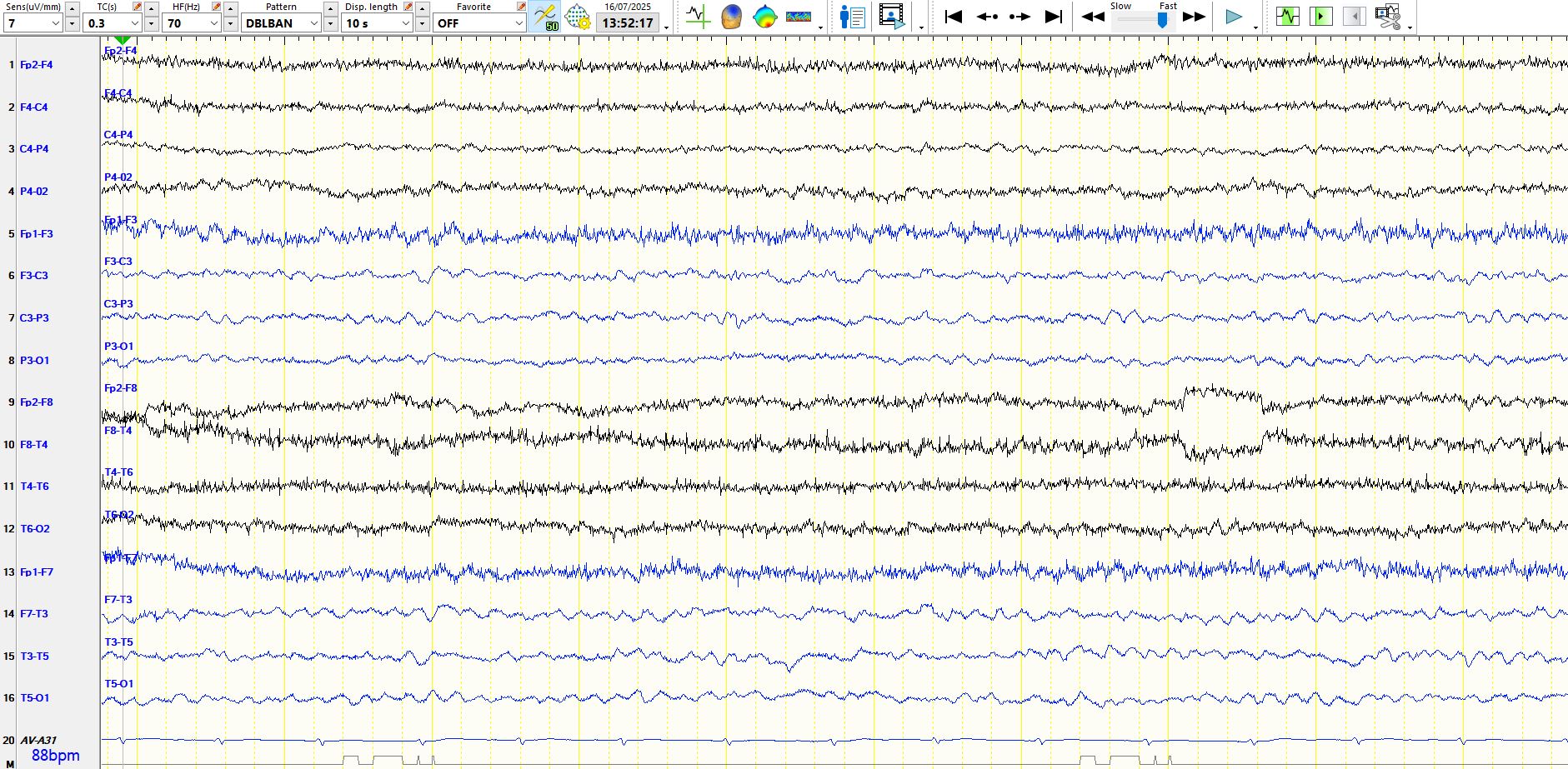
There is another important point about such asymmetries. Somewhat counterintuitively, hemispheric theta waves, delta waves and suppression are sometimes difficult to see on the common average referential montage, as the widespread appearance of theta or delta waves over one hemisphere is incorporated into the reference, obscuring or minimizing its appearance. This is a significant disadvantage of common average referential montages. The dramatic asymmetries sometimes missed by reading on this montage never cease to amaze me.
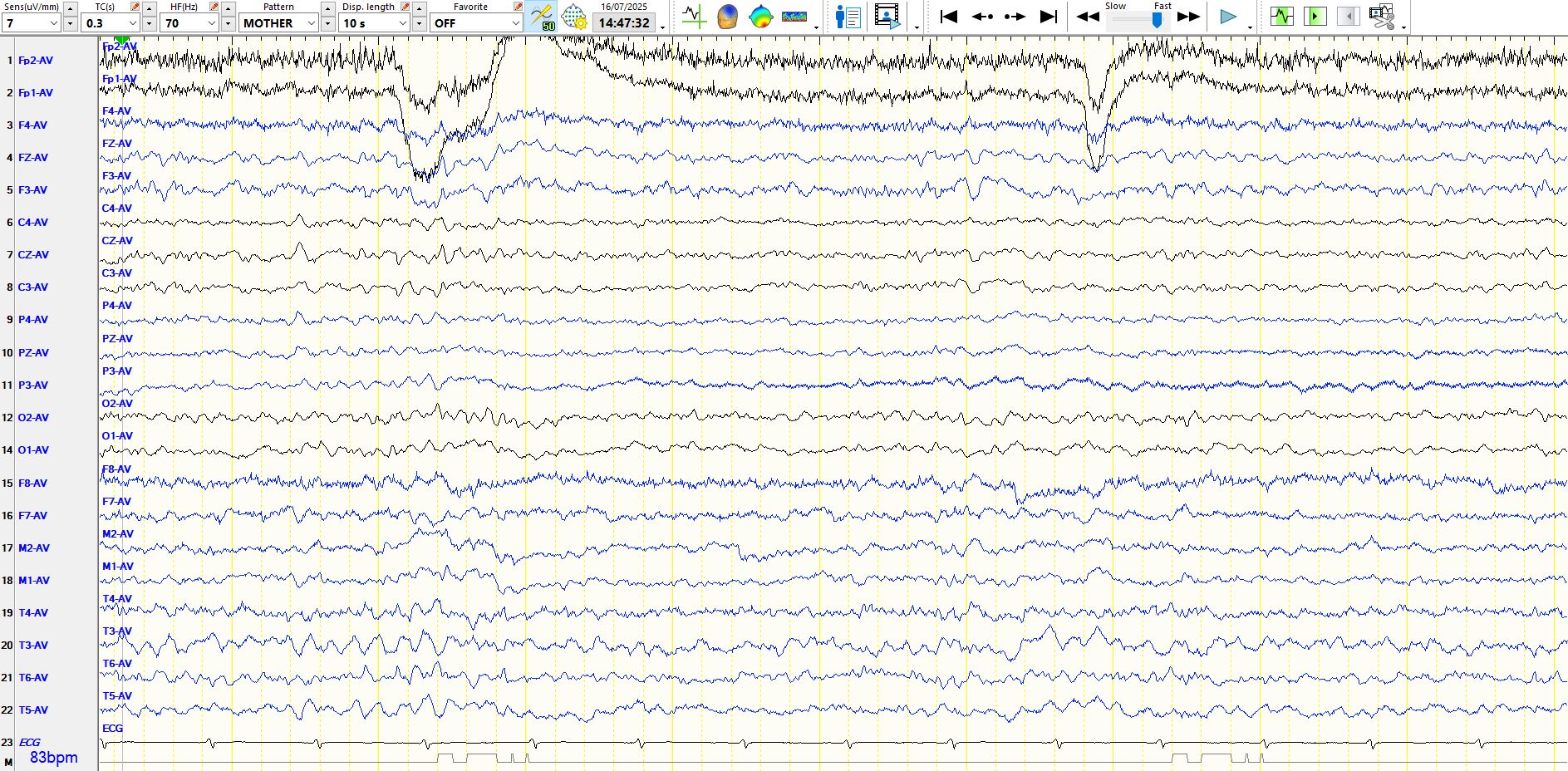
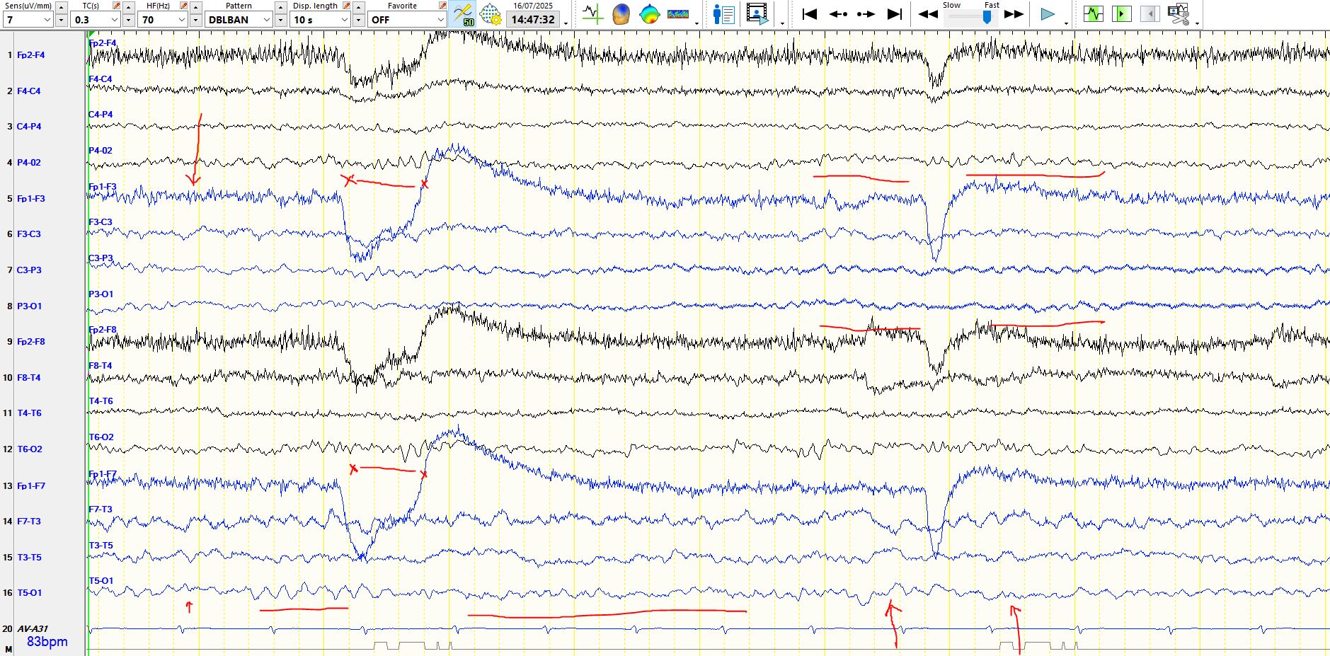
Have a look at the same page represented on the common average referential montage (top) and a bipolar montage (immediately above); you may appreciate how much easier it is to see the asymmetry of the theta (horizontal red lines), delta frequencies (red arrows) and alpha rhythms (red horizontal lines between the crosses) on the bipolar montage.
Standard teaching is that traditional referential montages are the preferred means of assessing asymmetries. Such referential montages include those to the ears or to the vertex (CZ). As a rule, I do not find these montages useful and, while I have a large suite of montages that I use, I abandoned these a long time ago. The reasons are best illustrated by the above epoch represented in these montages. Have a look at these:
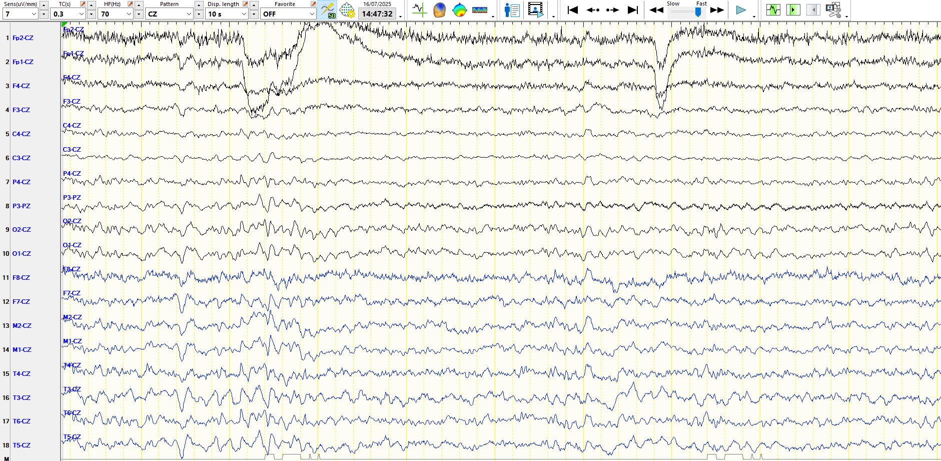
The above is the referential montage to CZ. Lots of laboratories use this. The problem is that the recording is so contaminated by the reference that other waveforms tend to be obscured; one's brain tends to focus on the gorilla walking front of the screen. The other disadvantages of this montage include its proximity to C4, C3, PZ, P4, P3, F4, FZ, F3, with the result that the deflections in these channels tend to be very low in amplitude when compared to the electrodes located more remotely. Finally, single electrode artifacts at CZ typically ruin the EEG at the time of their occurrence, in the same way that background rhythms from CZ tend to overwhelm the recording. Some laboratories use 0Z as the referential; there are similar advantages and disadvantages to CZ and since it is at the back of the head, it is only really suitable for patients who are having short EEGs.
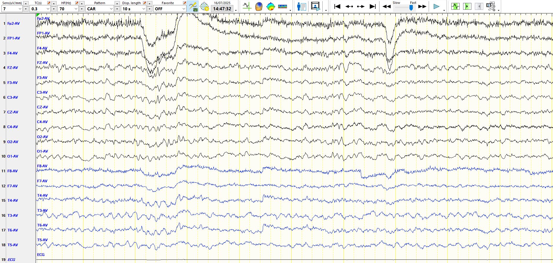
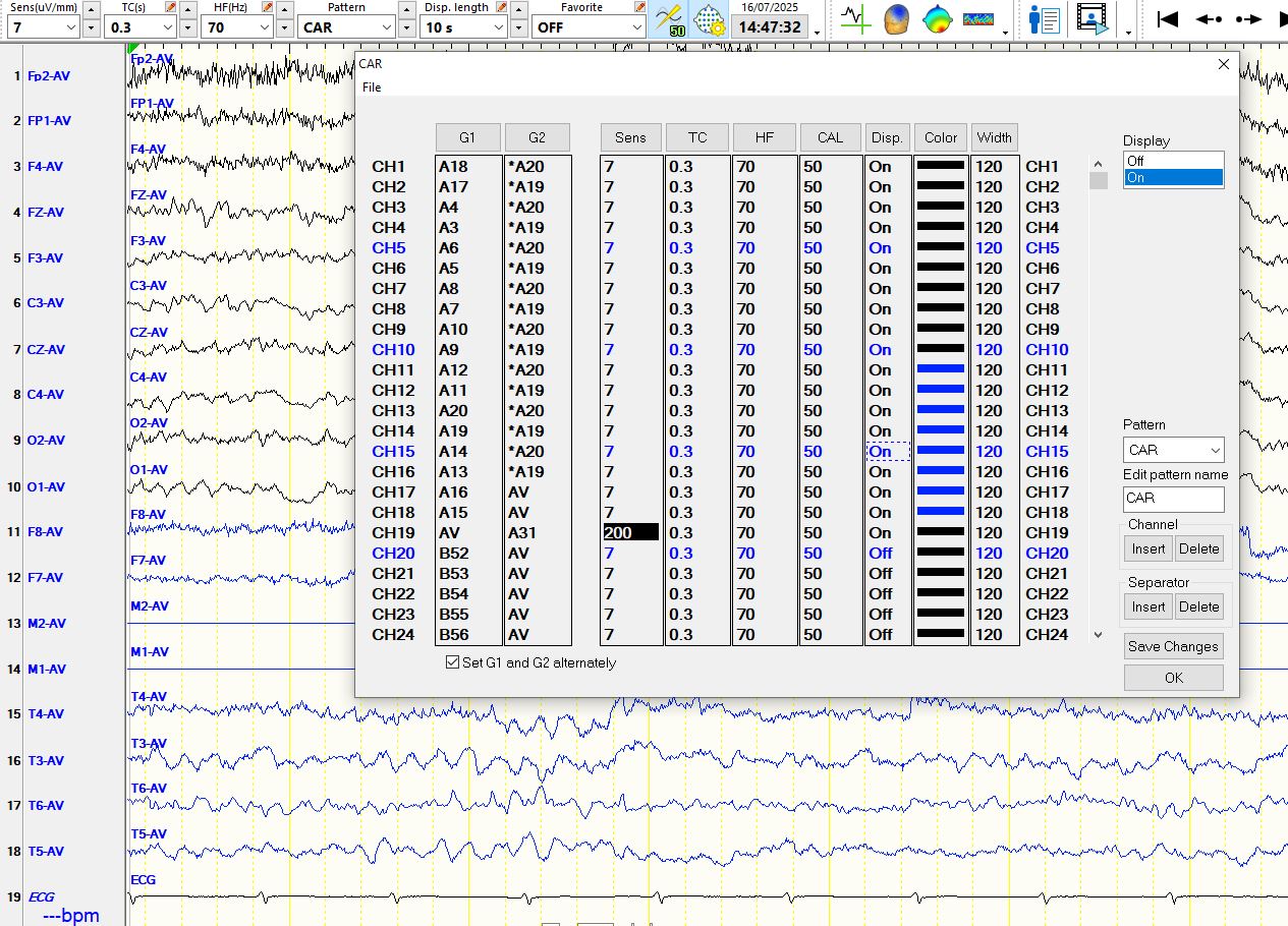
The above is the referential montage to the right and left mandibular notches, located immediately anteriorly to the ears, for the right and left hemispheric electrodes respectively. You can safely ignore the "AV" as the reference in each of the channels on the left. This is a metric headbox and I have manually changed the references (see image at the bottom, M2=20, M1=19). The asymmetries are a little easier to see in the above montage, but because M1 and M2 are located close to F8 and F7, there are cancellation effects resulting in "flatlining" of the channels representing at F8 and F7. Since spikes disproportionately frequently occur at F8, F7, M2 and M1, one may miss important abnormalities. If one is to use the ears, this requires electrodes applied to the ears. Such electrodes are fine to use for short recordings but are notoriously unreliable for more prolonged recordings. My dislike of such single or dual reference electrodes is that artefact either contaminates the full suite of channels or half of the channels. This is not to say that one should not use them and there is no harm in programming these into your software and using them as needed. Also, the common average referential montage is not immune to contamination by single electrode artifacts: if these are of sufficient amplitude, they too can ruin the EEG.
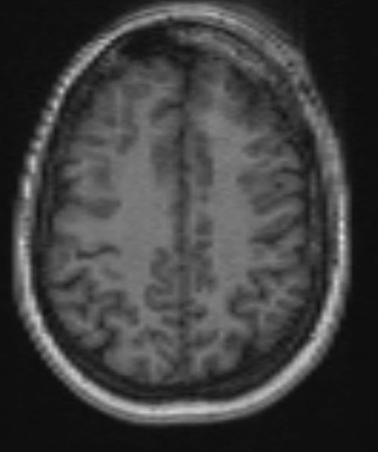
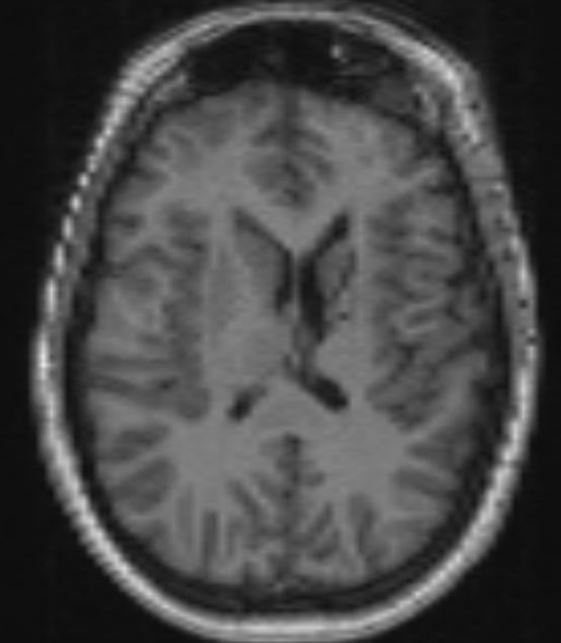
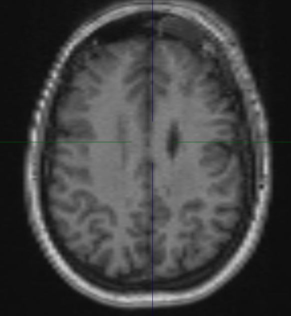
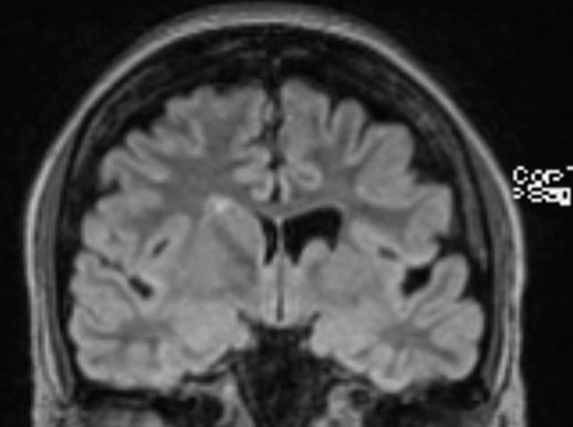
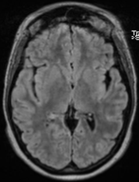
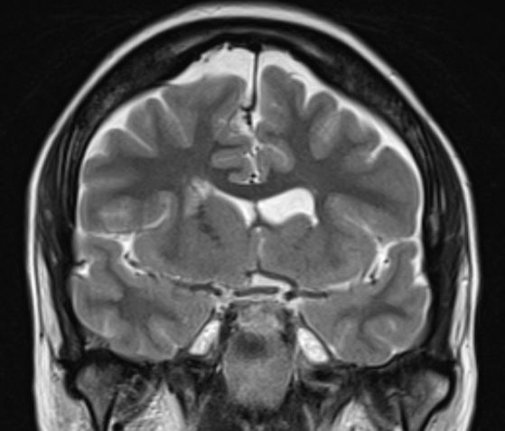
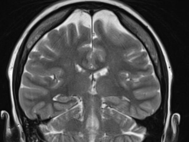
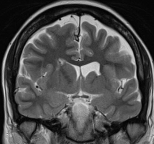
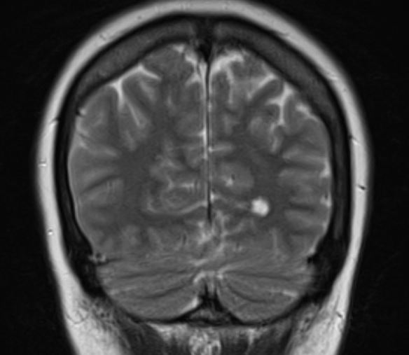
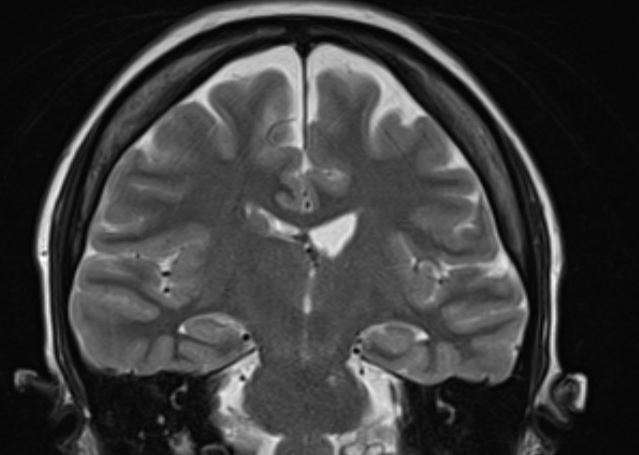
The structural correlates of the above EEG findings appear below. There is enlargement of the left-ventricular system, atrophy of the head, body and tail of the caudate nucleus and the left hemisphere appears modestly smaller in size than the right, most evident on axial sequences. The MRI was done while the patient was quite restless. As you can see from these pictures, the left occipital cortex and the left thalamus do not demonstrate any structural correlate to the asymmetry of alpha rhythms. However, we know from the EEG that the left hemisphere is markedly dysfunctional. Unsurprisingly, on functional MRI scans language is represented in the right hemisphere.