A colleague's question
Jul 21, 2025Re this post, Frontal lobe seizures versus functional seizures? Teenager
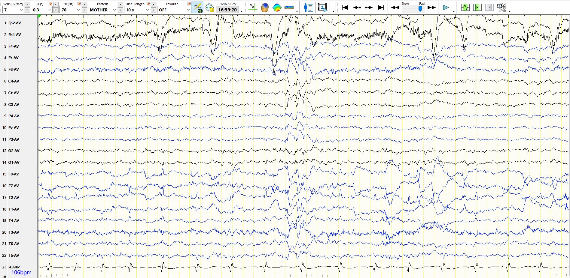
A colleague asks, "could this be phantom spike-and-wave?"
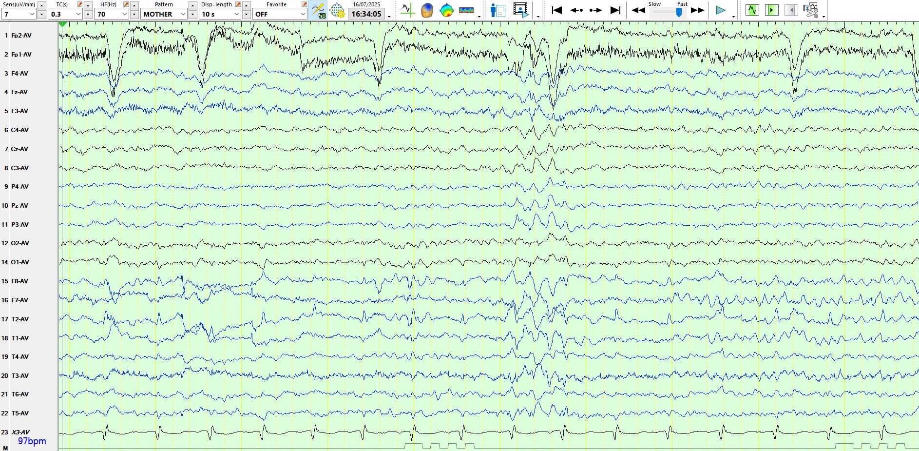
The short answer is no. Why? Phantom spike-and-wave (also known as Six Hz spike-and-slow-wave) typically has slow waves that are no more than moderate in amplitude and typically lie in the 4-7 Hz frequency range. By contrast, the waves in the above post are approximately 3-3.5 Hz and the slow waves are high in amplitude. Furthermore, Phantom spike-and-wave discharges are typically posteriorly predominant or anteriorly predominant, while the discharges in the above post primarily reside over the central regions.
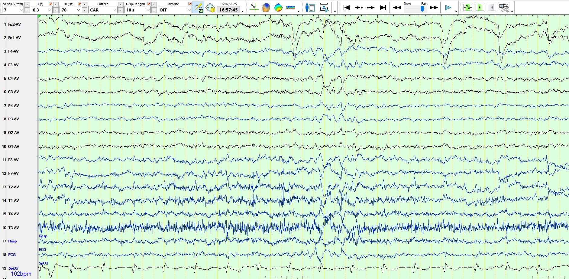
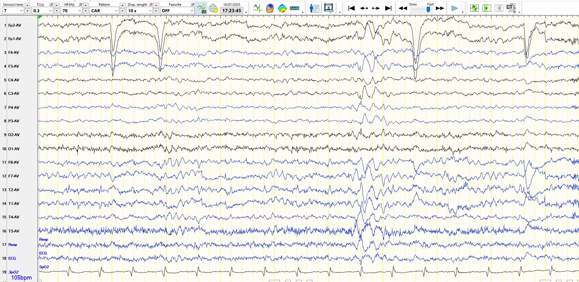
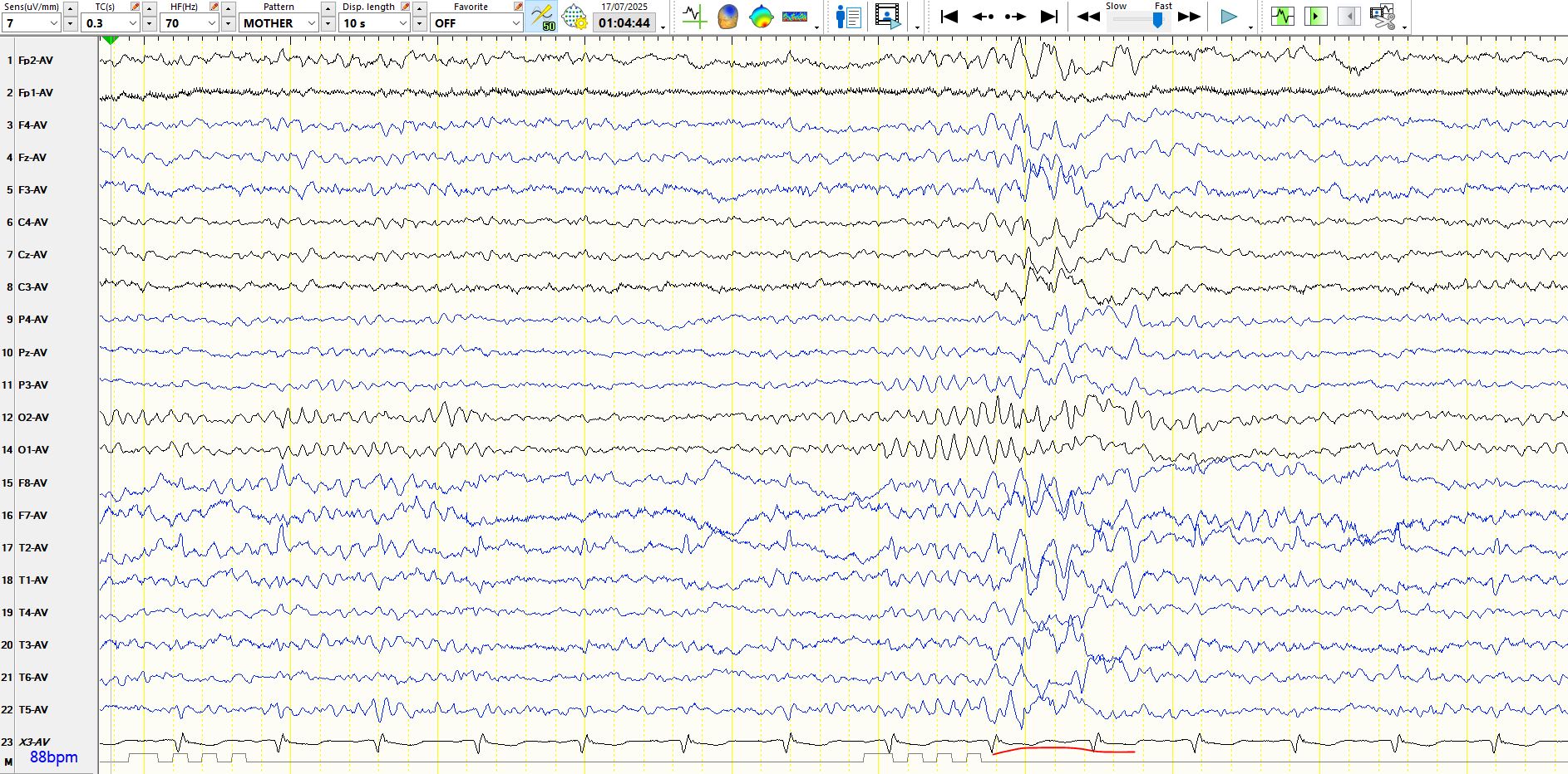
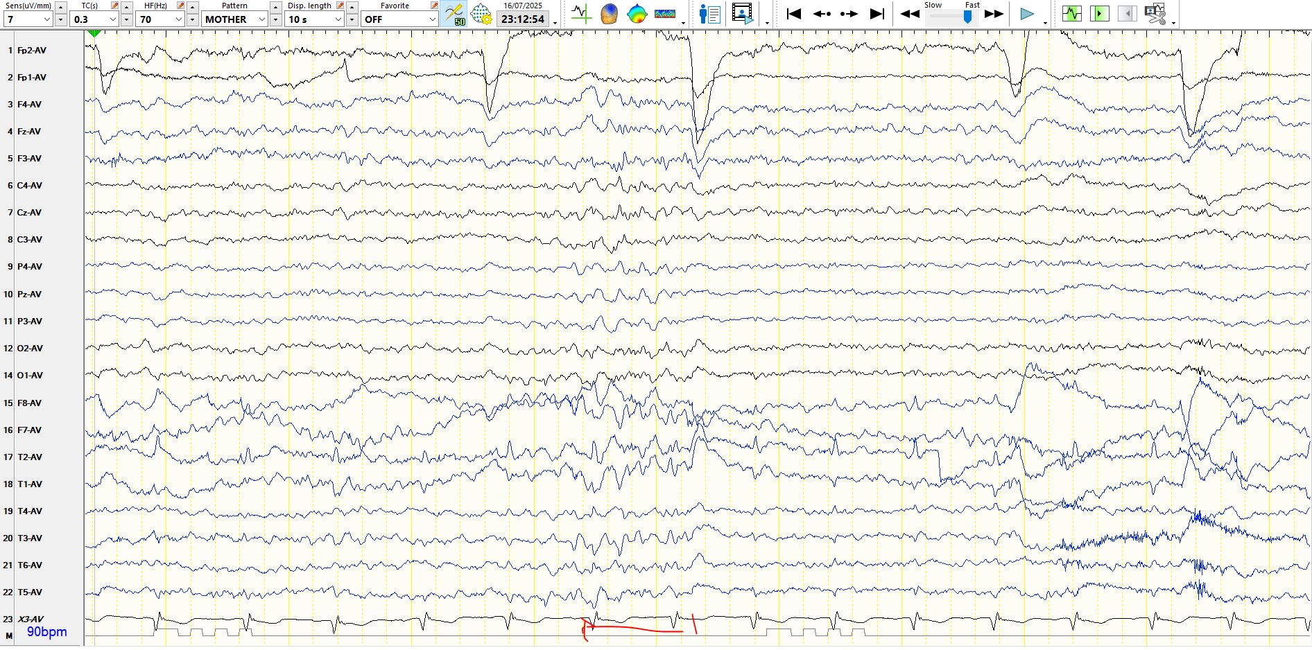
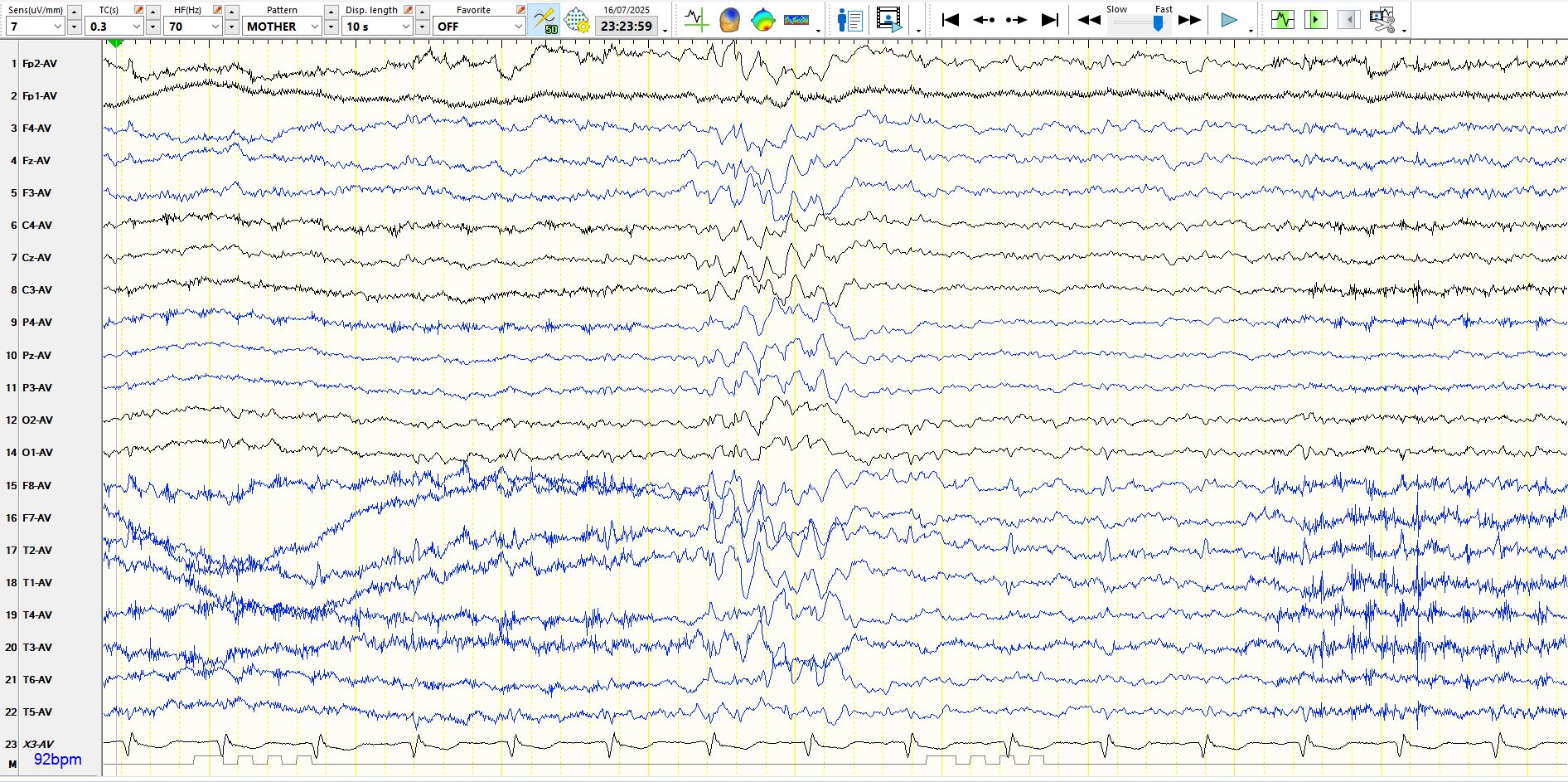
Here are some phantom spike-and-wave discharges from last week. The patient is a young adult.
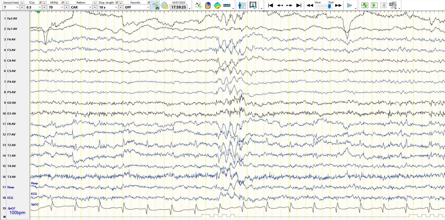
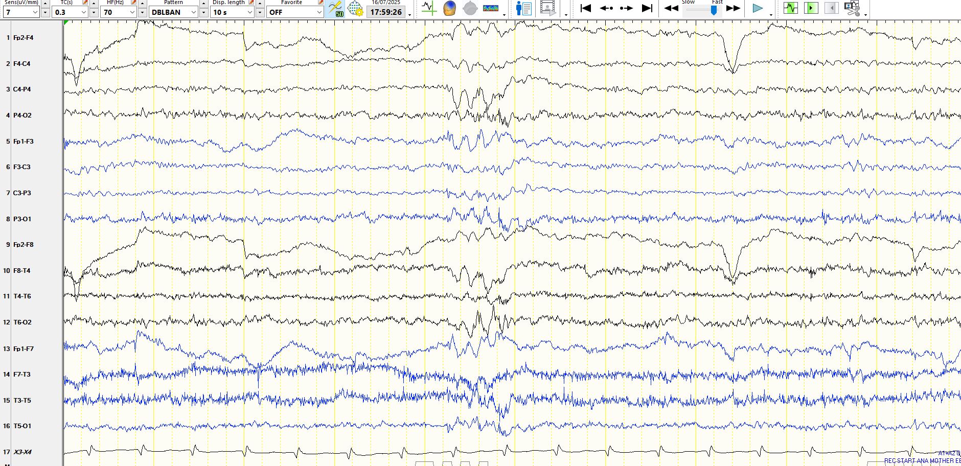
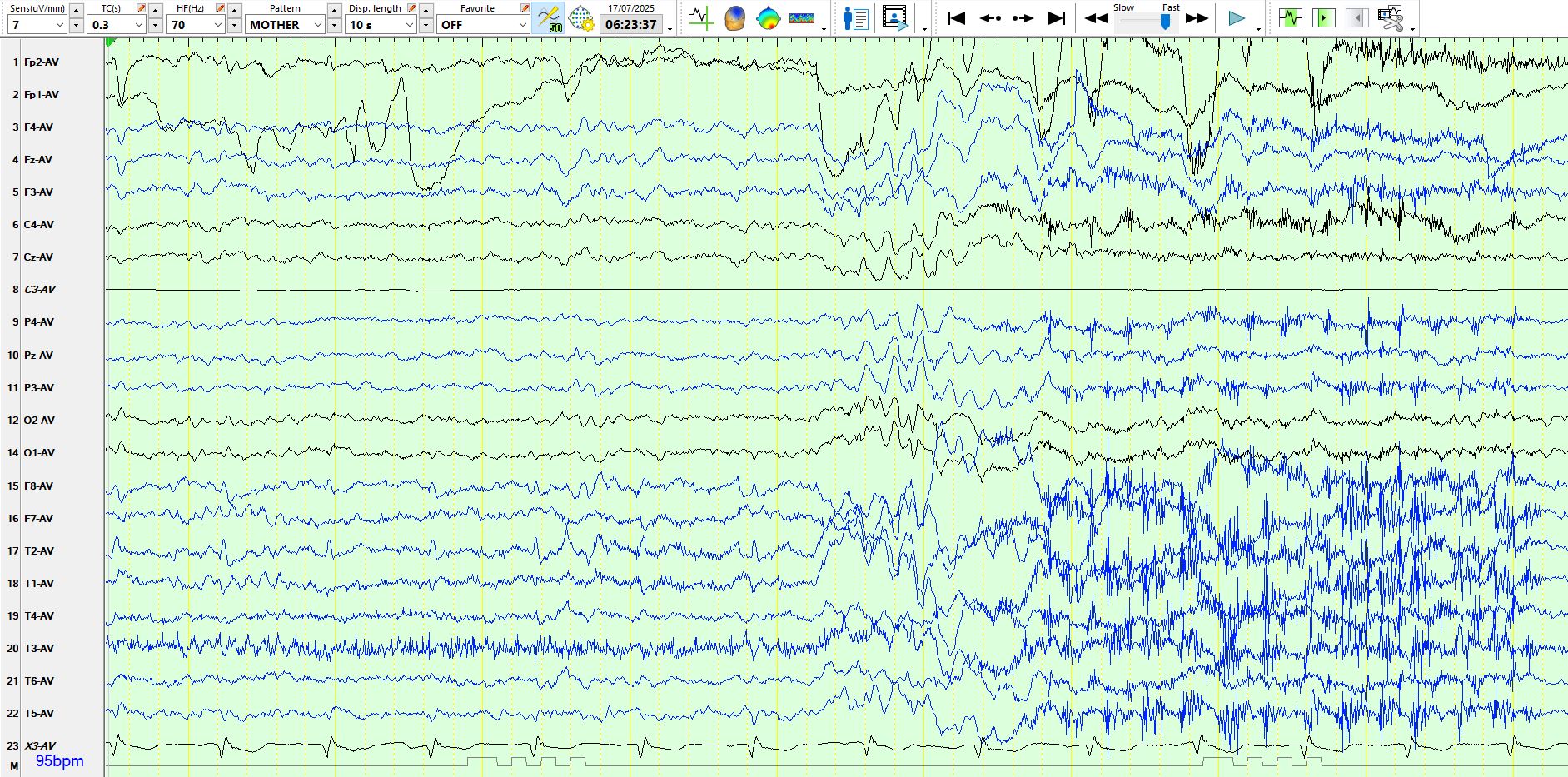
Here is the above page represented on the bipolar AP montage:
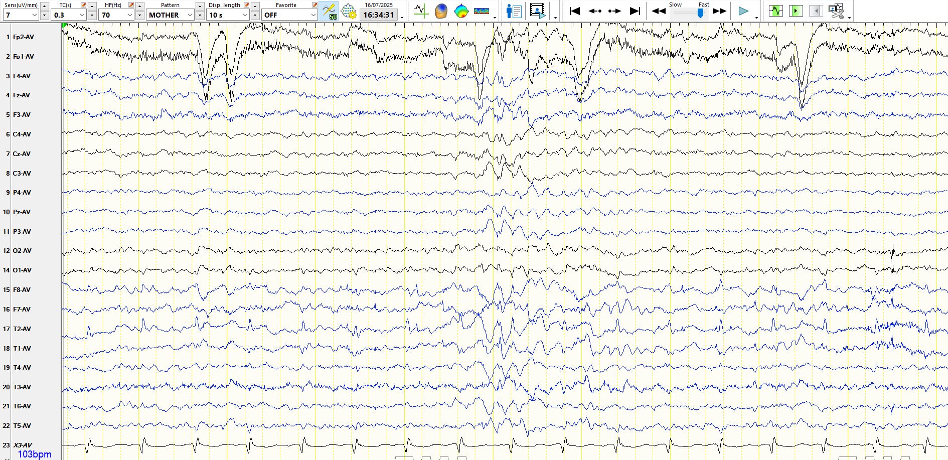
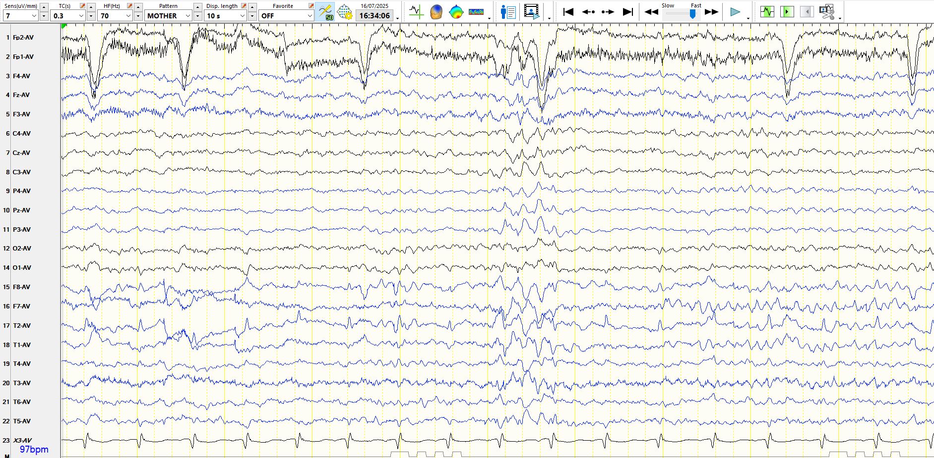
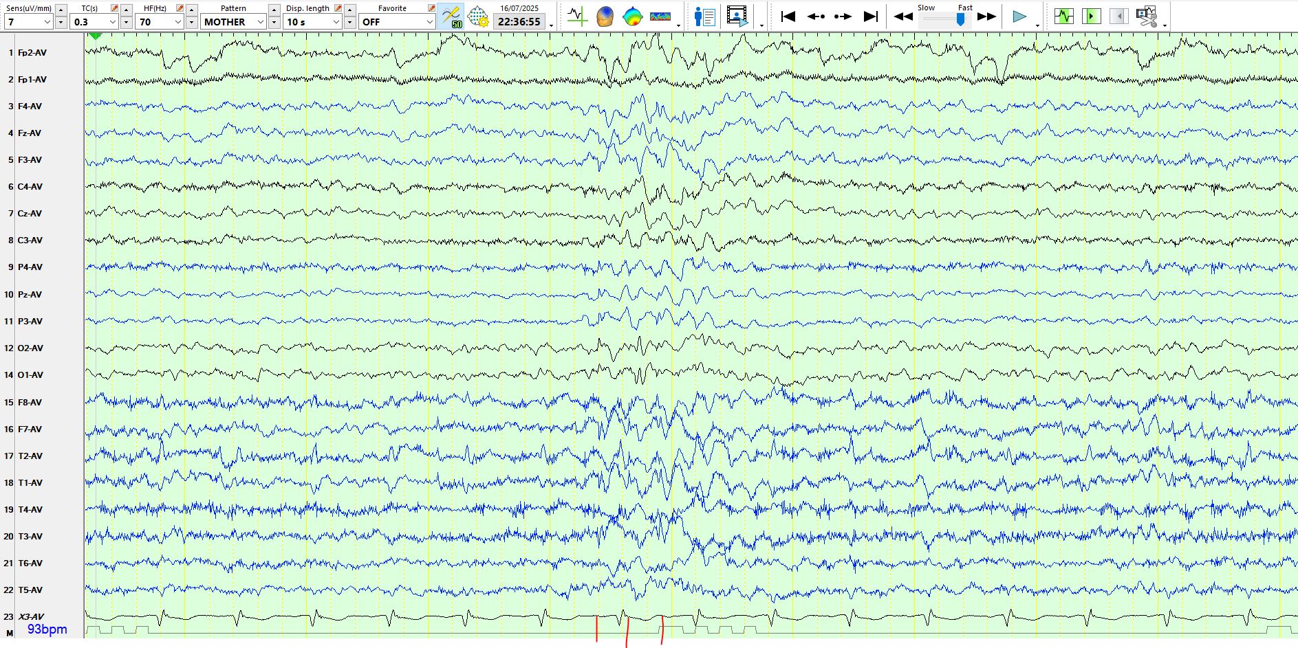
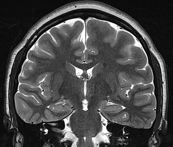
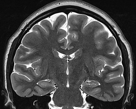
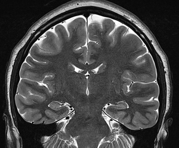
The above waves appear exclusively while awake and during very light sleep, but none are seen during deeper sleep, including stage II. For what it's worth, there is no history to suggest generalised epilepsy; The history provided by patient and the family suggests right temporal lobe epilepsy of 16 years duration; The patient is entirely unaware of seizures. The father's cousin (mid 50's) has epilepsy, manifest with GTCS without warning, since childhood. The inter-ictal EEG recording is normal. The patient has had 4 recorded seizures lasting 1-3.5 minutes, all of which appear over the right mid-temporal region (T4) on scalp EEG recordings; neither the patient, other patients nor the nursing staff noticed anything when she had these seizures, which were discovered on systematic scrutiny of the EEG recording. The patient and the family think that the seizures are well controlled, but that memory is poor. This likely is a consequence of intermittent amnestic seizures, as these appear on the scalp EEG recordings without a change in the antiseizure medication. The MRI scan of the brain is normal.