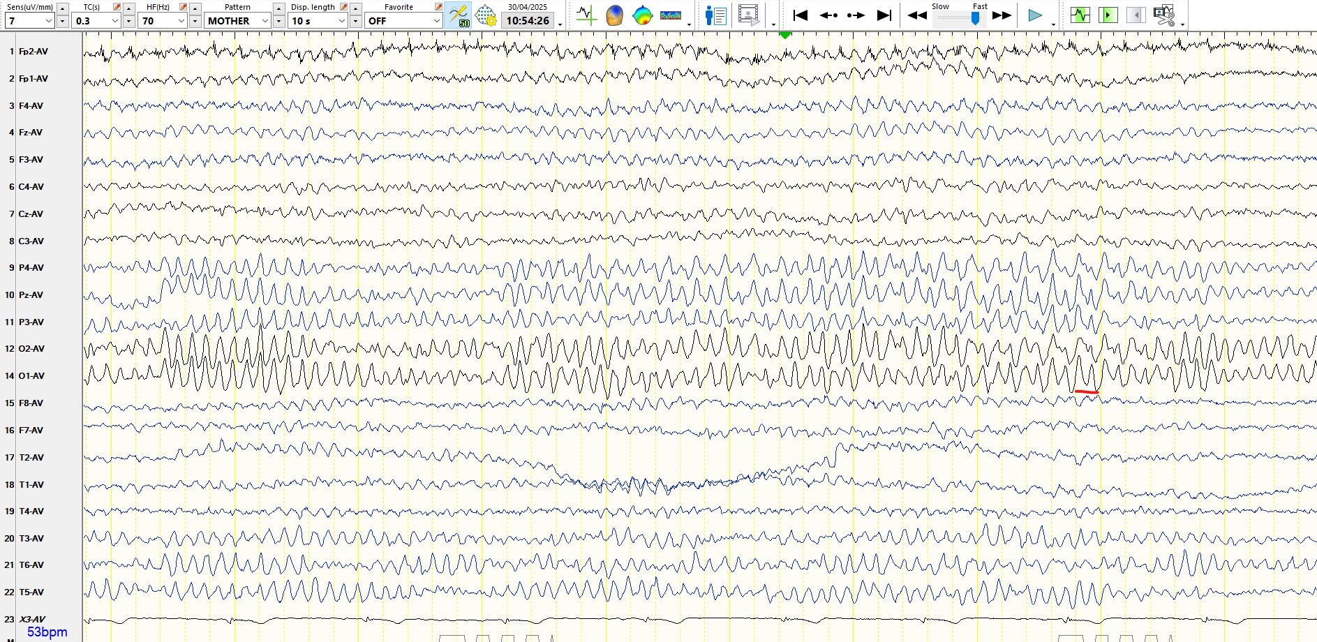
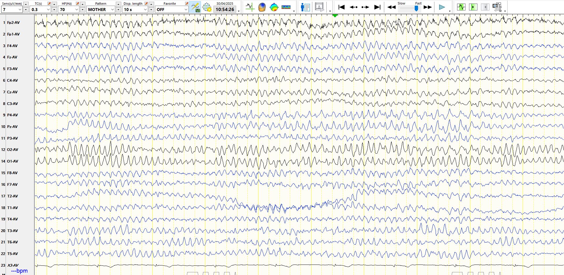
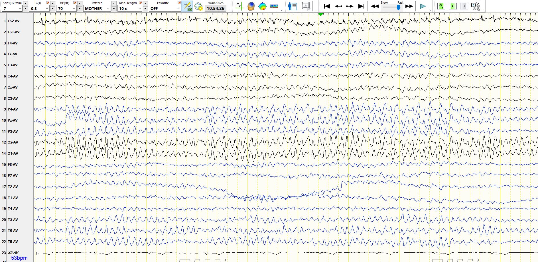
71y, known with epilepsy
May 16, 2025Is the following an example of a seizure, spiking in the parietal region, slow alpha variant, drowsiness or sleep? The first 2 pages represent 20 consecutive seconds. Subsequent epochs are identical to the 2nd epoch below, other than changes in montage or settings. Have a careful look at the first 2 pages first.
1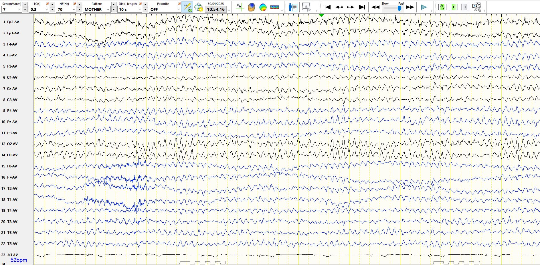
3. In the following epoch, the occipital and parietal derivations have been removed from the common average reference used in the previous epoch
4. The following represents the previous page, but in the anterior-posterior bipolar montage:

5. Previous epoch represented in a coronal montage below:
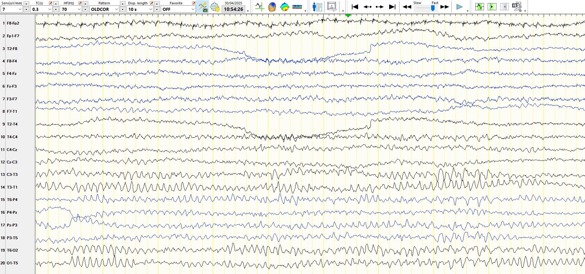
The above recordings are normal. In all of the above epochs there are slow eye movements, indicative of drowsiness. In figure 3 the background rhythms progressively slow, creating the illusion of an evolving seizure, especially as there are sharply contoured waveforms intermixed with the slower frequency discharges. As can be seen below, the 2 sequential alpha waves at 01 are perfectly synchronous with a slower waveform at O2, which is then a harmonic of the alpha rhythms. The patient becomes increasingly drowsy and there is slowing of the alpha rhythms. This slowing of the alpha rhythms is reversed in the last second of the recording, synchronous with disappearance of the anterior alpha, as the patient becomes more alert. Theta waves appear at the vertex as the patient becomes drowsy but also disappears as drowsiness resolves 1 second from the end.
Fig. 3, from above