
A sleeping adult; identify the wave
Sep 16, 2025Spend some time looking at the first page and try and answer the question; the wave of interest has been arrowed. Which of the following are correct? The wave represents:
1. An asymmetric F-wave
2. A right prefrontal Interictal epileptiform discharge
3. A generalized interictal epileptiform discharge
4. A small, sharp spike
5. A right temporal inter-ictal epileptiform discharge
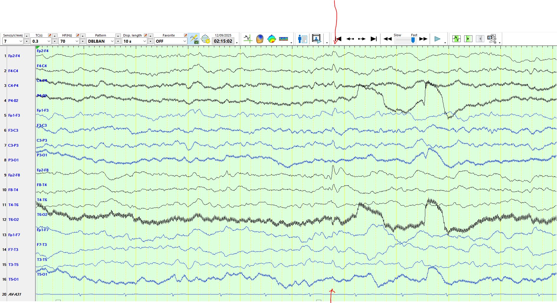
From the above image, you might deduce that the waveform is primarily electro-negative at FP2-F4, but the clue lies in the artifactual derivation, channel 12. The deflection in channel 12 is in the opposite direction to that seen in the anterior leads. This strongly suggests a surface electro-positive discharge over the right temporal region, rather than an electronegative discharge over the right frontal region. You can confirm or refute this hypothesis by looking at other montages of the same discharge:
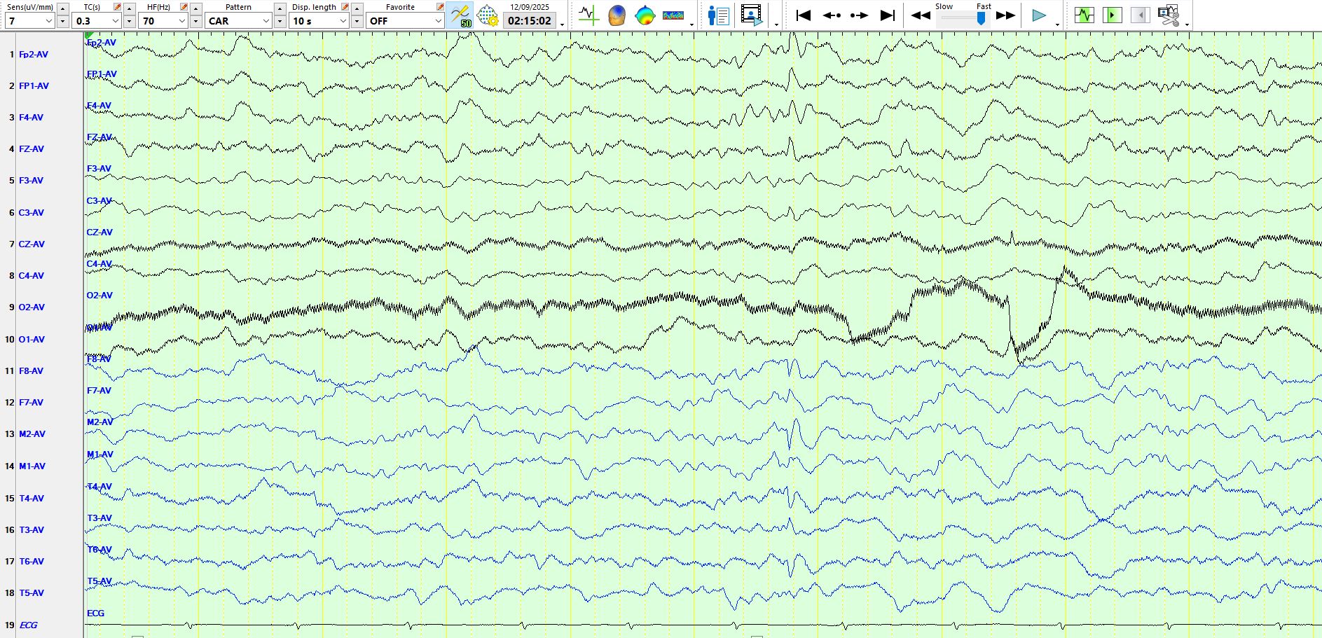
The referential montage demonstrates that there is an electropositive at discharge at M2-F8-T4-T6. Furthermore, this is followed by an inverted slow wave, which is therefore also surface positive. The second component of the spike-and-wave, which is electro-negative, gives rise to the upward deflection on the bipolar AP montage, seen right at the top.
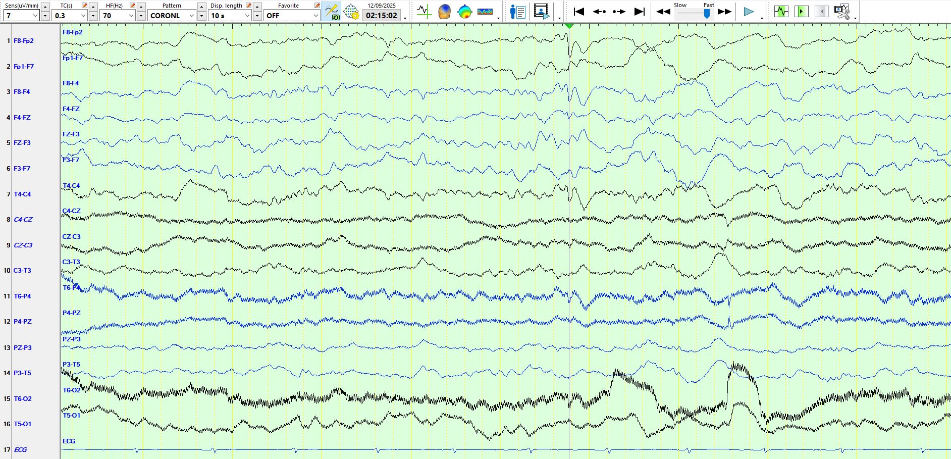
The coronal montage provides additional corroborative evidence for this conclusion of a surface electro positive spike over the right temporal region.
Bottom line?
1. It is best to review waves of interest on different montages
2. Electropositive spikes are sometimes present on the EEG; in some cases, they may be the only spikes
3. Artifactual electrodes may contain useful information
Positive sharp waves in the EEG of children and adults - PubMed