
31y, seizures
May 17, 2025Seizures consist of pins and needles in the right hand, a "pulling" feeling and videos show piano playing movements with the right hand, which she reports as being involuntary and with retained awareness. The following epochs are representative of her inter-ictal state. Identify the inter-ictal abnormalities. Electrode artifacts have been turned off and removed from the common average reference
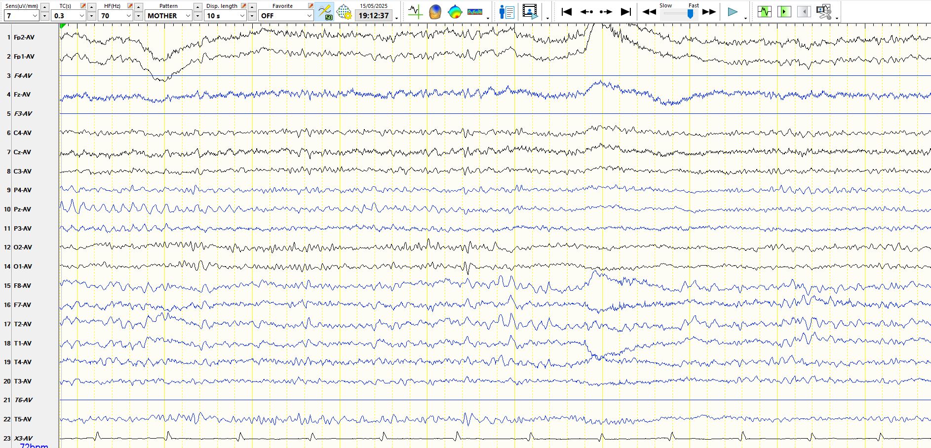
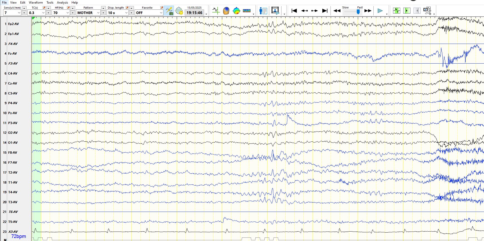
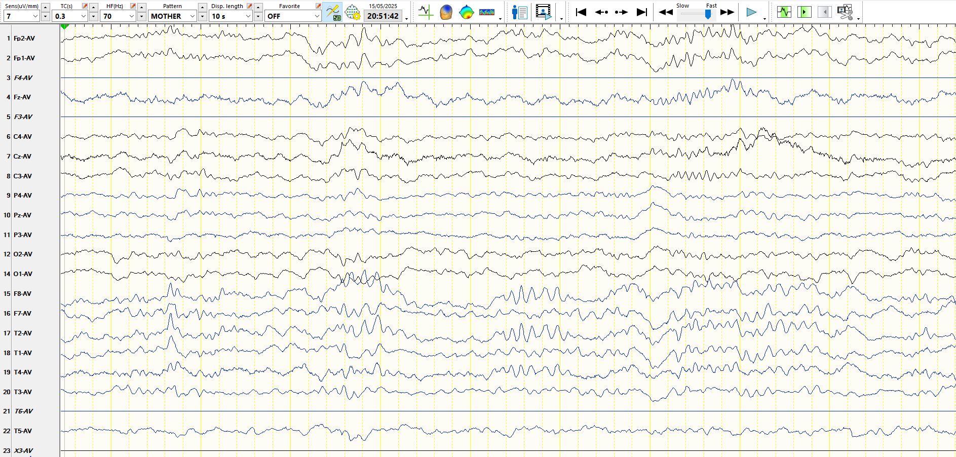
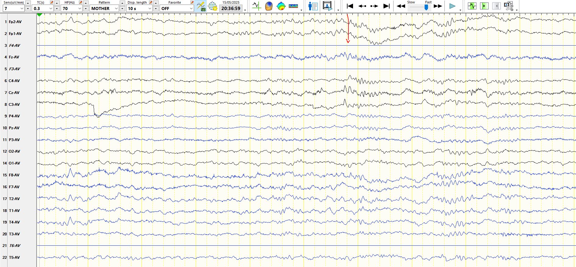
Above: in the 1st 7s there is evolving drowsiness. There is parietal alpha in the 1st second, followed by a slow eyeblink, followed by resolution of parietal alpha, followed by slower alpha frequencies in the temporal regions in the middle few seconds. Drowsiness then ends after there is an oblique movement of the eyes at 7s (downwards and to the left; see channels 1, 2, 15-18). This is followed by arousal, with the disappearance of the slower frequencies and the appearance of faster alpha frequencies in P4 and temporal regions at 8-10s (compare parietal alpha frequency in the first second with the last (where it is even faster), and likewise in the temporal regions across the 10s) The low amplitude sharply contoured wave appearing to the right of the sixth ECG beat is part of the background and is not a spike. It is followed by a few theta waves, best seen in the frontal derivations, representing part of the evolution of mild drowsiness.
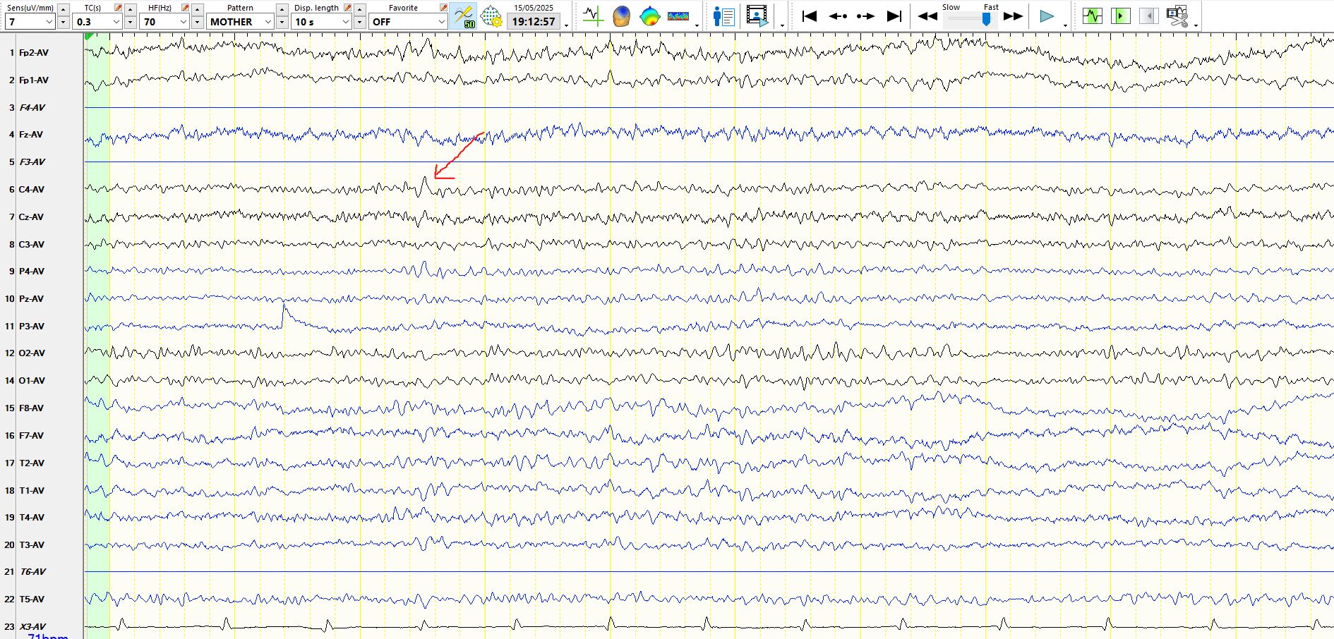
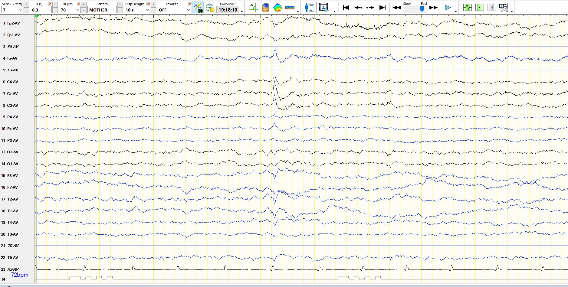
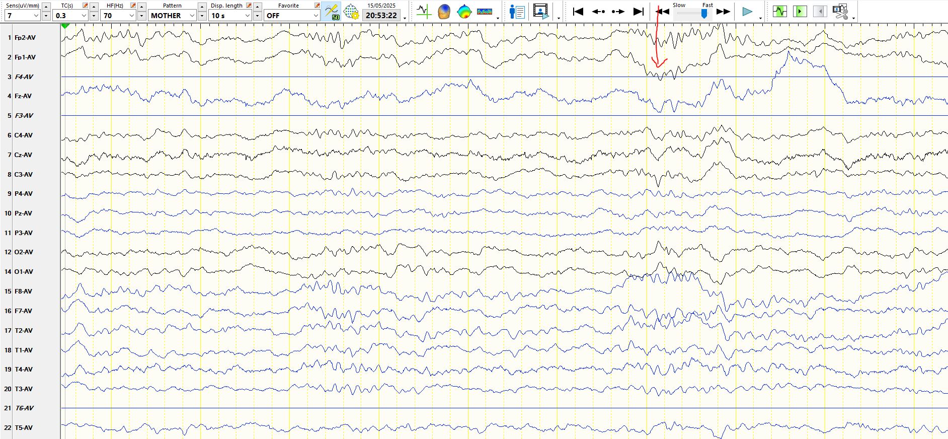
In the above, the asymmetric bilateral frontal theta wave (arrowed), indicates mild drowsiness, with subsequent progressive slowing of alpha rhythms in O2 and O1 (indicating evolving drowsiness) and with cessation of these alpha frequencies in O2 and O1 about 3 seconds from the end. This is followed by the appearance of slow eye movements at 7s. The slowing occipital alpha frequencies then disappear completely
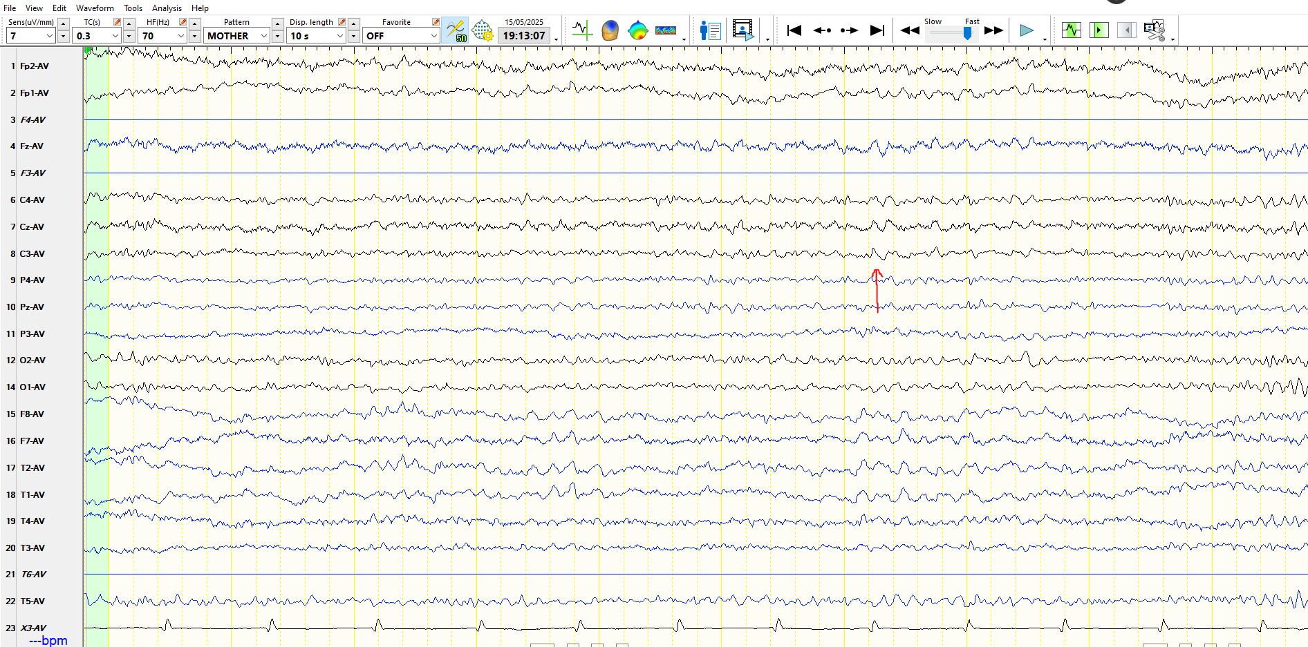
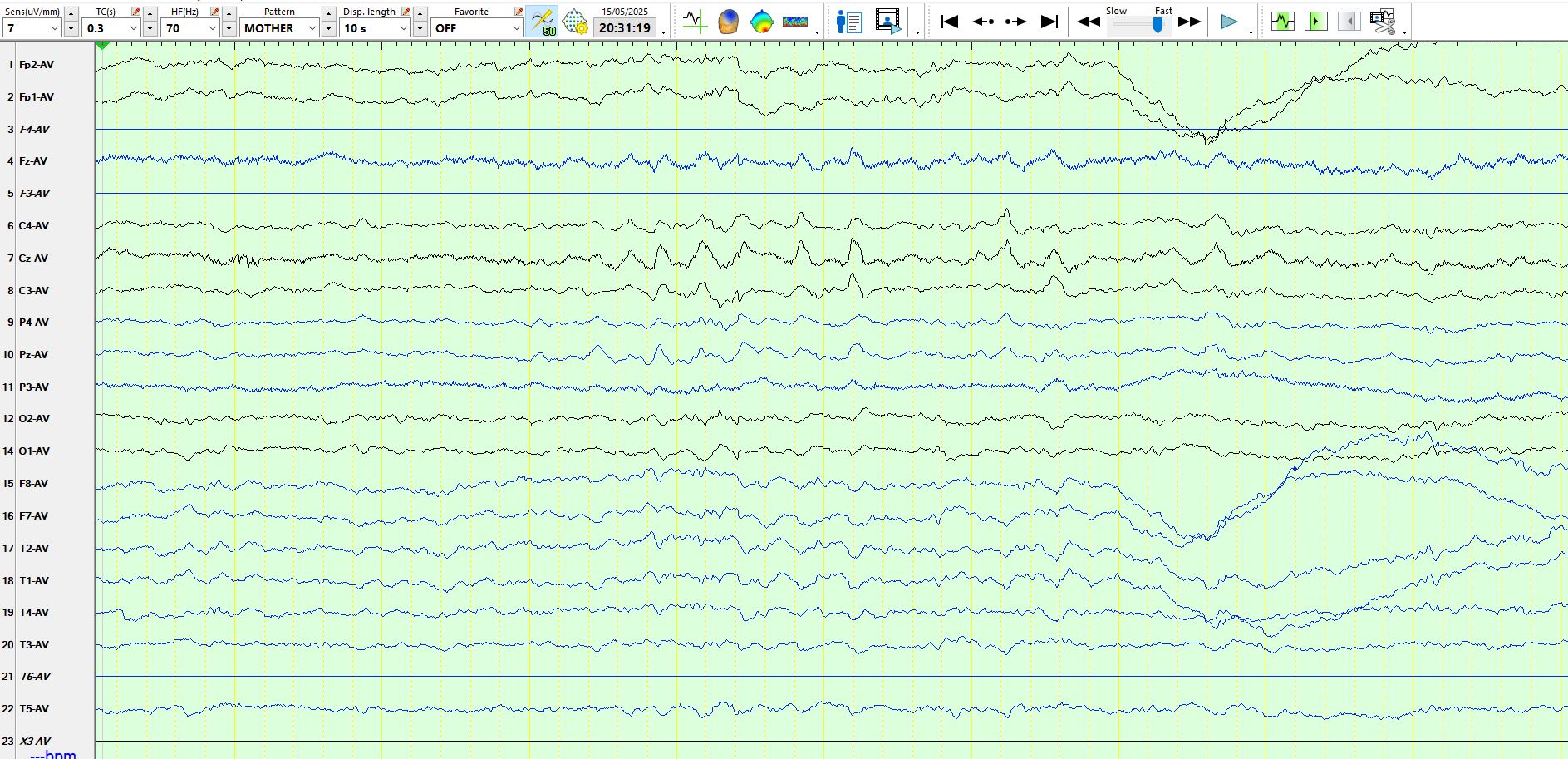
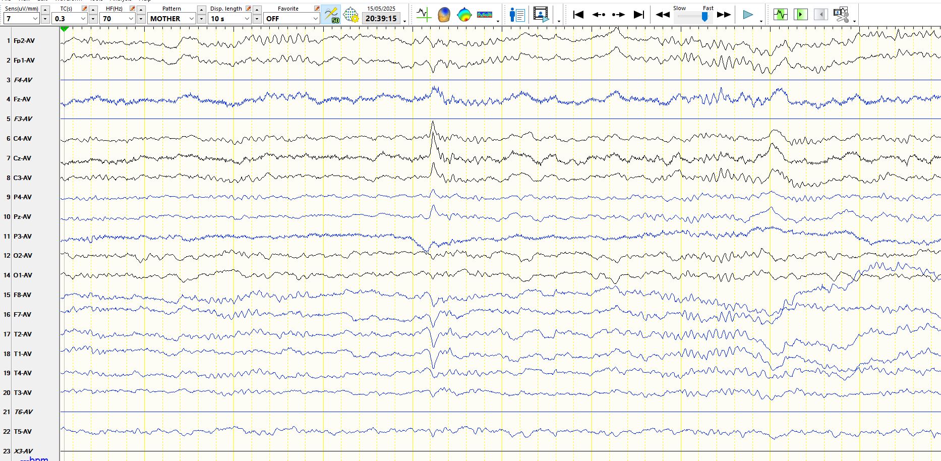
In the above, during the progressive evolution of drowsiness over 7s, central theta rhythms become apparent (arrowed) a few seconds after bilateral temporal theta waves. Two seconds from the end there is relative arousal, as generalized faster frequencies appear, with alpha in the occipital regions in the final few hundred milliseconds, but still with slow eye movements in the final two seconds (FP2 and FP1, F8 and F7, M2 and M1)
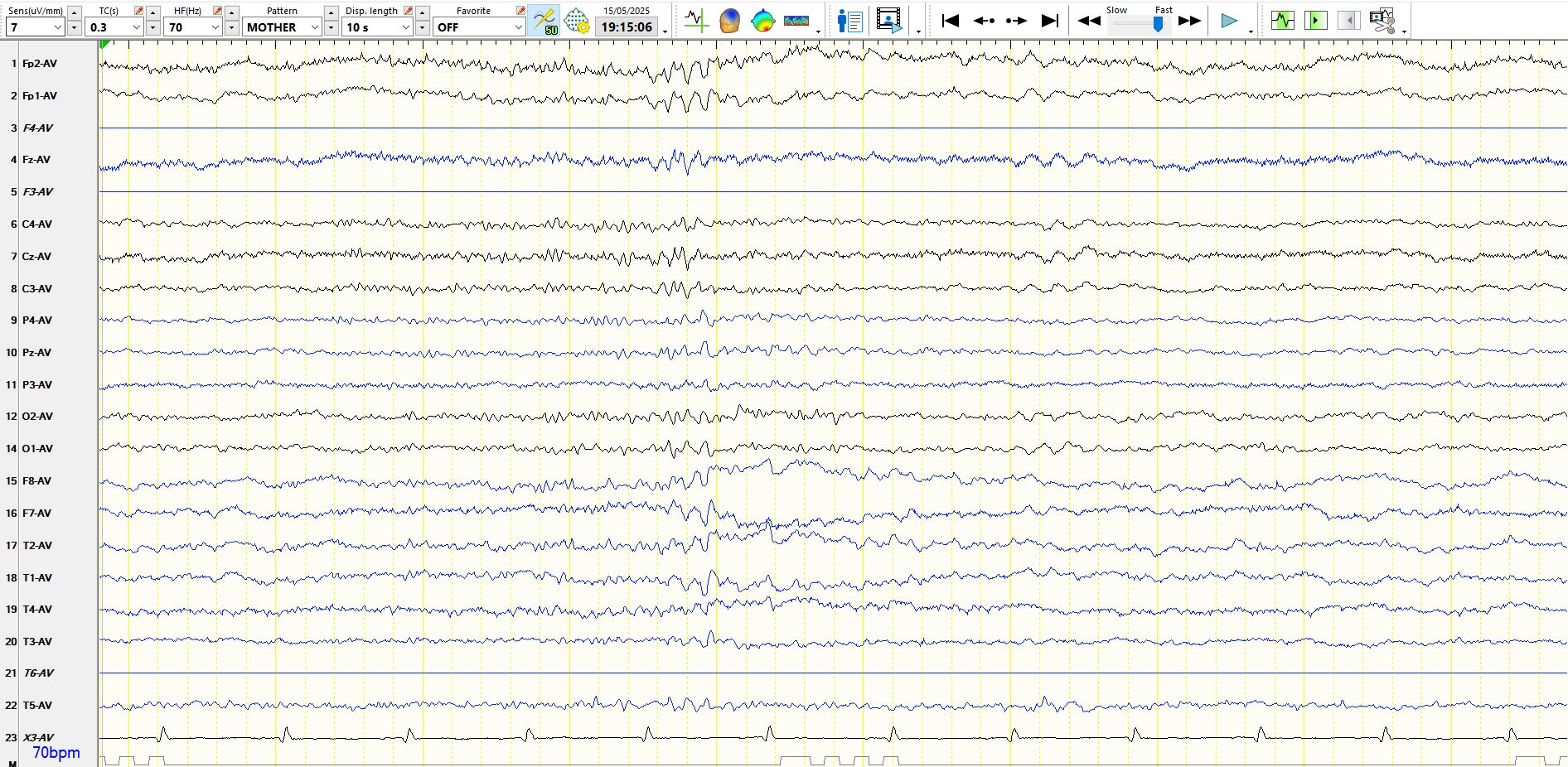
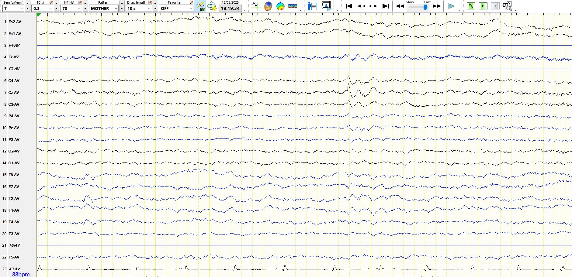
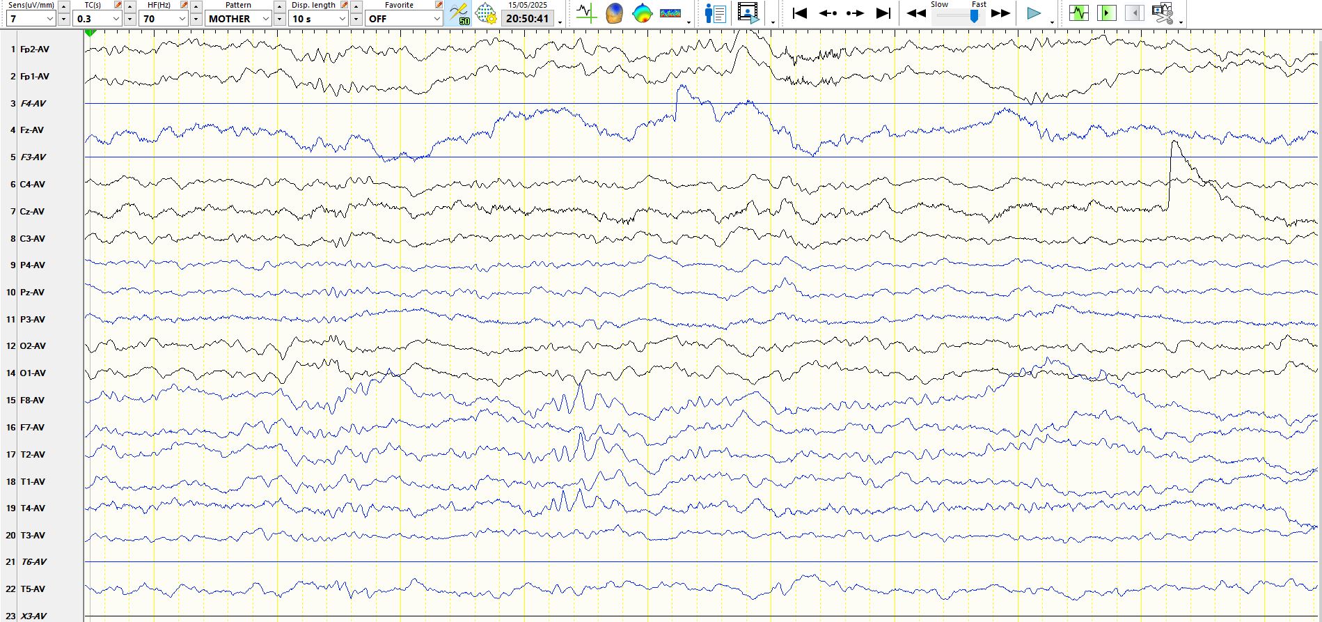
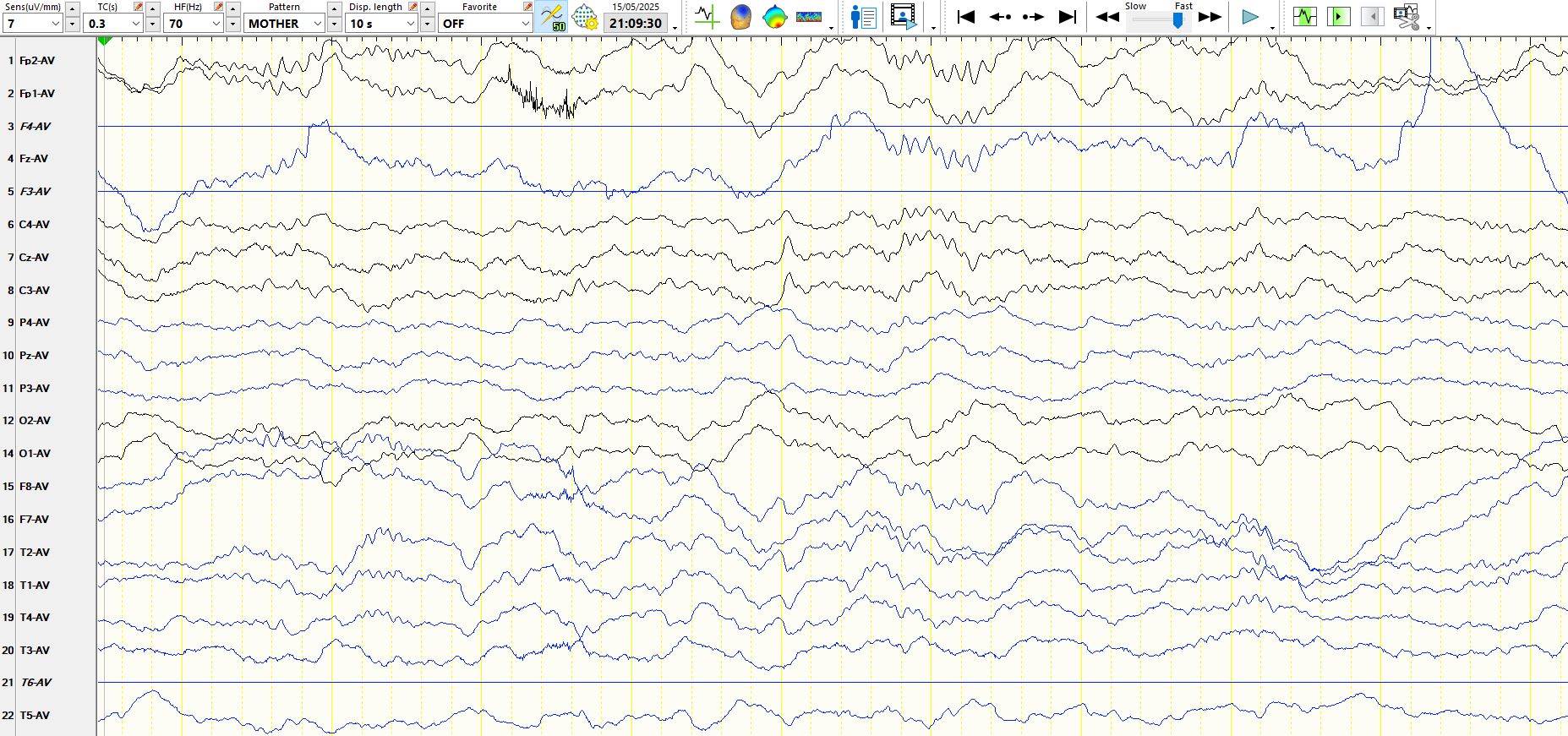
In the above 10s there is evolving drowsiness. The background rhythms slow subtly in the first 4 seconds, followed by anterior propagation and slowing of the alpha (indicative of drowsiness), immediately followed by a slow lateral eye movement 5s from the beginning (F8, F7 of opposite polarity0, ditto M2 and M1), Alpha frequencies then disappear, theta waves appear and there are slow eye movements in FP1 and Fp2 near the end of the page. Hence, these 10s represent progressive drowsiness.
Initial drowsiness (no alpha, no EMG, no eyeblinks) for the first 5s. The appearance of beta and alpha frequencies in the temporal regions heralds arousal, followed my movement and EMG artefact in the final 1s
Stage II Sleep, Vertex wave in the middle of the page, sleep spindles (Aadefining feature of stage II sleep) are more distinct below
Another V-wave
Asymmetric, temporal alpha in Stage II sleep (normal)
Same as above, but with an asymmetric normal sharply contoured theta wave of Stage II sleep in F8, F7, M2 and M1 at 1.5s
Stage II sleep, but here is an electropositive V-wave (arrowed)
Frontal spindles and a V-wave, different in shape to those above, evolution to Stage III sleep
Sequential V-waves, contrast shape of these with V-waves further above
Probable very low amplitude V-wave, followed by normal central beta of Stage II sleep. Sleep spindles at 2s (frontal) and 8s (widely seen), Stage II sleep.
Another V-wave and spindles
The following represents arousal from stage II sleep, followed by the seizure described above. The seizure lasts about 15s.
. 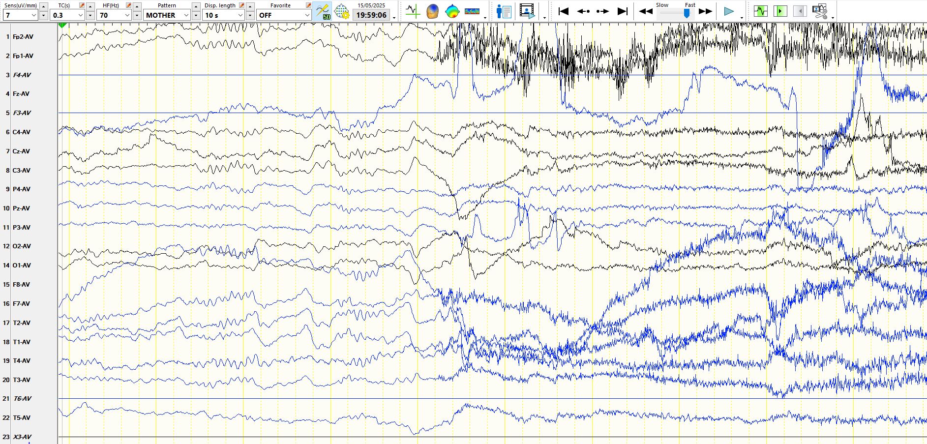
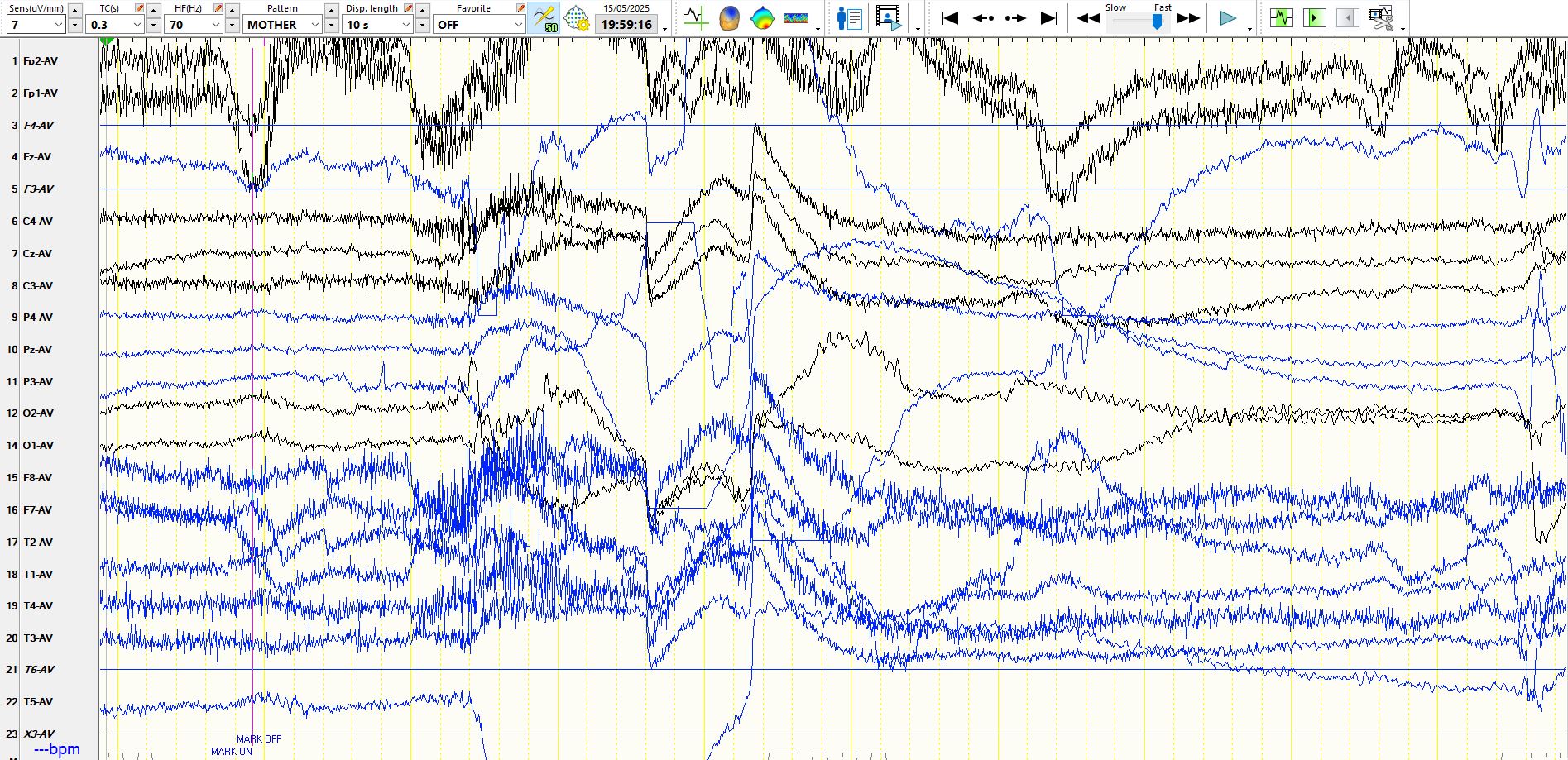
The EEG in this patient is normal inter-ictally and during seizures. The diagnosis is of focal aware seizures - the EEG is most often normal during such seizures, including those involving the convexities
Clinical and electroencephalographic features of simple partial seizures - PubMed
There are biological reasons for this
Relationship Between EEG Electrode and Functional Cortex in the International 10 to 20 System - PubMed
MRI: old ischaemic lesion, left post-centra gyrus
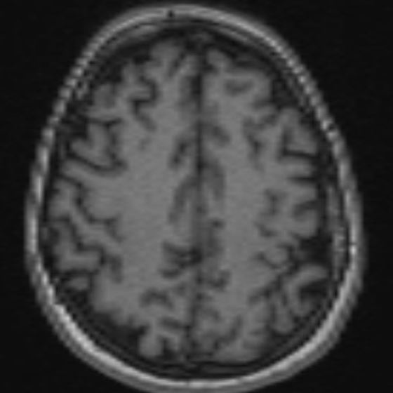
Anatomical location confirmed on fMRI