
11y, seizures, flaps hands and arms and talks during seizures
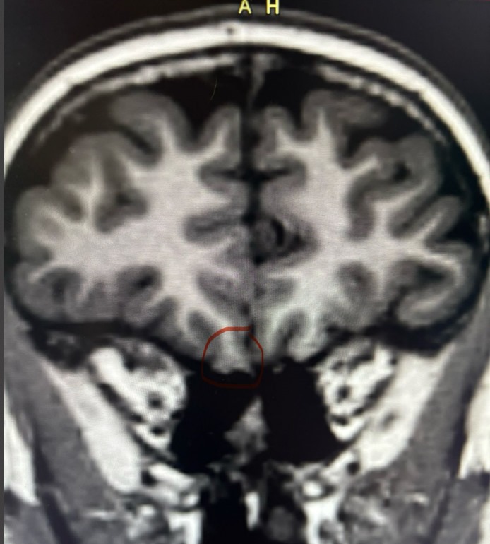
May 19, 2025Long-term monitoring demonstrated no inter-ictal abnormalities during wakefulness and sleep. Similarly, the EEG was normal during ssizures. A confident diagnosis of frontal lobe epilepsy was made, based on careful analysis of the history and the video findings (> 90% seizures in sleep). Multiple MRI scans had been reported as normal. A repeat high resolution 3T MRI scan was reported as normal. Repeated scrutiny eventually disclosed following probable focal cortical dysplasia
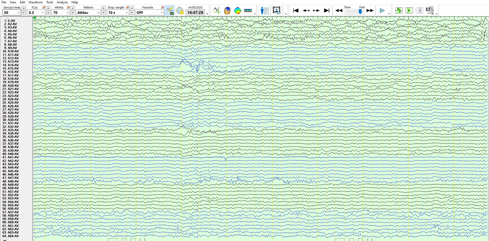
Sub dural electrodes: Alpha rhythms in electrodes 57, 58, located over the right occipital convexity 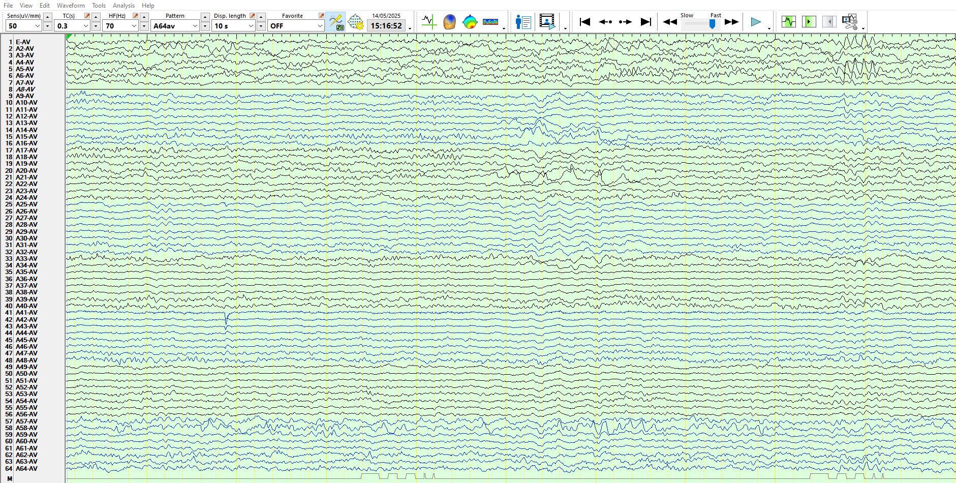
The patient is awake. There are rare spikes at 15, 21, located adjacent to one another in the right inferior, lateral frontal region. Little surprise that the scalp EEG recording was normal during wakefulness
Intracranial EEG substrates of scalp EEG interictal spikes - PubMed
Rare spikes at 19 and independently at 34,33, 20
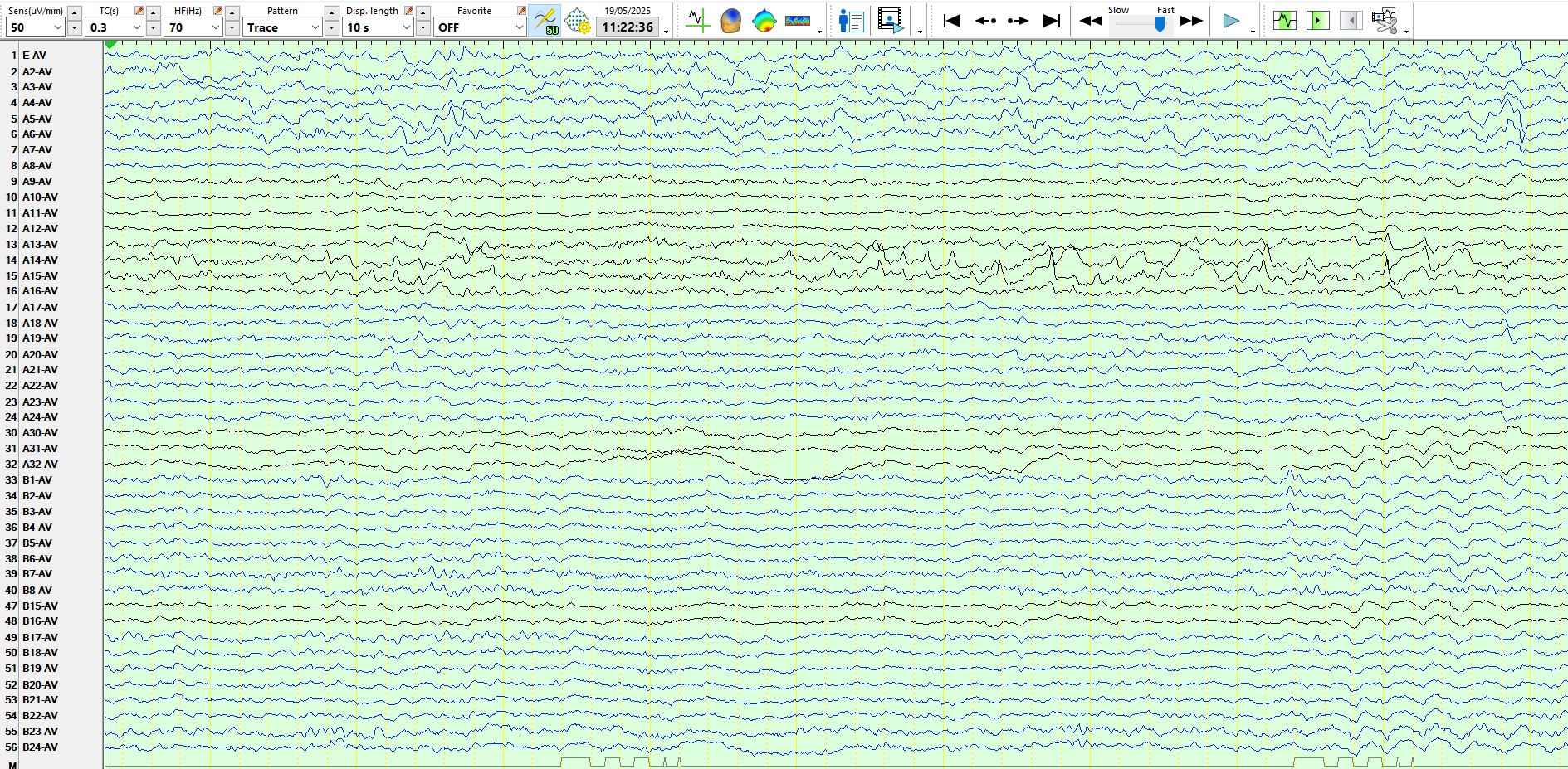
Trains of spikes and theta waves at 15,14,13 (The inferior aspect of the right frontal convexity). Not likely to be seen on scalp EEG
Normal alpha rhythms located over the right inferior and lateral temporal region during wakefulness (2-7). This "normal alpha" is also seen on scalp EEG recordings, as referred to elsewhere on these posts.
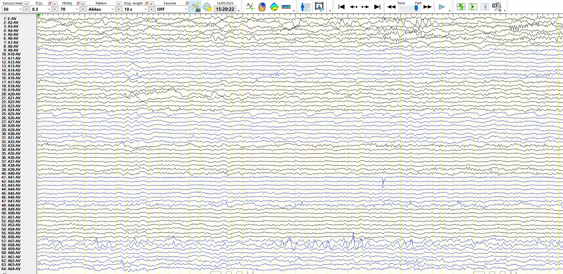
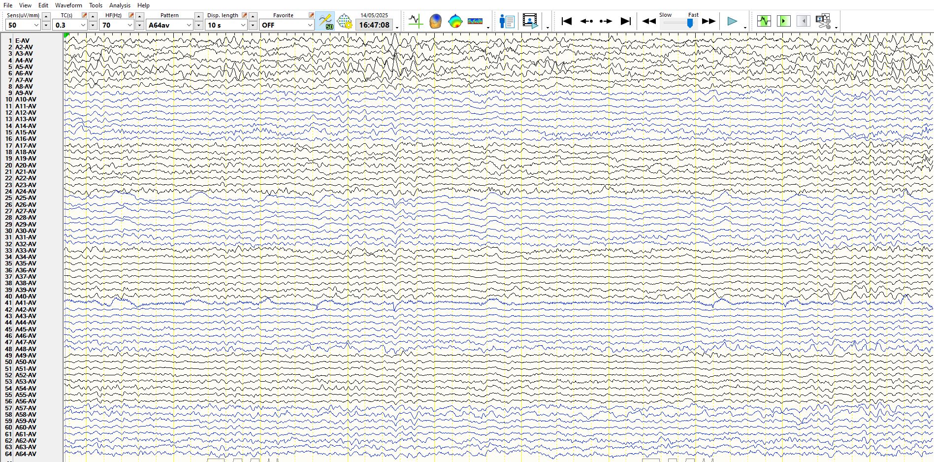
Subdural EEG, awake, background rhythms are normal, single spike at a single electrode located over the right inferior, lateral frontal region (channel 15), no chance of seeing this on scalp EEG, given its highly restricted spatial extent
Normal spindles and Vertex waves, 26-32, located over the right central convexity
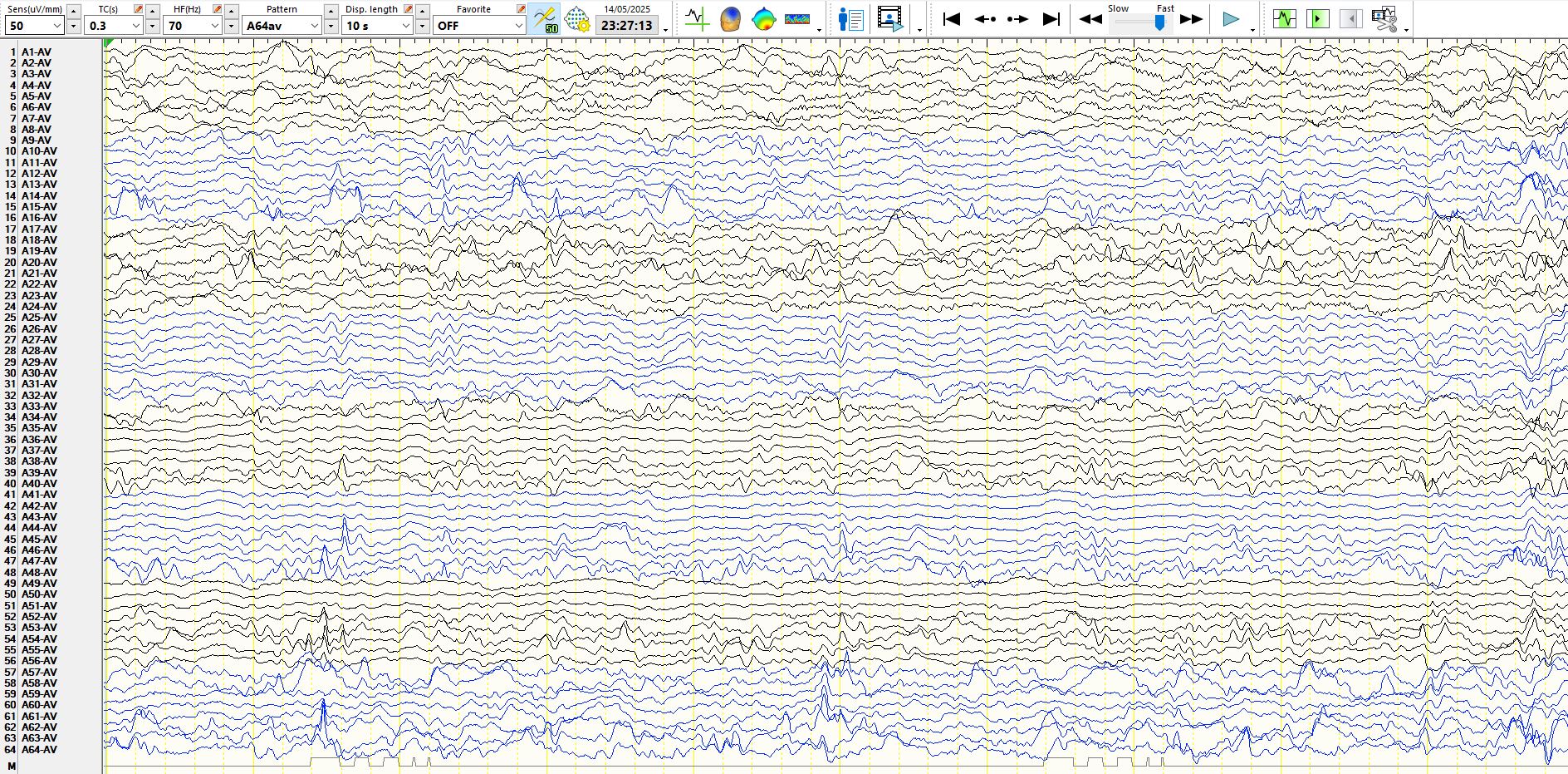
Abnormal subdural EEG, spikes and delta waves during wakefulness
Intracranial EEG substrates of scalp EEG interictal spikes - PubMed
Spikes in sleep at 18, 19
spike at 63, 62 during sleep
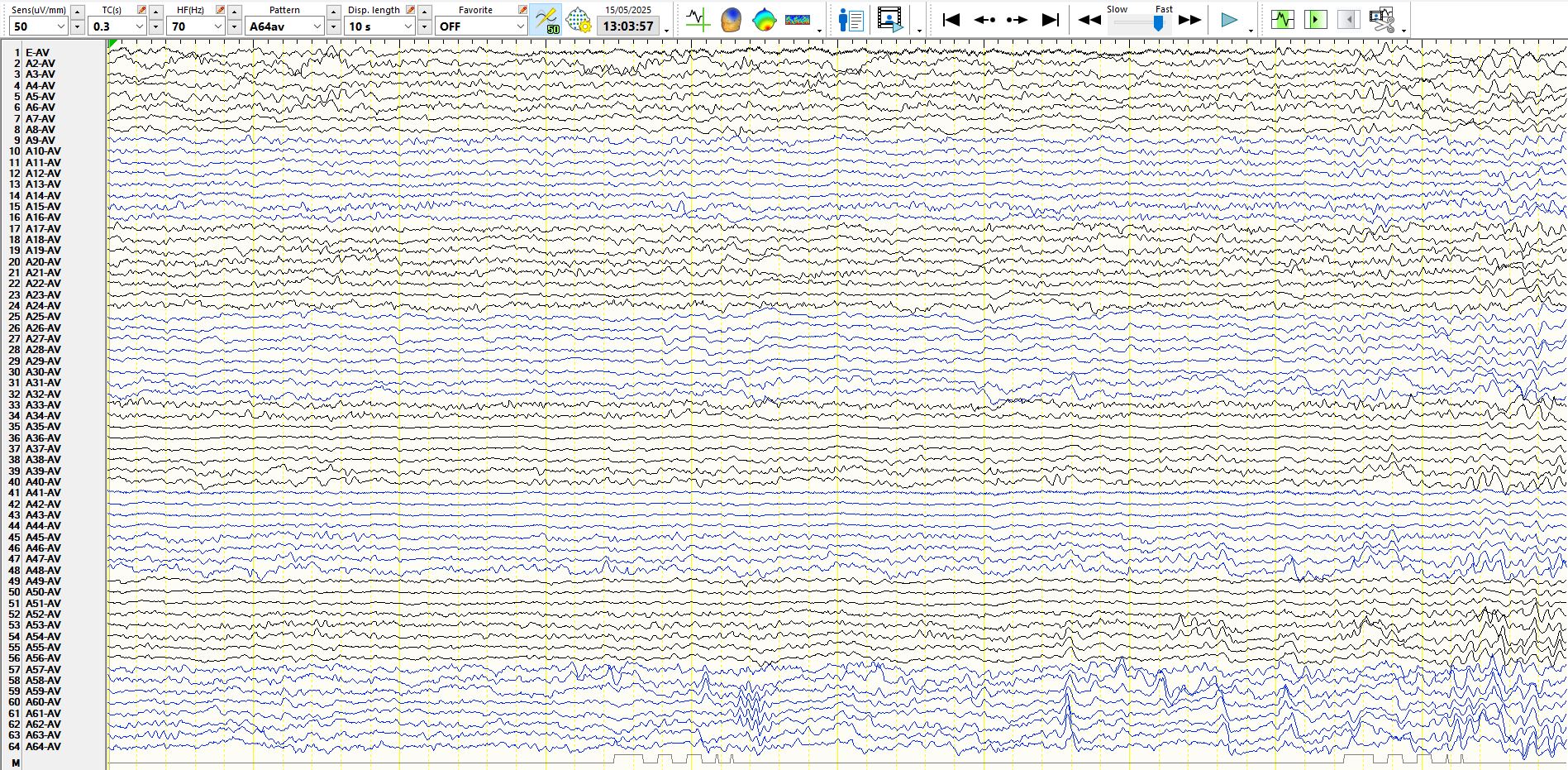
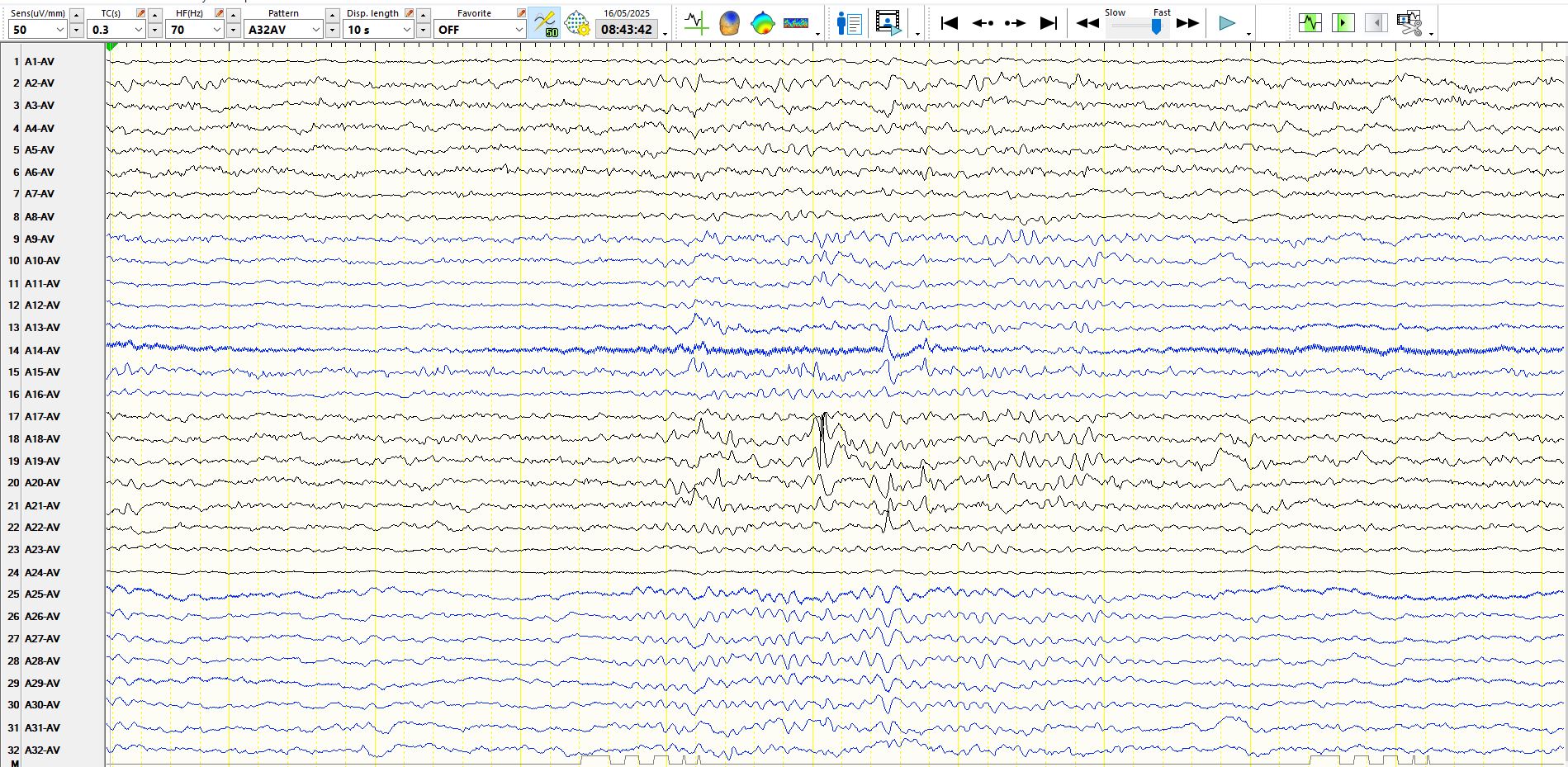
Localised spike on the left of the page, not likely to be seen on scalp EEG. Notice the more widely synchronous spike on the right at about 8s; such discharges may summate sufficiently to be seen on scalp EEG. Note the dipole at electrodes 9-16 (electropositive at 9, 10, electronegative 12-16). Even though the scalp EEG was normal during wakefulness and sleep, at times there were frequent, multifocal right frontal spikes during sleep on subdural EEG
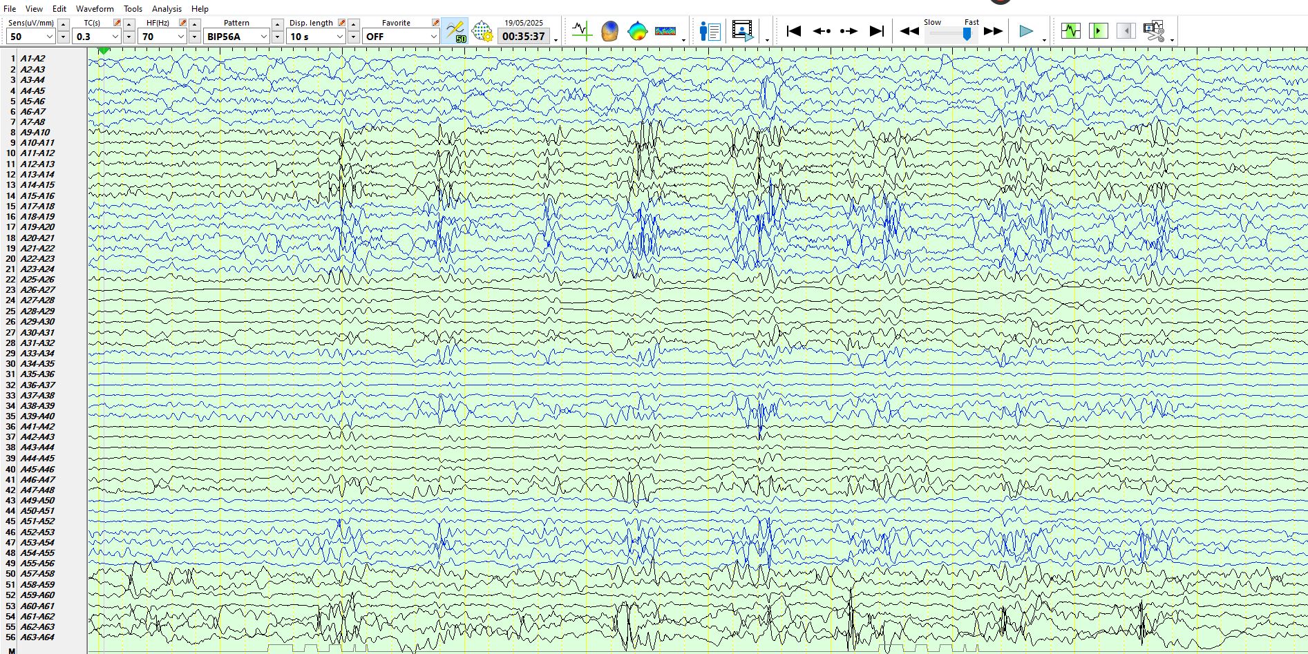
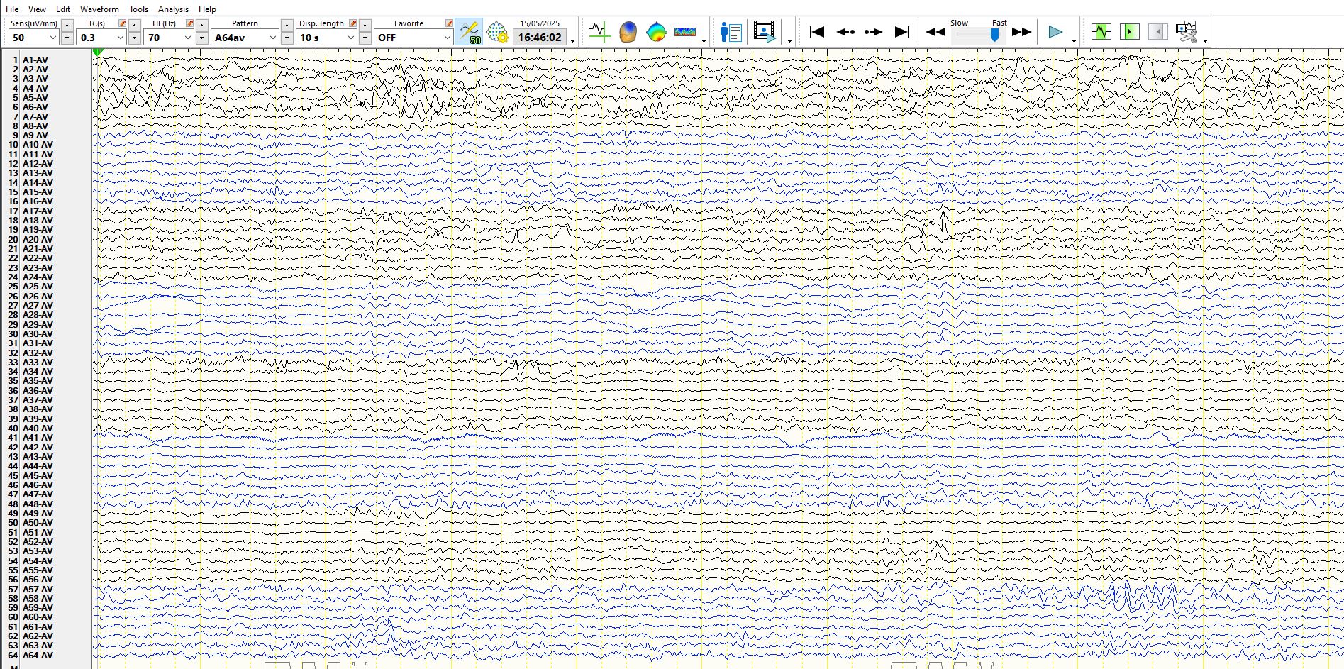
Remarkably even these high amplitude widely synchronous, frequent, periodic (at times) polyspikes in sleep are not seen on scalp EEG
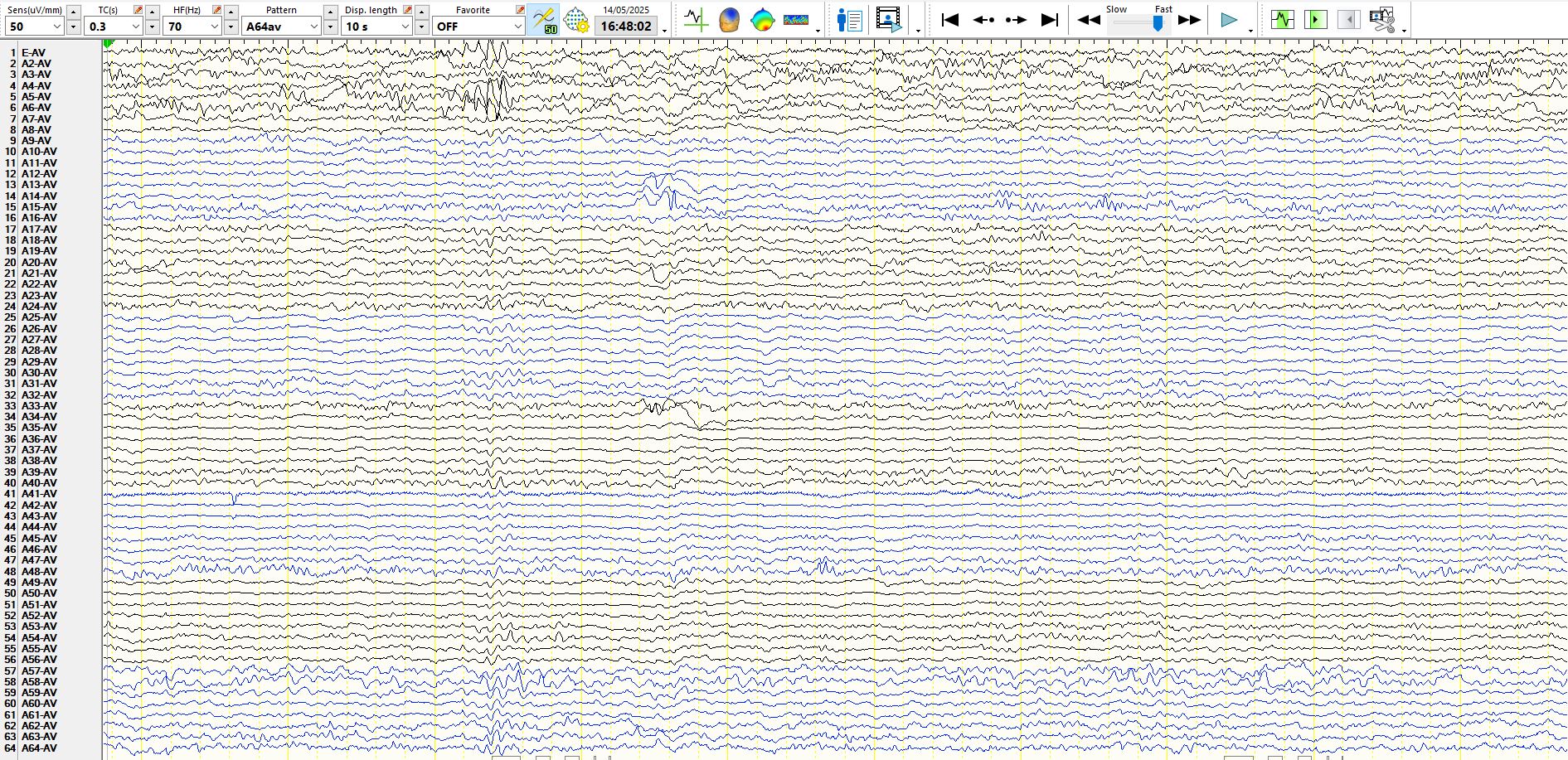
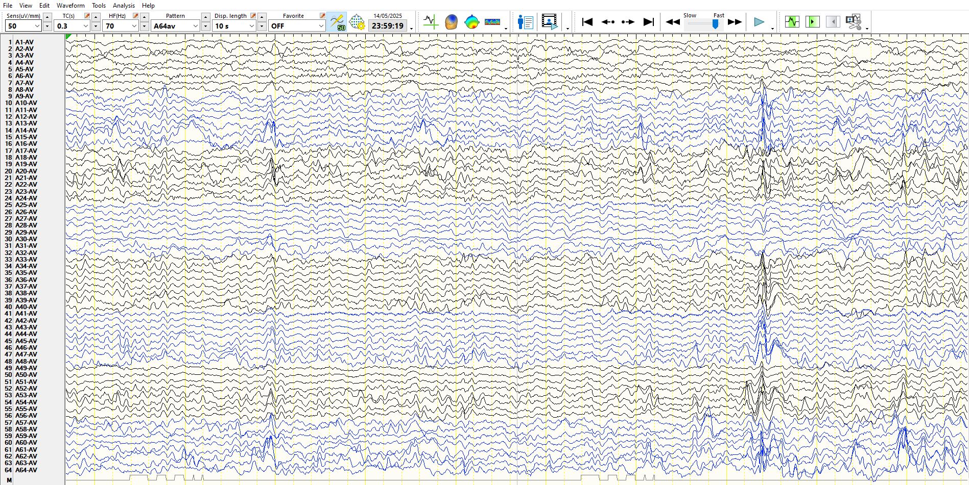
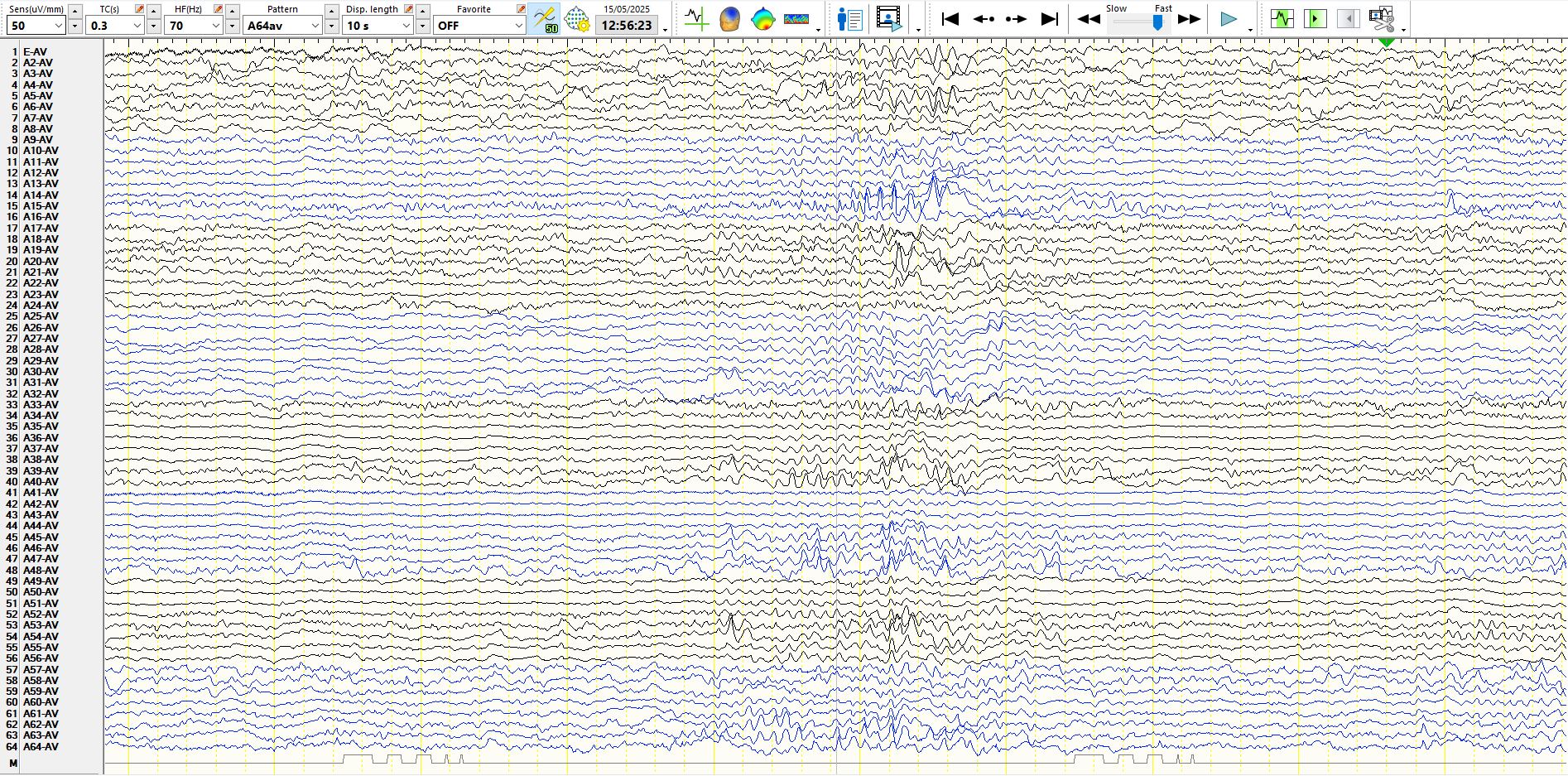 Highly localised, probable electrographic seizure on subdural EEG at 15, 14, located over the right inferior aspect of the lateral frontal region, no chance of seeing this on scalp EEG. Notice the gradual increase in amplitude, slowing in frequency and the subsequent slow-wave. This is unlike any background rhythm seen during the recording in the same region.
Bottom line?
The scalp EEG recording may be normal in someone with epilepsy, both during the inter-ictal and during seizures, despite there being a plethora of spikes on subdural EEG